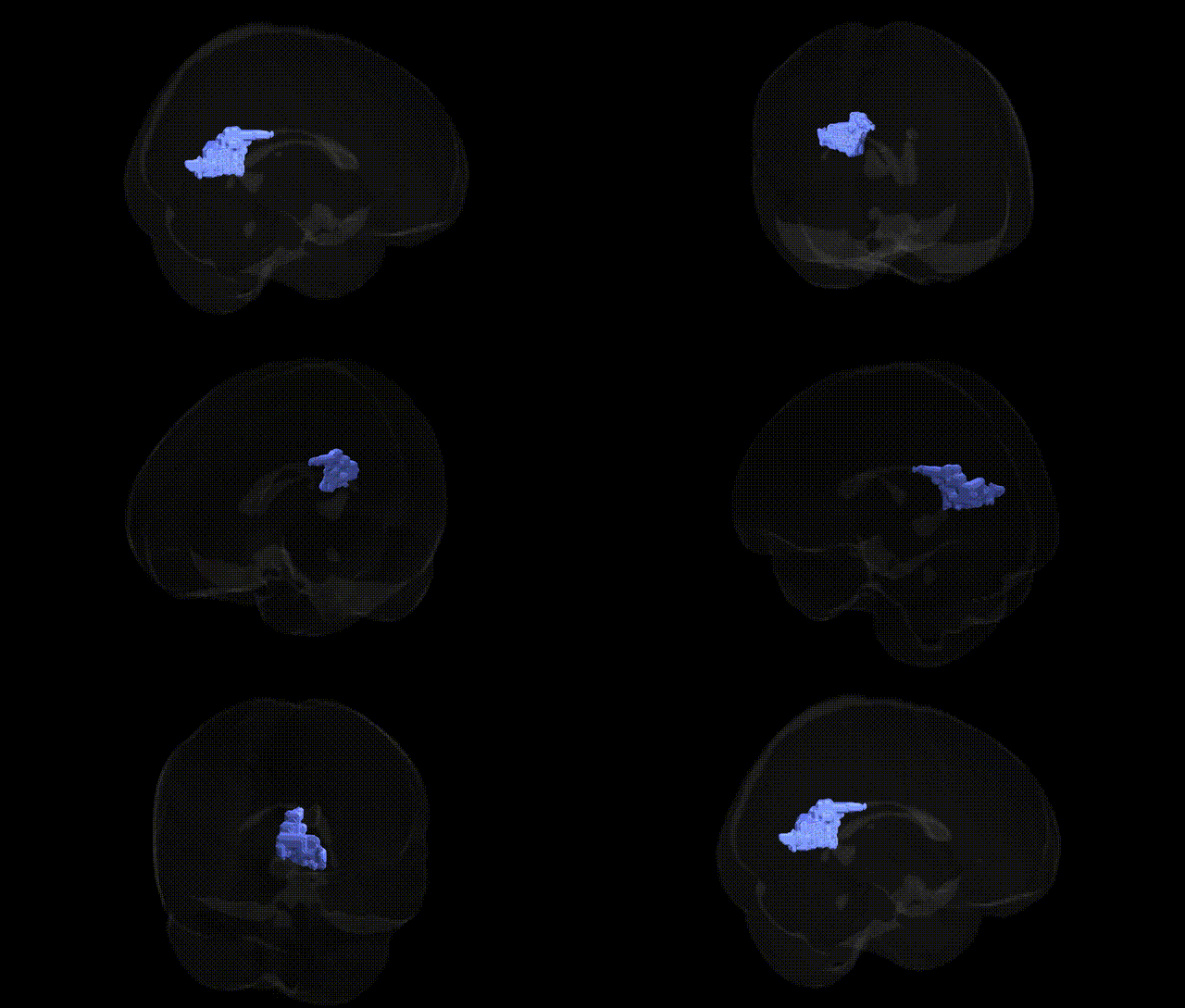
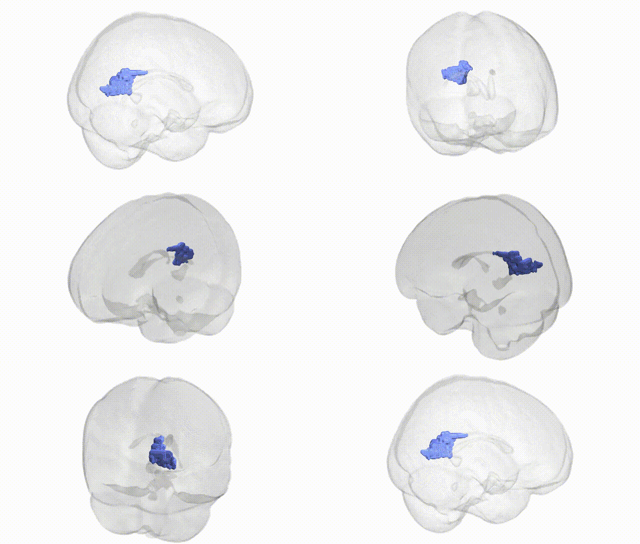
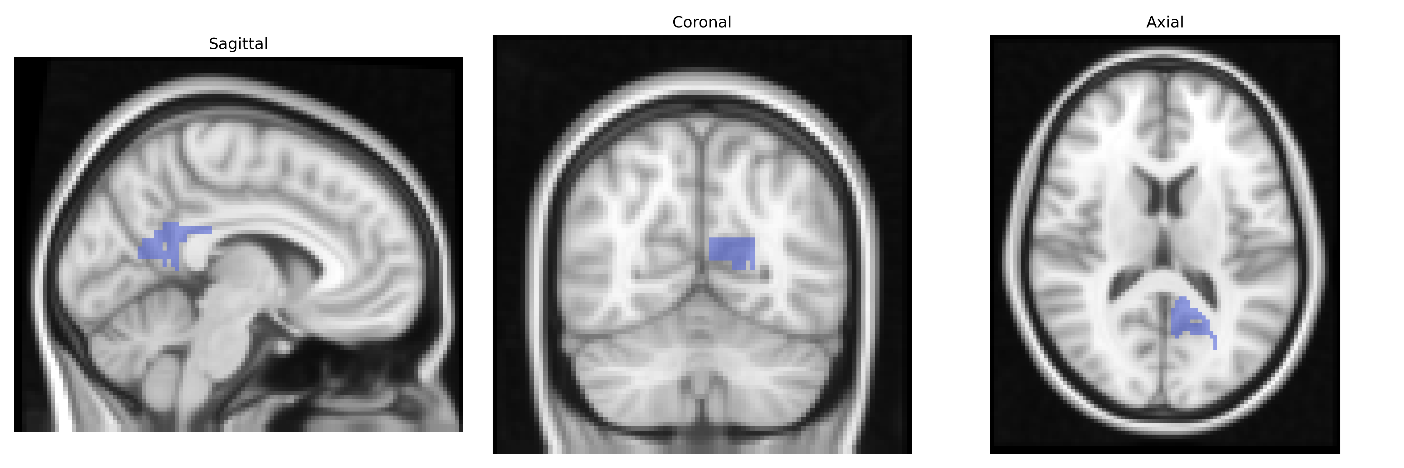
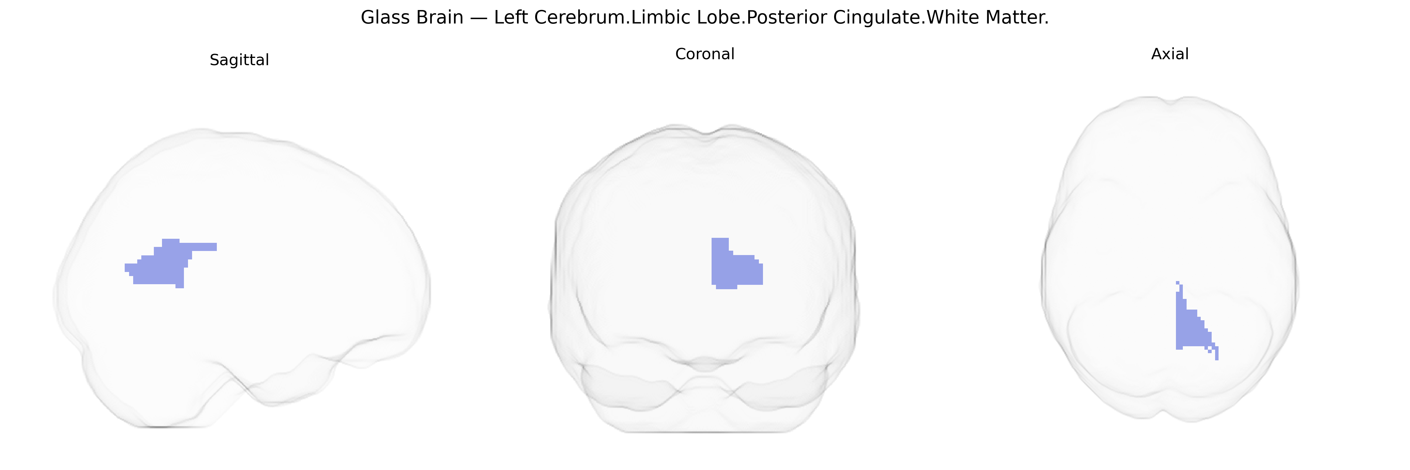
The bilateral Left Cerebrum.Limbic Lobe.Posterior Cingulate.White Matter corresponds to the subcortical myelinated fiber pathways underlying the posterior segment of the cingulate gyrus within the limbic lobe of the left cerebral hemisphere, with homologous regions in the right hemisphere. This white matter region supports connectivity between posterior cingulate cortex and distributed networks including medial parietal, medial prefrontal, and medial temporal structures, contributing to functions such as internally directed cognition, memory processing, and aspects of the default mode network. As a predominantly association fiber zone, it carries long-range cortico-cortical projections and forms part of larger tracts such as the cingulum bundle, integrating limbic and heteromodal association areas. There is no direct Wikipedia article for the exact Talairach label; a closely related structure is the Posterior cingulate cortex.
The bilateral Left Cerebrum Limbic Lobe Posterior Cingulate White Matter region, as defined in the Talairach 2 mm atlas, is part of the posterior cingulate cortex (PCC) connectivity hub, and genetic associations are typically inferred from imaging genetics and GWAS of PCC structure, function, or network-level measures rather than this specific white-matter parcel. Variants in APOE (especially APOE ε4) and genes involved in amyloid processing and tau pathology (e.g., CLU, CR1, PICALM, BIN1) have been linked through GWAS and imaging studies to altered PCC volume, metabolism, connectivity, and early Alzheimer’s disease–related hypometabolism, reflecting vulnerability of PCC white matter tracts within the default mode network. Polygenic risk scores for Alzheimer’s disease, schizophrenia, and major depressive disorder show associations with posterior cingulate functional connectivity and white-matter microstructure, implicating synaptic, immune, and myelination-related genes (including loci near CACNA1C, GRIN2A, and complement pathway genes). Large imaging GWAS (e.g., UK Biobank) have identified common variants influencing white-matter integrity metrics (FA, MD) in cingulum bundle and adjacent PCC-associated tracts, pointing to genes involved in axonal guidance, oligodendrocyte function, and neurodevelopment (such as NRG1/ERBB signaling and cell-adhesion genes), which indirectly map onto this Talairach-defined region. Overall, genetic findings emphasize that PCC white matter is a convergence zone where neurodegenerative and psychiatric risk variants manifest as changes in connectivity and microstructure, but fine-grained, atlas-specific genetic associations for the exact “Left Cerebrum.Limbic Lobe.Posterior Cingulate.White Matter” label remain limited, with evidence largely derived from broader PCC or cingulum-based phenotypes.
Overview generated by GPT-4o (2026).
Region ID: 662
Hemisphere: bilateral
Atlas: Talairach labels 2mm
Full Quality Version: Download MP4
Full Quality Version: Download MP4
Wali Sidiqyar*, Gaurav Rudravaram*, Elyssa M. McMaster, Trent M. Schwartz, Adam M. Saunders, Kurt G. Schilling, Bennett A. Landman "Introducing SPINS: A Shared Public Visualization Library of Neuroanatomical Structures." Medical Imaging with Deep Learning- short paper
This resource is licensed under CC0 1.0 Universal (Public Domain).