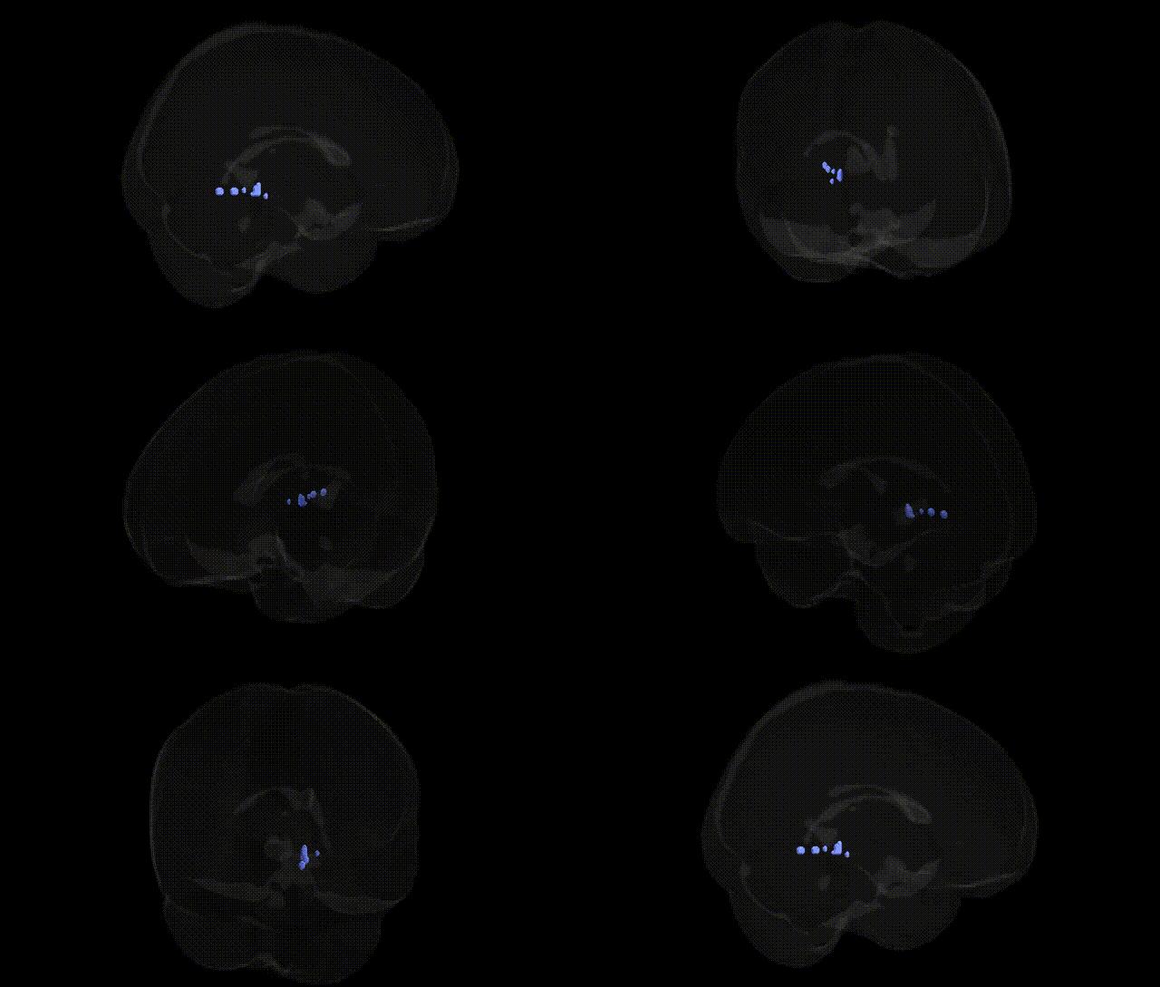
The bilateral Left Cerebrum.Limbic Lobe.Sub-Gyral region, as defined in the Talairach 2 mm atlas, refers to subcortical white matter located beneath the cortical gyri of the limbic lobe in the left hemisphere, encompassing fiber pathways that interconnect key limbic structures such as the cingulate gyrus, parahippocampal region, and related medial temporal and frontal areas. Functionally, this sub-gyral limbic white matter supports the integration and transmission of information involved in emotion, memory, motivation, and autonomic regulation by linking cortical limbic areas with deeper structures including the hippocampus, amygdala, and hypothalamus. Damage or dysfunction in this region can disrupt limbic circuitry, potentially affecting emotional processing, episodic memory, and affective regulation. There is no direct Wikipedia article for this specific Talairach label; a closely related structure is the Limbic system.
The bilateral left cerebrum limbic lobe sub-gyral territory in the Talairach 2 mm atlas primarily overlaps medial temporal and limbic structures (dominated by hippocampal and parahippocampal subcortical gray matter), so genetic associations largely reflect findings for these canonical limbic regions rather than the specific “sub-gyral” label. GWAS and imaging‑genetics studies have repeatedly linked common variants in genes such as APOE (especially ε4), BIN1, CLU, CR1, ABCA7, SORL1, TOMM40, and MS4A cluster genes to structural differences and neurodegeneration in medial temporal limbic areas in Alzheimer’s disease. Schizophrenia and bipolar disorder GWAS—including variants in CACNA1C, ZNF804A, MIR137, and multiple synaptic and immune‑related loci—have been associated with altered limbic volumes and connectivity, particularly in hippocampal/parahippocampal segments encompassed by this label. Large consortia (e.g., ENIGMA) have identified polygenic influences on hippocampal and amygdala volume, with genome‑wide significant loci near HRK, ASTN2, DPP4, and FAF1, and broad polygenic risk scores for major depression, PTSD, and anxiety disorders show correlations with limbic morphometry and function. Candidate gene and GWAS data for stress and emotion‑related traits (e.g., variants in BDNF, FKBP5, SLC6A4, CRHR1, and COMT) have been associated with altered activation and microstructure in medial limbic circuitry during fear, memory, and reward processing, while addiction‑related loci (e.g., OPRM1, CHRNA5–CHRNA3–CHRNB4) show imaging‑genetic links to limbic responses to drug cues. Overall, genetic associations for this Talairach region reflect its inclusion in the broader medial temporal limbic network implicated in neurodegenerative disease, psychosis, mood and anxiety disorders, stress reactivity, and memory‑related traits, rather than region‑specific GWAS signals uniquely tied to the “sub-gyral” label.
Overview generated by GPT-4o (2026).
Region ID: 336
Hemisphere: bilateral
Atlas: Talairach labels 2mm
Full Quality Version: Download MP4
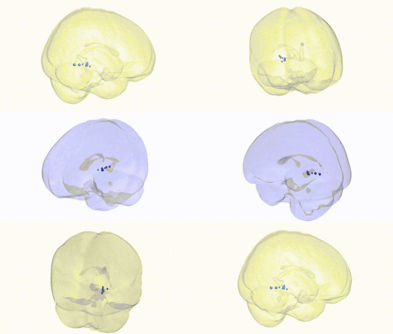
Full Quality Version: Download MP4
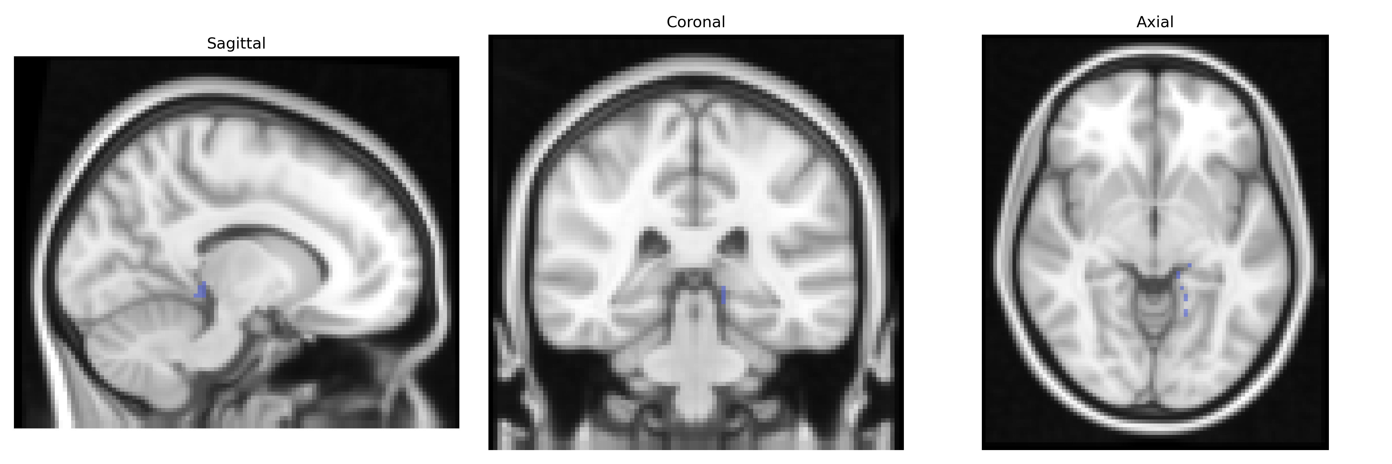
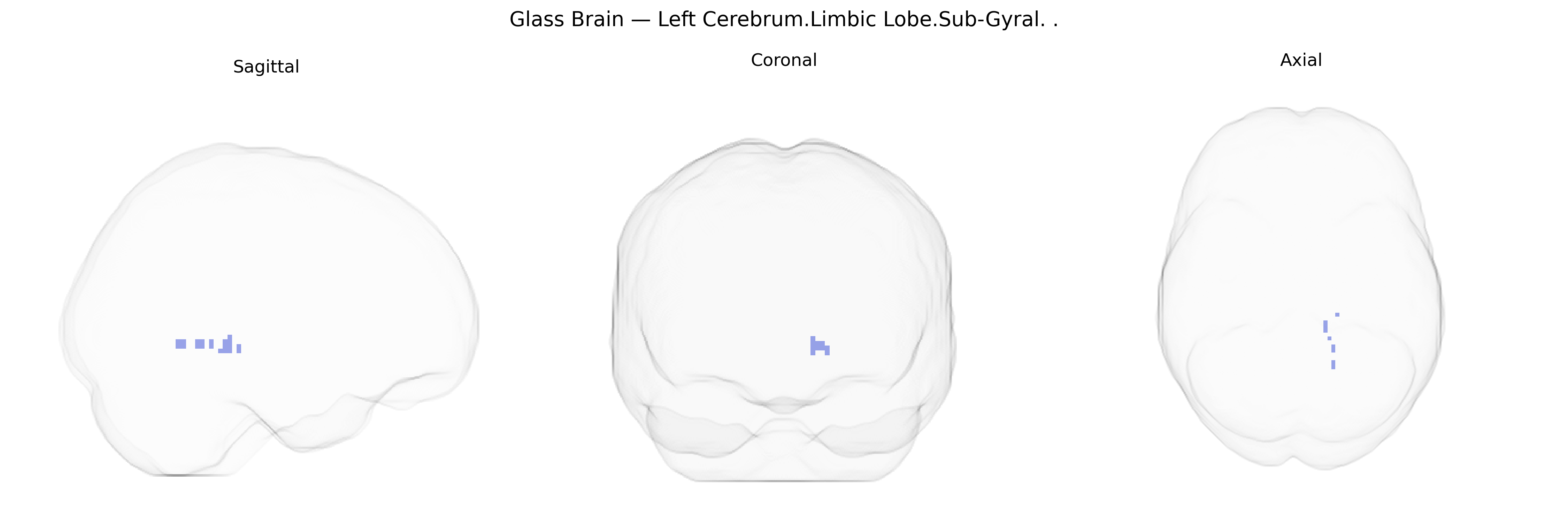
Wali Sidiqyar*, Gaurav Rudravaram*, Elyssa M. McMaster, Trent M. Schwartz, Adam M. Saunders, Kurt G. Schilling, Bennett A. Landman "Introducing SPINS: A Shared Public Visualization Library of Neuroanatomical Structures." Medical Imaging with Deep Learning- short paper
This resource is licensed under CC0 1.0 Universal (Public Domain).