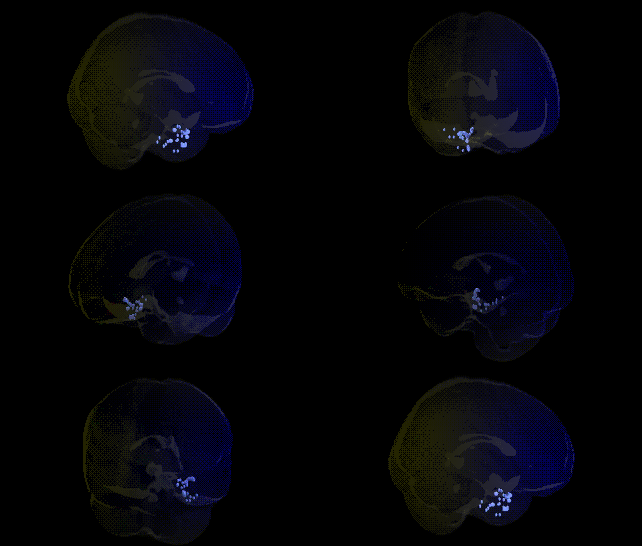
The bilateral Left Cerebrum.Limbic Lobe.Uncus corresponds to the left uncus, a hook-shaped medial temporal lobe structure forming part of the parahippocampal gyrus and closely associated with the limbic system. It overlies the amygdala and is involved in olfactory processing, emotional evaluation of sensory input, and aspects of memory, particularly through its connections with the hippocampus and other limbic structures. The uncus is a landmark in neuroanatomy because of its proximity to critical medial temporal structures and its clinical relevance in uncal herniation, where medial temporal tissue can compress the brainstem. There is no direct link for “Uncus” as a standalone article; a related and encompassing structure is the Parahippocampal gyrus.
The left limbic lobe uncus, a medial temporal structure encompassing anterior parahippocampal and amygdaloid regions, is frequently implicated in genetic studies of epilepsy, neuropsychiatric disorders, and memory-related traits, although few GWAS target it specifically as a stand-alone ROI. Structural and functional variation in the uncus falls within broader medial temporal or hippocampal–amygdala phenotypes in imaging genetics, where common variants in genes such as BDNF, APOE, and those within the MAPT region have been associated with medial temporal lobe volume, connectivity, and susceptibility to Alzheimer’s disease and other dementias that affect uncinate and parahippocampal regions. GWAS of focal epilepsies and temporal lobe epilepsy have identified polygenic risk involving loci near SCN1A, GRIN2A, LGI1, and other synaptic/ion channel genes, conditions in which the uncus is a frequent site of seizure onset or structural abnormality. Psychiatric genetics studies of anxiety, depression, bipolar disorder, and schizophrenia have linked risk variants (e.g., in CACNA1C, ANK3, and multiple immune-related loci from the MHC region) to altered medial temporal and amygdalo-hippocampal structure and reactivity, networks that include the uncus and uncinate fasciculus. GWAS of brain imaging phenotypes from large cohorts (e.g., UK Biobank, ENIGMA) report associations between polygenic scores for cognitive ability, neuroticism, and PTSD with volumetric and cortical thickness measures in limbic temporal regions overlapping the uncus, suggesting that common genetic variation influencing synaptic plasticity, neurodevelopment, and neuroinflammation contributes to inter-individual differences in uncus morphology and function, which in turn modulate risk for seizure disorders, memory dysfunction, and affective psychopathology.
Overview generated by GPT-4o (2026).
Region ID: 17
Hemisphere: bilateral
Atlas: Talairach labels 2mm
Full Quality Version: Download MP4
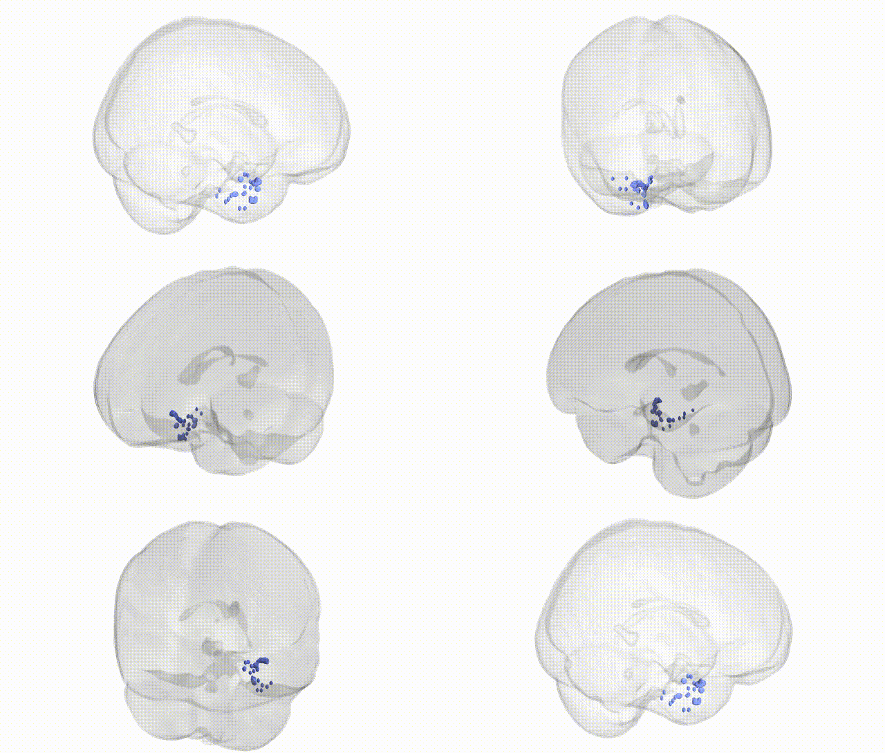
Full Quality Version: Download MP4
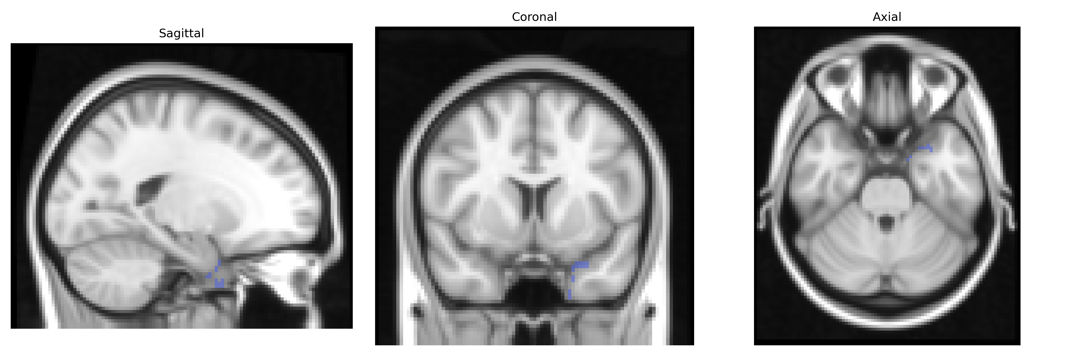
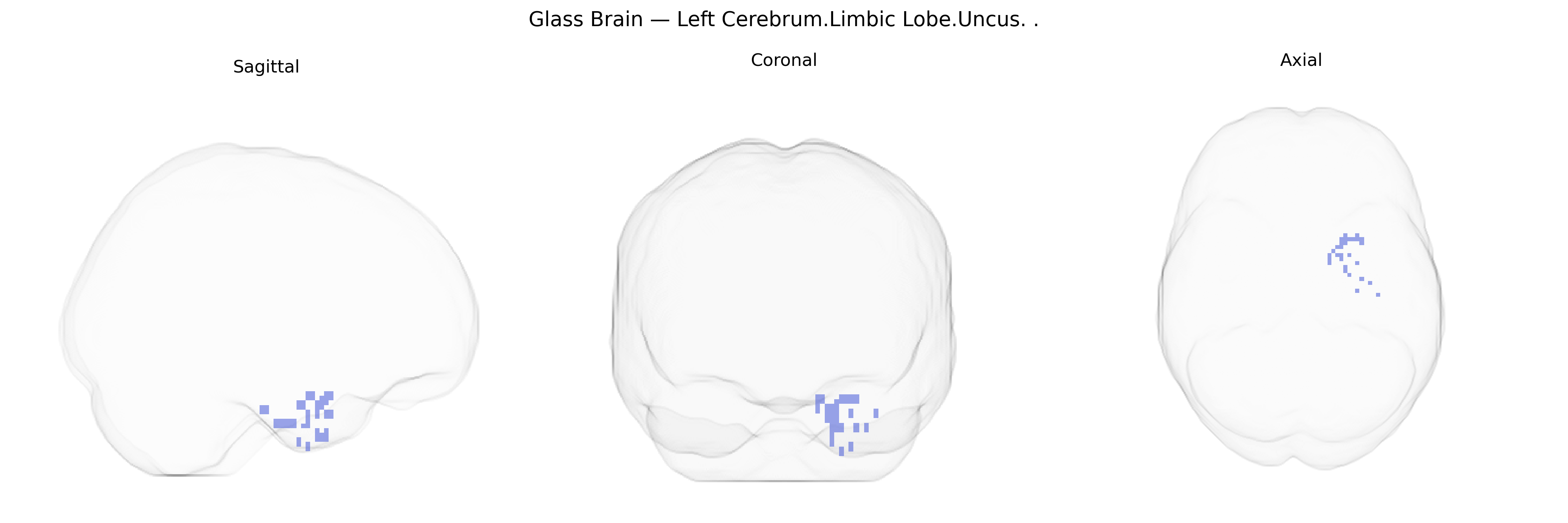
Wali Sidiqyar*, Gaurav Rudravaram*, Elyssa M. McMaster, Trent M. Schwartz, Adam M. Saunders, Kurt G. Schilling, Bennett A. Landman "Introducing SPINS: A Shared Public Visualization Library of Neuroanatomical Structures." Medical Imaging with Deep Learning- short paper
This resource is licensed under CC0 1.0 Universal (Public Domain).