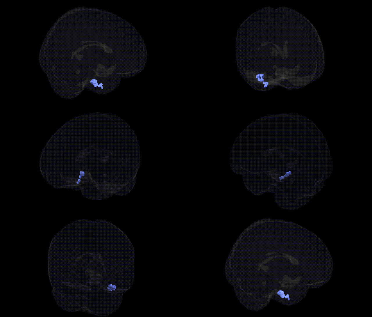
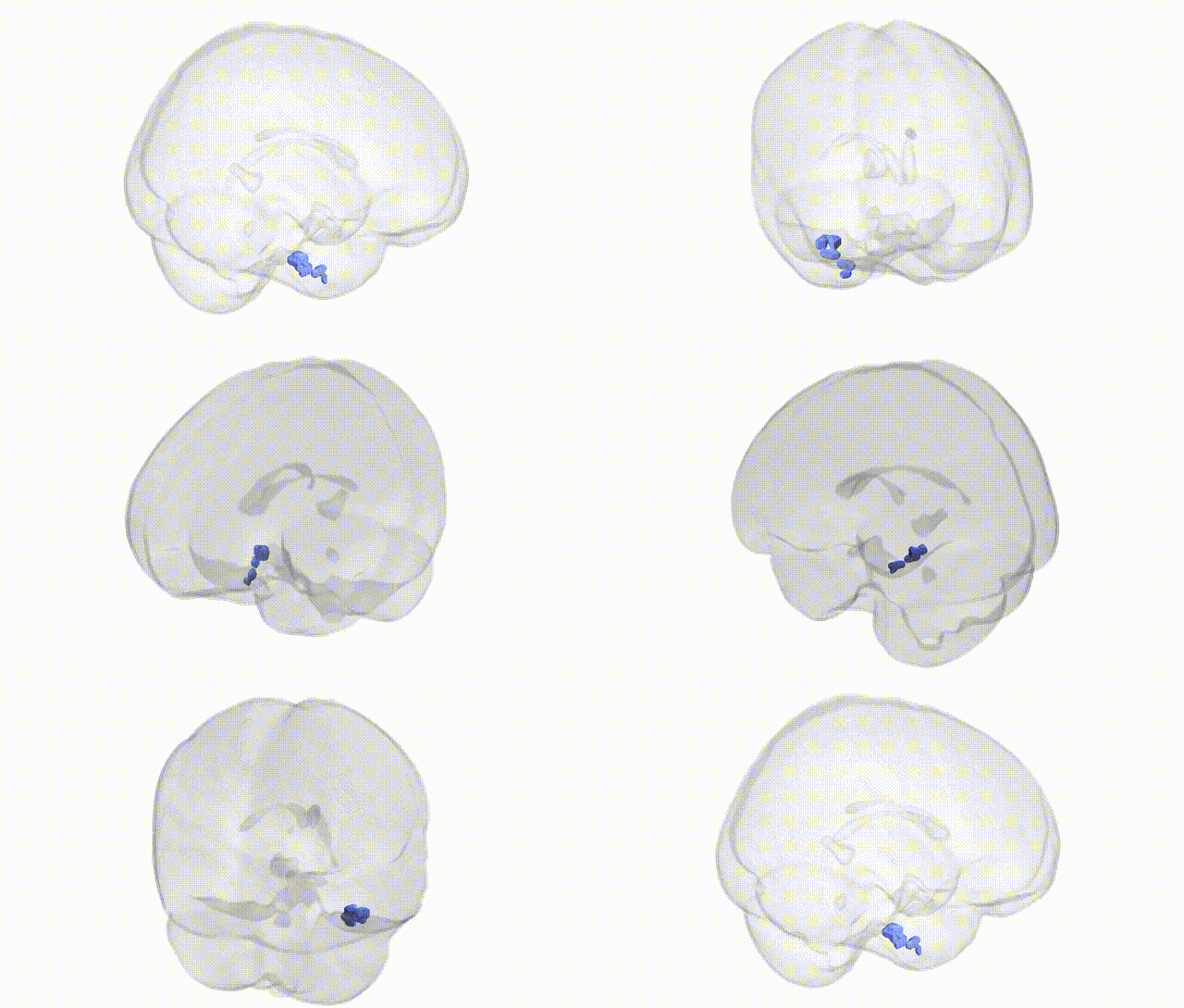
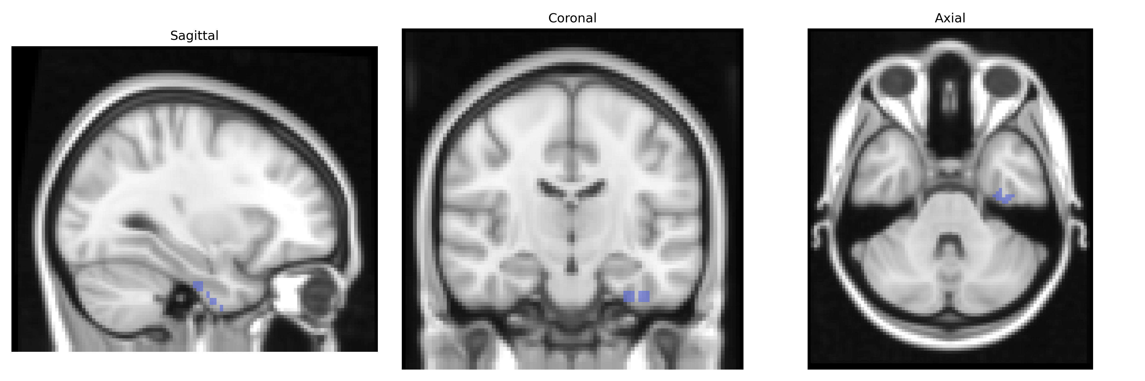
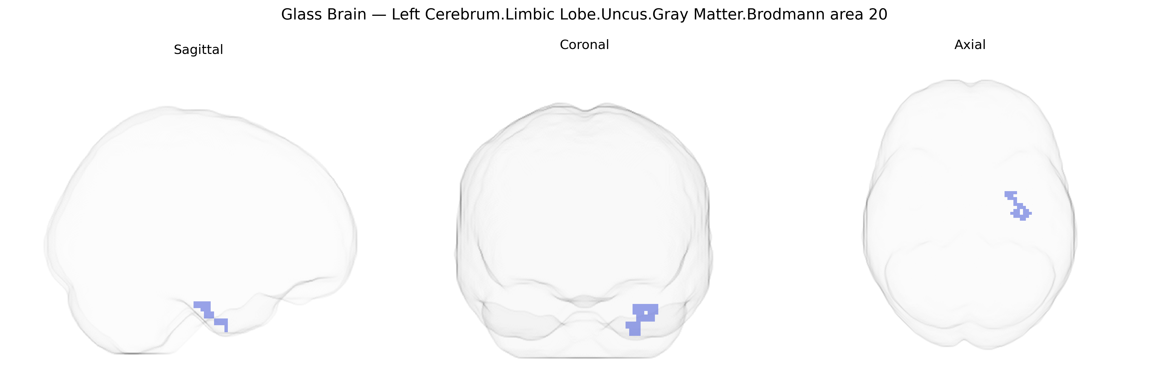
The bilateral Left Cerebrum.Limbic Lobe.Uncus.Gray Matter.Brodmann area 20 region corresponds to cortical territory in the anterior medial temporal lobe where the uncus (a medial protrusion of the parahippocampal gyrus) overlies and neighbors portions of the temporal association cortex classified cytoarchitectonically as Brodmann area 20. Functionally, this region participates in high-level visual object recognition and semantic processing (BA20) while being closely integrated with limbic structures involved in olfaction, memory, and emotional evaluation (uncus, including its relationships to the amygdala and hippocampal formation). Through extensive reciprocal connections with the hippocampus, amygdala, and other temporal and frontal association areas, this territory supports the linkage of complex visual inputs to stored representations and affective significance, contributing to processes such as recognition of familiar objects, contextual memory, and the emotional coloring of sensory experiences. There is no direct link for this exact composite region; a closely related structure is the Uncus.
The bilateral left cerebrum limbic lobe uncus (gray matter) overlapping Brodmann area 20 lies at the anterior medial temporal cortex, integrating aspects of high-level visual processing with limbic functions such as memory, emotion, and olfactory processing, and genetic associations largely reflect broader medial/anterior temporal and limbic circuitry rather than this precise Talairach label. Genome-wide association studies implicating nearby medial temporal and anterior ventral temporal regions highlight variants in genes involved in synaptic plasticity, neurodevelopment, and glutamatergic signaling (e.g., BDNF, GRIN2B, and other neurotrophin/PSD-related genes), with structural and functional measures of medial temporal lobe volume or cortical thickness often serving as imaging endophenotypes. Risk loci for temporal lobe epilepsy frequently implicate hippocampal–amygdala–uncal networks and genes involved in neuronal excitability (e.g., SCN1A and other ion channel genes), and medial/anterior temporal abnormalities—sometimes including uncus involvement—are common in genetic epilepsies. In Alzheimer’s disease, APOE and polygenic risk scores correlate with medial temporal atrophy and connectivity changes that can extend to entorhinal and uncinate regions, while schizophrenia and major depressive disorder GWAS point to distributed polygenic influences on limbic–temporal circuits rather than single-region specificity, with genes related to synaptic function (e.g., CACNA1C, GRM3, and others) repeatedly implicated in network-level dysfunction encompassing anterior temporal cortex. Autism spectrum and social cognition GWAS also show polygenic effects on temporal and limbic regions, including fusiform and anterior temporal areas adjacent to BA20, although fine-grained localization to the uncus/BA20 combination remains limited and most genetic findings are interpreted at the broader medial/anterior temporal or limbic network level rather than this exact Talairach-defined parcel.
Overview generated by GPT-4o (2026).
Region ID: 16
Hemisphere: bilateral
Atlas: Talairach labels 2mm
Full Quality Version: Download MP4
Full Quality Version: Download MP4
Wali Sidiqyar*, Gaurav Rudravaram*, Elyssa M. McMaster, Trent M. Schwartz, Adam M. Saunders, Kurt G. Schilling, Bennett A. Landman "Introducing SPINS: A Shared Public Visualization Library of Neuroanatomical Structures." Medical Imaging with Deep Learning- short paper
This resource is licensed under CC0 1.0 Universal (Public Domain).