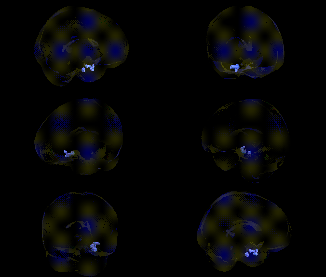
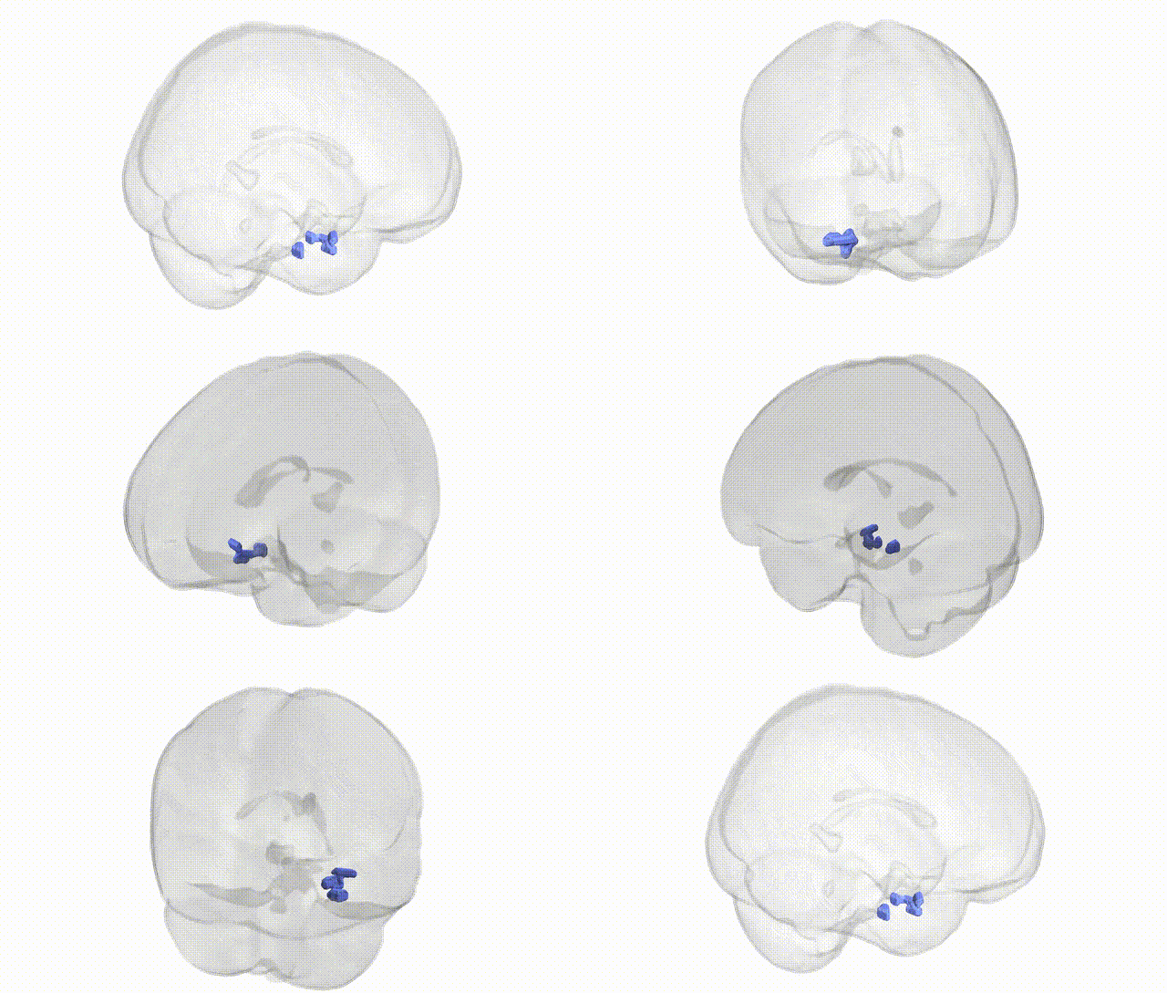
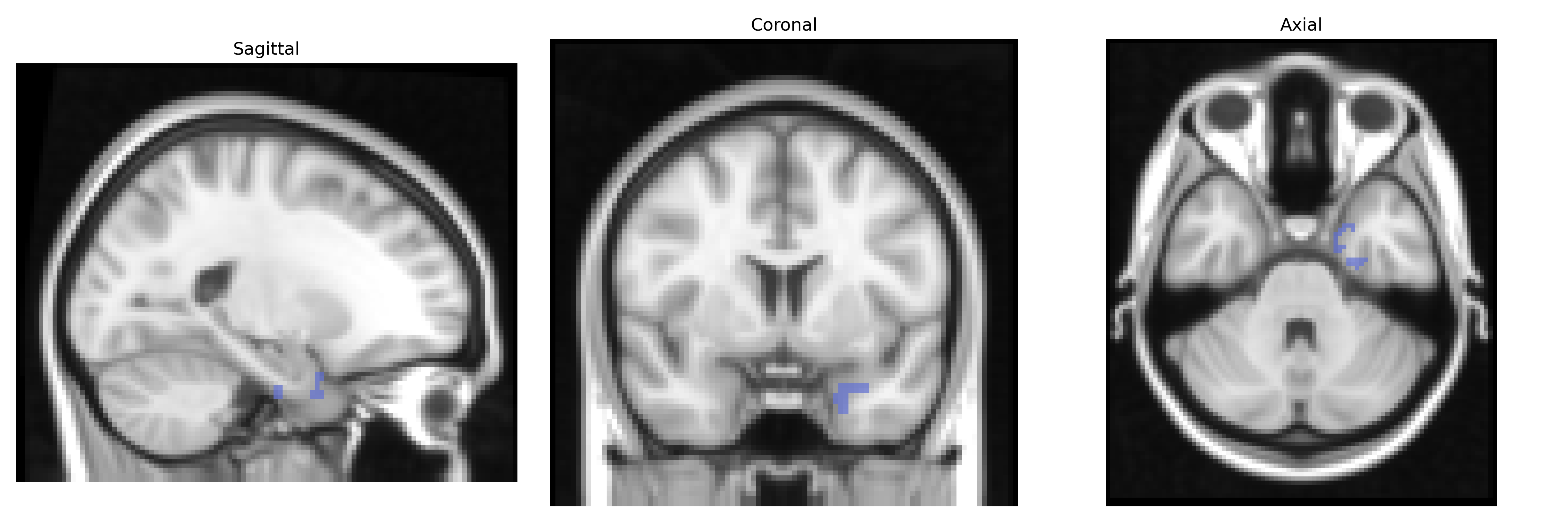
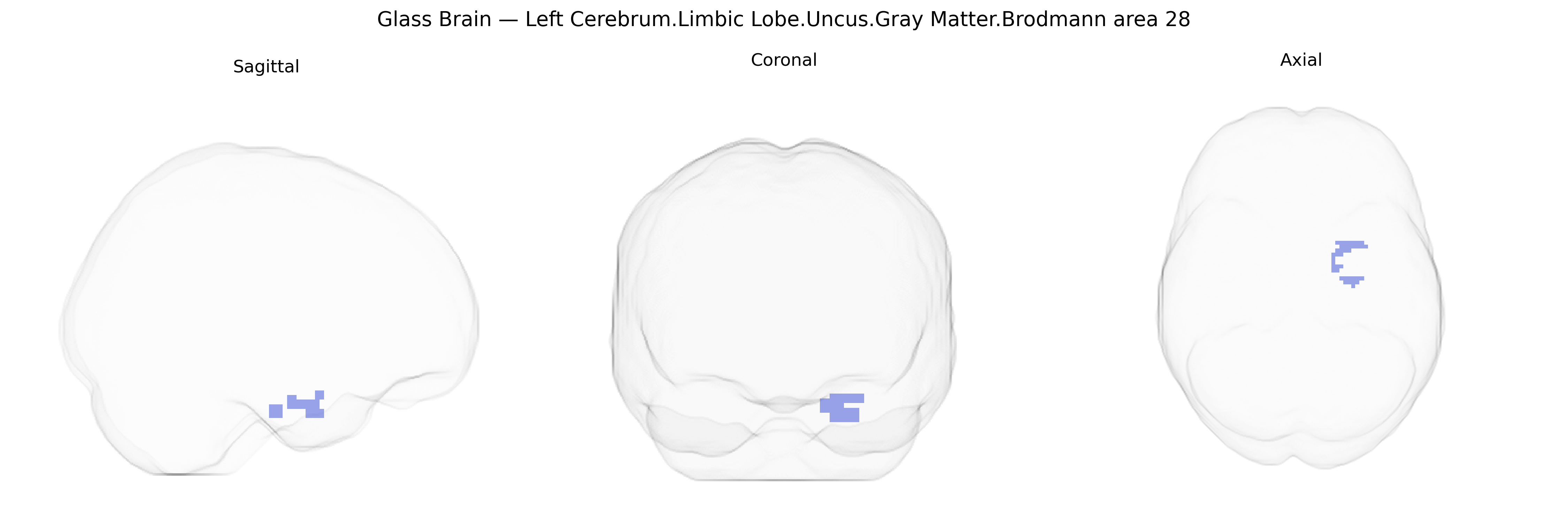
The bilateral Left Cerebrum.Limbic Lobe.Uncus.Gray Matter.Brodmann area 28 corresponds to the entorhinal cortex located in the medial temporal lobe, specifically within the anterior parahippocampal gyrus forming part of the uncus. Brodmann area 28 serves as a principal interface between neocortical association areas and the hippocampal formation, receiving highly processed multimodal sensory input and providing major output pathways to the dentate gyrus, hippocampus, and subiculum. This region is critically involved in episodic and spatial memory, navigation, and the consolidation of long-term memories, and it plays an early and central role in neurodegenerative processes such as Alzheimer’s disease, where entorhinal layer II neurons are particularly vulnerable. Histologically, BA28 is characterized by a distinct laminar organization with relatively sparse granular cells compared to nearby cortices, reflecting its transitional allocortical architecture. Entorhinal cortex
The bilateral left cerebrum limbic lobe uncus (Brodmann area 28, entorhinal cortex) is a medial temporal structure central to memory and emotion, with genetic associations largely inferred from imaging genetics and GWAS of hippocampal–entorhinal circuitry rather than region-specific studies. Variants in genes related to Alzheimer’s disease risk and tau/amyloid pathology (e.g., APOE, BIN1, CLU, PICALM, SORL1, and MAPT) show robust associations with entorhinal and medial temporal cortical atrophy, accelerated thinning, and neurodegeneration, reflecting the region’s role as an early site of Alzheimer’s pathology. Large neuroimaging GWAS have linked loci involved in neurodevelopment and synaptic function (such as BDNF, CELF4, and genes in glutamatergic and GABAergic pathways) to volumetric and thickness measures across the medial temporal lobe, including entorhinal and adjacent uncus/BA28 territory. Genetic studies of temporal lobe epilepsy implicate variants in ion channel and synaptic genes (e.g., SCN1A, GABRA2, GRIN2A, LGI1), consistent with seizure foci frequently involving the uncus and adjacent hippocampus. Psychiatric GWAS for major depressive disorder, schizophrenia, bipolar disorder, and anxiety-related traits have repeatedly highlighted polygenic influences on medial temporal and limbic structures, with risk alleles in synaptic, neurodevelopmental, and stress-response genes (e.g., CACNA1C, GRIN2A, CRHR1) associated with structural and functional alterations in entorhinal–uncal circuits. More recently, large-scale brain morphology GWAS (such as ENIGMA and UK Biobank) have identified multiple common variants influencing entorhinal/medial temporal surface area and thickness, many in loci regulating neuronal differentiation, axon guidance, and cortical patterning (e.g., genes near DLG2, MEF2C, and EPH receptor pathways), underscoring that genetic influences on this Brodmann area 28 region are strongly tied to neurodegenerative, epileptic, and affective–psychotic phenotypes via shared limbic and memory-related circuitry rather than isolated, region-specific loci.
Overview generated by GPT-4o (2026).
Region ID: 94
Hemisphere: bilateral
Atlas: Talairach labels 2mm
Full Quality Version: Download MP4
Full Quality Version: Download MP4
Wali Sidiqyar*, Gaurav Rudravaram*, Elyssa M. McMaster, Trent M. Schwartz, Adam M. Saunders, Kurt G. Schilling, Bennett A. Landman "Introducing SPINS: A Shared Public Visualization Library of Neuroanatomical Structures." Medical Imaging with Deep Learning- short paper
This resource is licensed under CC0 1.0 Universal (Public Domain).