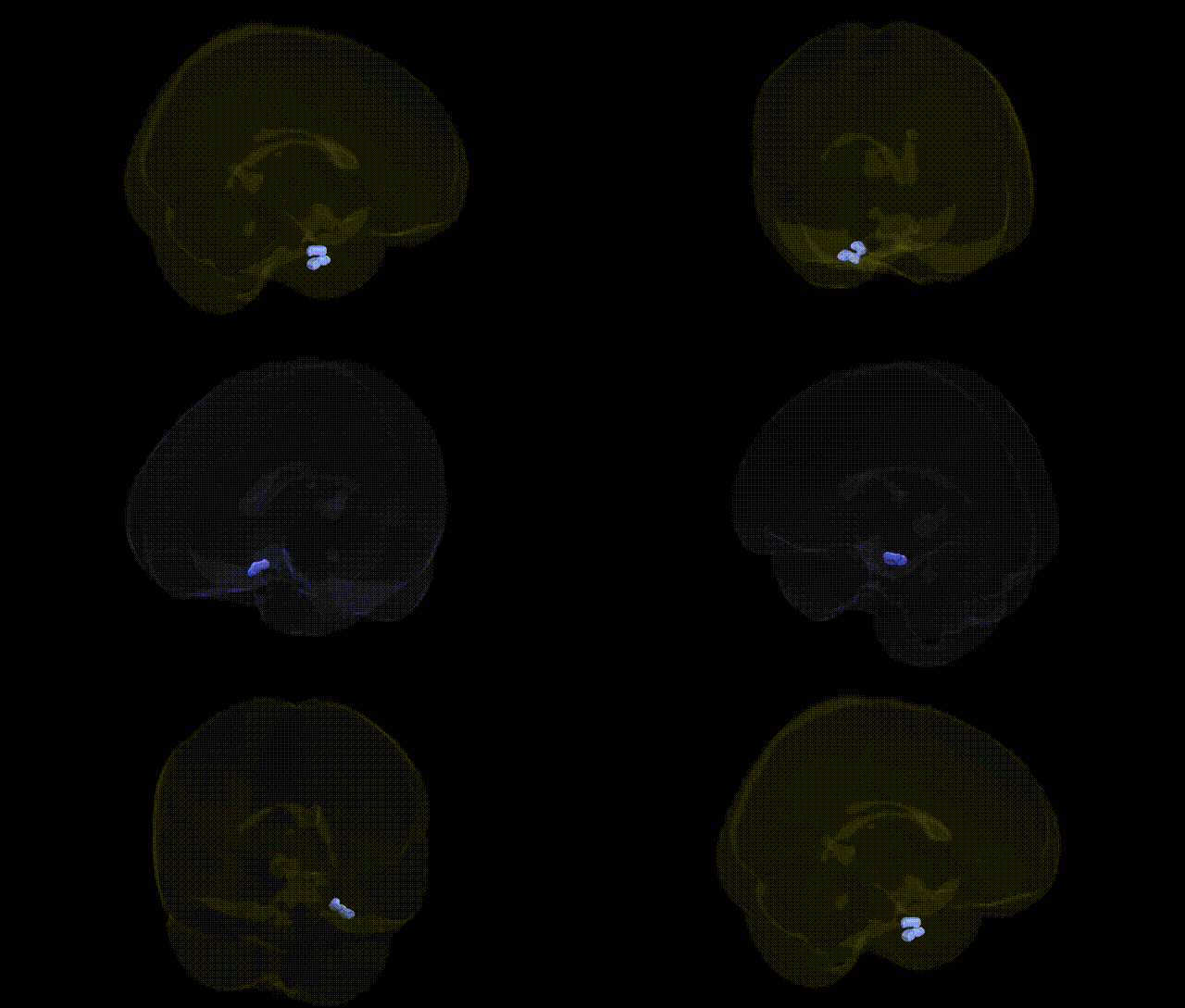
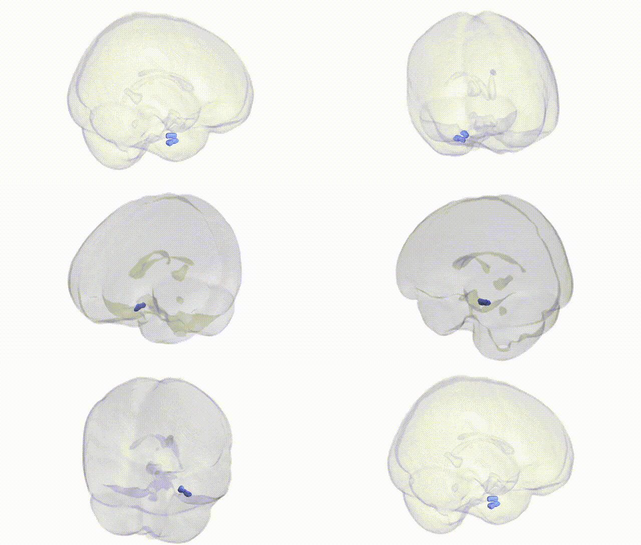
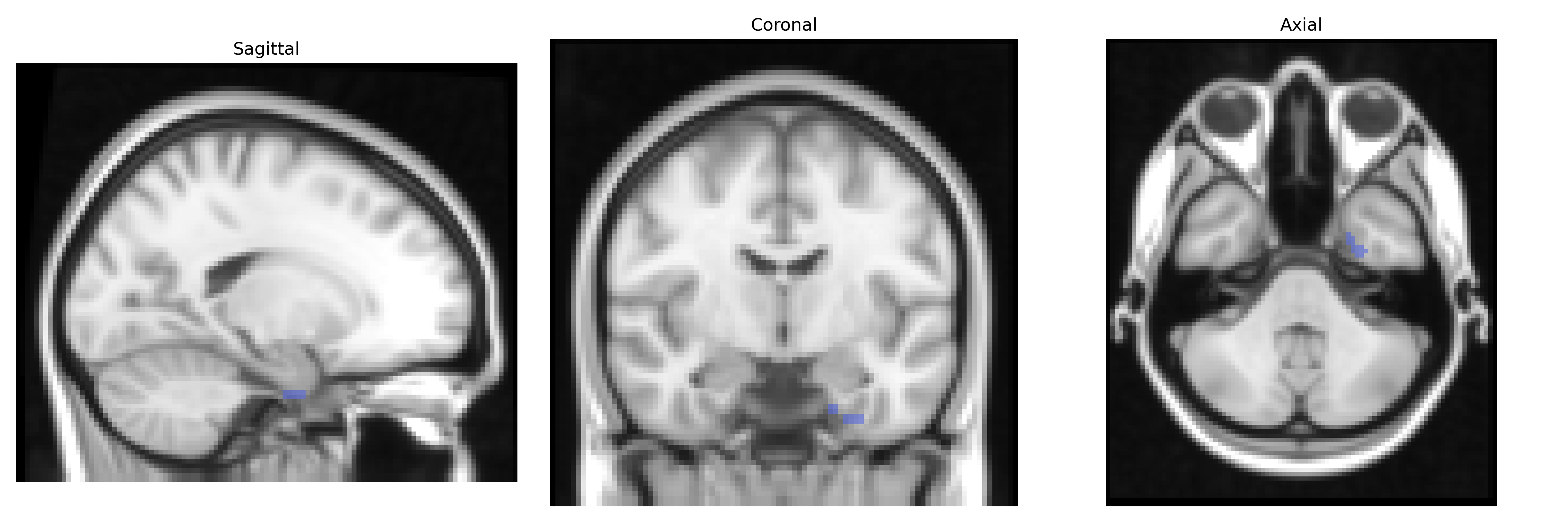
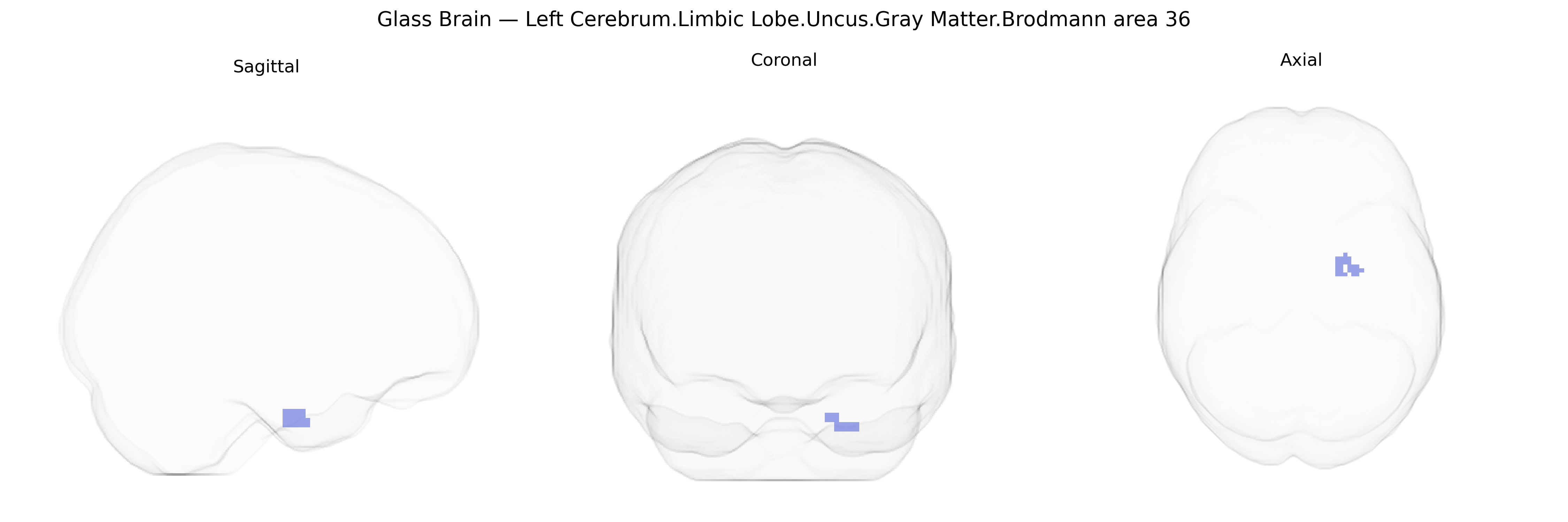
The bilateral Left Cerebrum.Limbic Lobe.Uncus.Gray Matter.Brodmann area 36 corresponds to cortex within the parahippocampal gyrus adjacent to the uncus, forming part of the medial temporal lobe’s perirhinal and parahippocampal regions. Brodmann area 36 is heavily implicated in higher-order visual association and memory processes, particularly familiarity-based recognition, multimodal object processing, and integration of sensory information with stored mnemonic representations. Its location within the limbic circuitry associates it with emotional valence tagging of sensory inputs and interaction with the hippocampal formation for episodic memory encoding and retrieval. The region receives input from visual association areas and projects to hippocampal and amygdalar structures, supporting the interface between perceptual processing and declarative memory. There is no direct link for Brodmann area 36; see the related region Parahippocampal gyrus.
The left uncus in Brodmann area 36, part of the medial temporal and parahippocampal/limbic network, is repeatedly implicated in genetic studies of episodic memory, emotional processing, and psychiatric vulnerability. GWAS of MRI-derived cortical thickness and surface area have linked common variants in genes such as BDNF (e.g., Val66Met), APOE (particularly ε4), and loci near KIAA0319, SLC39A8, and MAPT to structural differences in adjacent medial temporal and parahippocampal regions that encompass BA36, often in the context of Alzheimer’s disease risk, age-related memory decline, and general cognitive performance. Imaging genetics work has associated variation in serotonin transporter (SLC6A4) and COMT with functional activation and gray matter in medial temporal/uncal regions during affective and memory tasks, and schizophrenia and major depression GWAS have identified polygenic risk scores that correlate with reduced medial temporal/uncal volumes, implicating synaptic and neurodevelopmental pathways (e.g., CACNA1C, GRIN2A). Additionally, rare variants and GWAS loci involved in temporal lobe epilepsy (such as LGI1 and other synaptic genes) have been linked to structural and functional alterations in the uncus and nearby parahippocampal cortex, highlighting BA36 as a genetically sensitive node for memory, emotional regulation, and seizure susceptibility, even though few studies isolate this Brodmann area specifically rather than the broader medial temporal lobe.
Overview generated by GPT-4o (2026).
Region ID: 75
Hemisphere: bilateral
Atlas: Talairach labels 2mm
Full Quality Version: Download MP4
Full Quality Version: Download MP4
Wali Sidiqyar*, Gaurav Rudravaram*, Elyssa M. McMaster, Trent M. Schwartz, Adam M. Saunders, Kurt G. Schilling, Bennett A. Landman "Introducing SPINS: A Shared Public Visualization Library of Neuroanatomical Structures." Medical Imaging with Deep Learning- short paper
This resource is licensed under CC0 1.0 Universal (Public Domain).