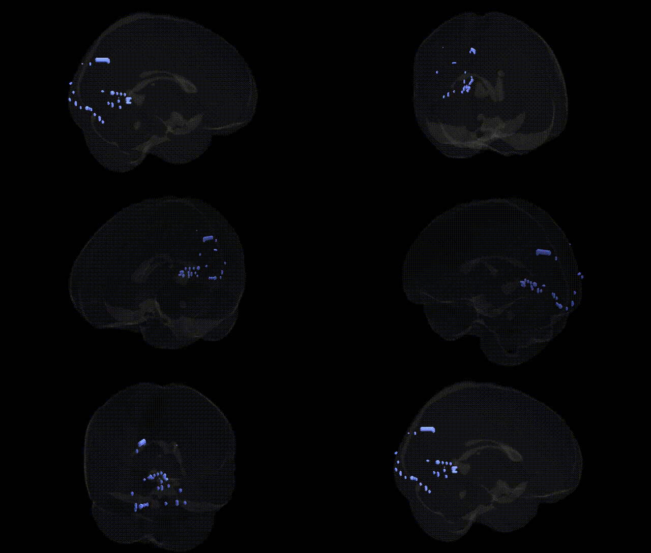
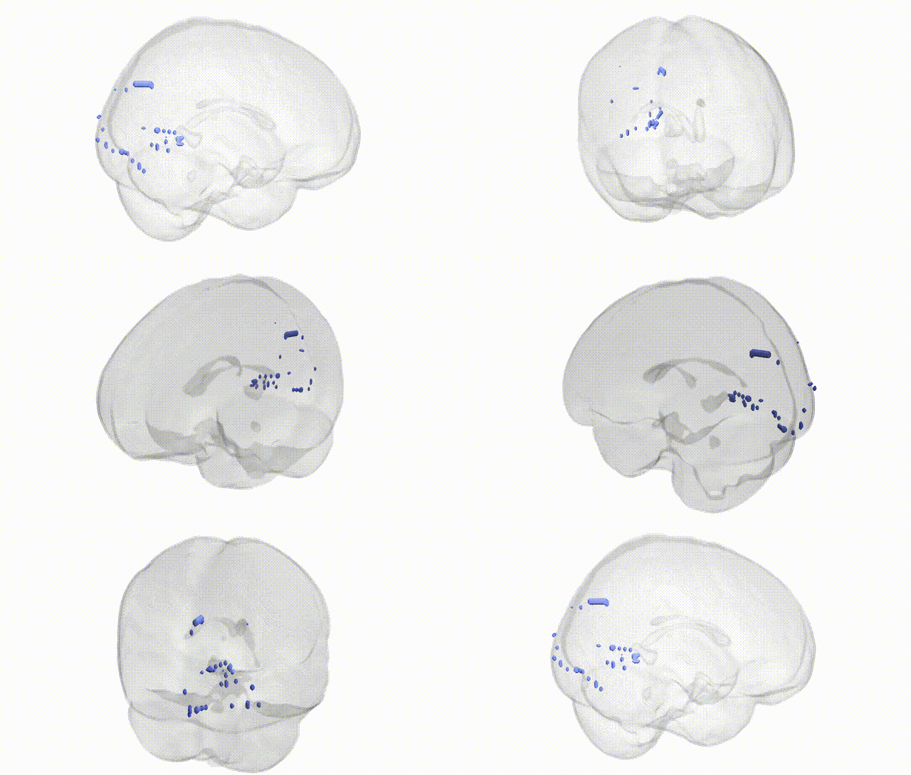
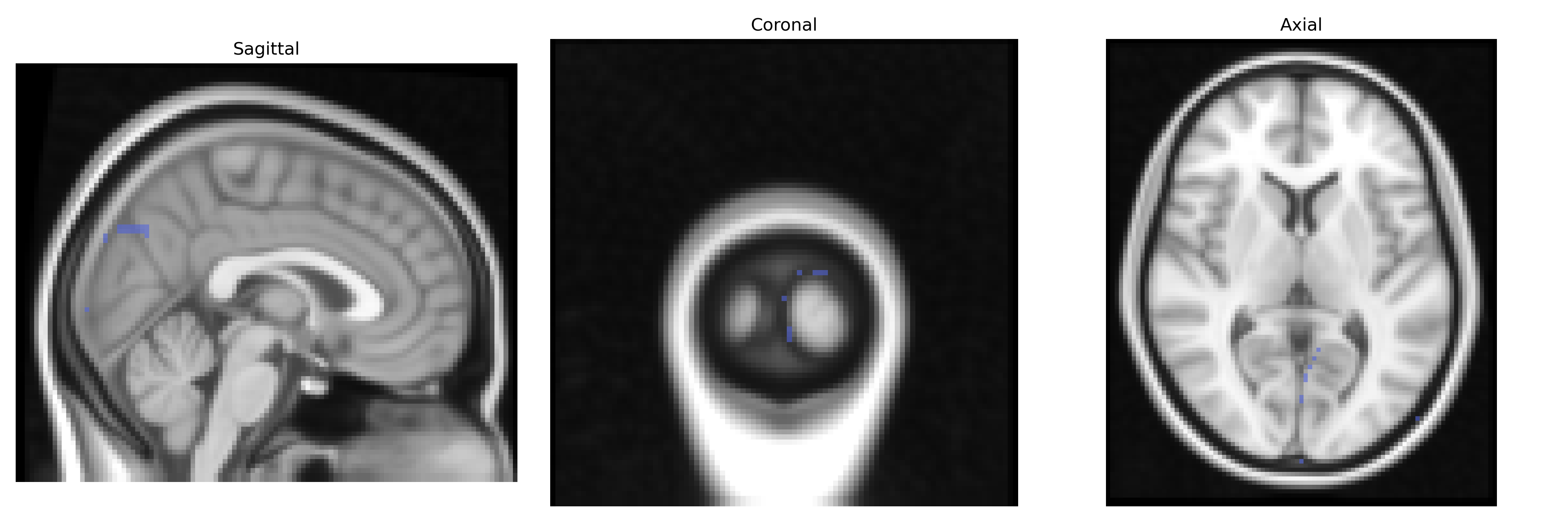
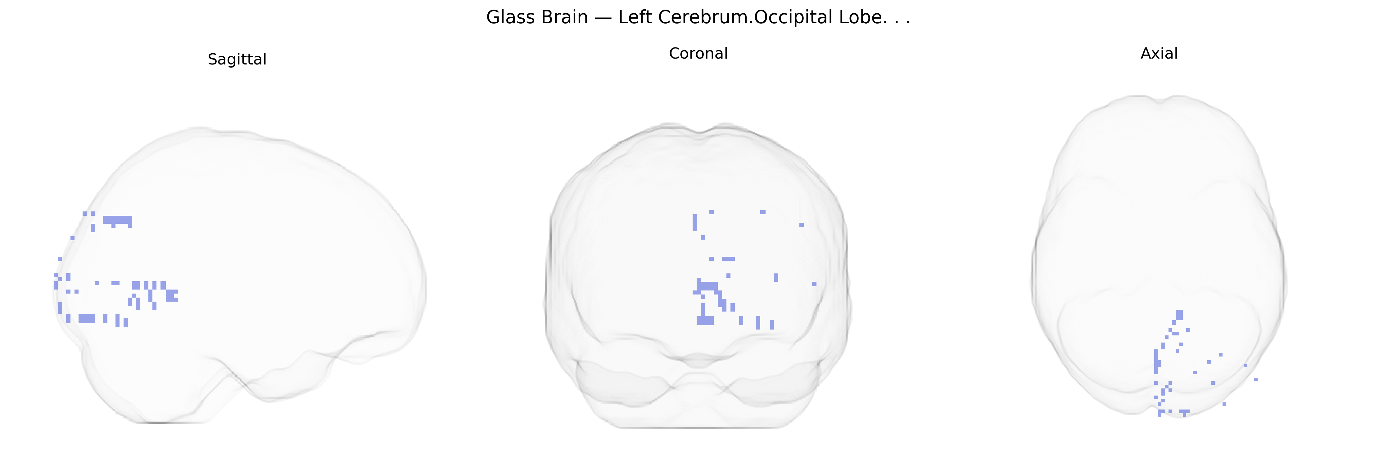
The bilateral Left Cerebrum.Occipital Lobe in the Talairach 2 mm Atlas refers to cortical tissue within the left cerebral hemisphere’s occipital lobe, encompassing primary and secondary visual areas that subserve early stages of visual processing. This region includes the calcarine cortex (primary visual cortex, or V1) and surrounding extrastriate areas involved in processing basic visual features such as orientation, motion, color, and spatial frequency, which are then integrated into higher-order visual representations. Functionally, the left occipital lobe contributes to visual perception and the encoding of visual information that can later be linked to language-related and semantic processes in adjacent and connected cortical regions. The occipital lobe’s organization is retinotopic, with neighboring neurons responding to neighboring locations in the visual field, and lesions here can produce characteristic visual field deficits (scotomas, hemianopia) contralateral to the damaged hemisphere.
Occipital lobe
The bilateral left cerebrum occipital lobe, as defined in the Talairach 2 mm atlas, encompasses primary and associative visual cortices, and genetic studies have largely implicated this region through its role in visual processing, cortical morphology, and neuropsychiatric and neurodevelopmental phenotypes. GWAS of brain imaging measures (e.g., ENIGMA and UK Biobank) have identified common variants in genes related to neurodevelopment and synaptic function—such as those in Wnt signaling pathways, cell-adhesion molecules, and axon guidance genes—that are associated with occipital cortical thickness, surface area, and volume, including variants near genes like TCF4, DLG2, and CELSR3 in some large-scale imaging-genetics analyses. Occipital lobe structure and function show heritability and have been linked to genetic risk for disorders involving visual perception and cortical organization, including migraine with aura (with implicated loci such as those in CACNA1A and other ion-channel or vascular regulation genes), occipital lobe epilepsy (rare pathogenic variants in ion channel and synaptic genes), and neurodevelopmental conditions such as autism spectrum disorder and dyslexia, where imaging-genetic work often reports altered occipital activation or morphology associated with polygenic risk scores. Additionally, schizophrenia and major depressive disorder GWAS have shown that higher polygenic risk correlates with subtle changes in occipital cortical measures, and candidate-gene and polygenic studies of visual hallucinations and visual working memory implicate occipital regions as downstream targets of broader genetic liability rather than region-specific genes. Overall, known genetic associations for the left occipital lobe reflect pleiotropic neurodevelopmental and synaptic pathways influencing global cortical architecture and visual system organization, rather than genes uniquely dedicated to this specific Talairach-defined area.
Overview generated by GPT-4o (2026).
Region ID: 246
Hemisphere: bilateral
Atlas: Talairach labels 2mm
Full Quality Version: Download MP4
Full Quality Version: Download MP4
Wali Sidiqyar*, Gaurav Rudravaram*, Elyssa M. McMaster, Trent M. Schwartz, Adam M. Saunders, Kurt G. Schilling, Bennett A. Landman "Introducing SPINS: A Shared Public Visualization Library of Neuroanatomical Structures." Medical Imaging with Deep Learning- short paper
This resource is licensed under CC0 1.0 Universal (Public Domain).