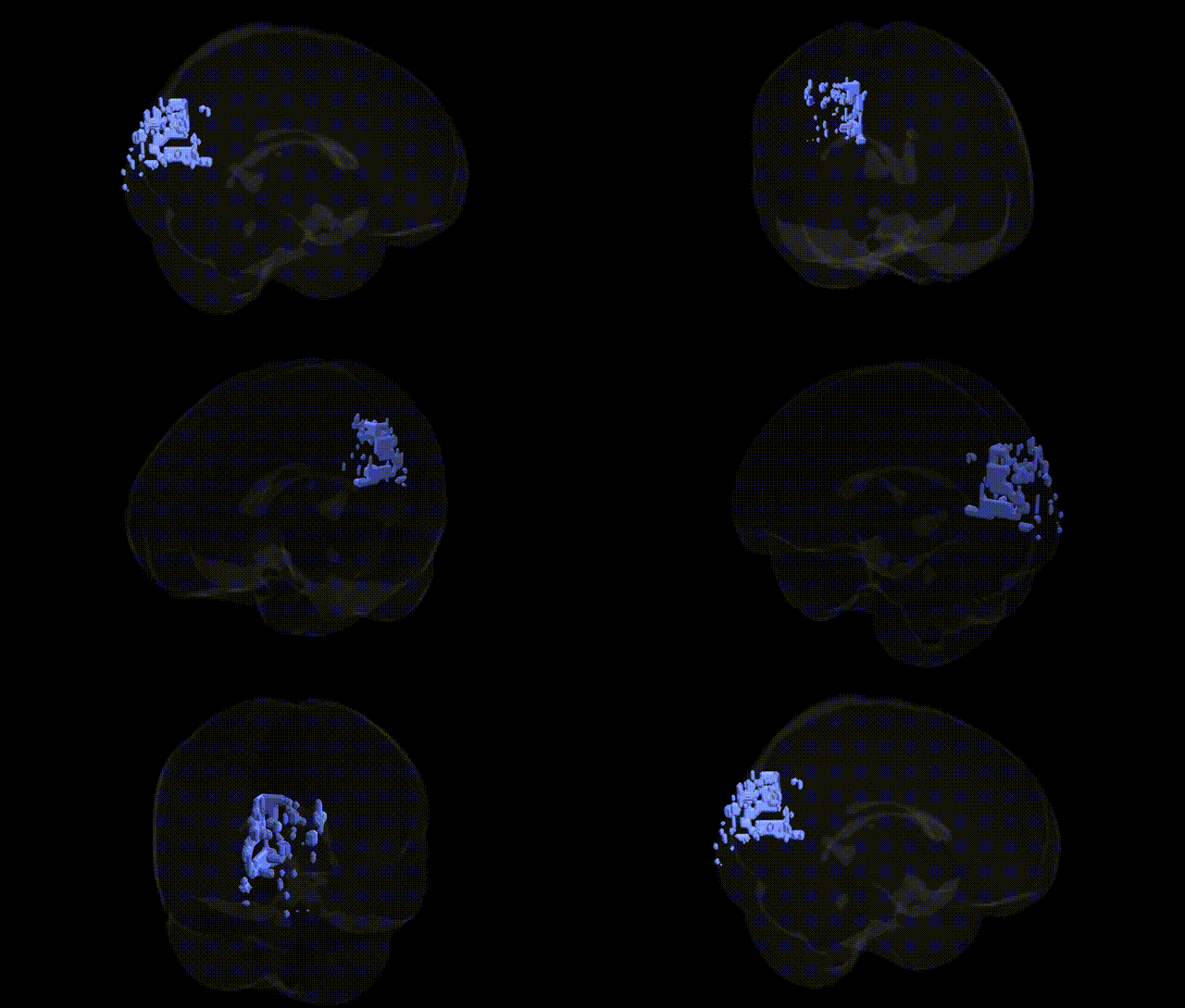
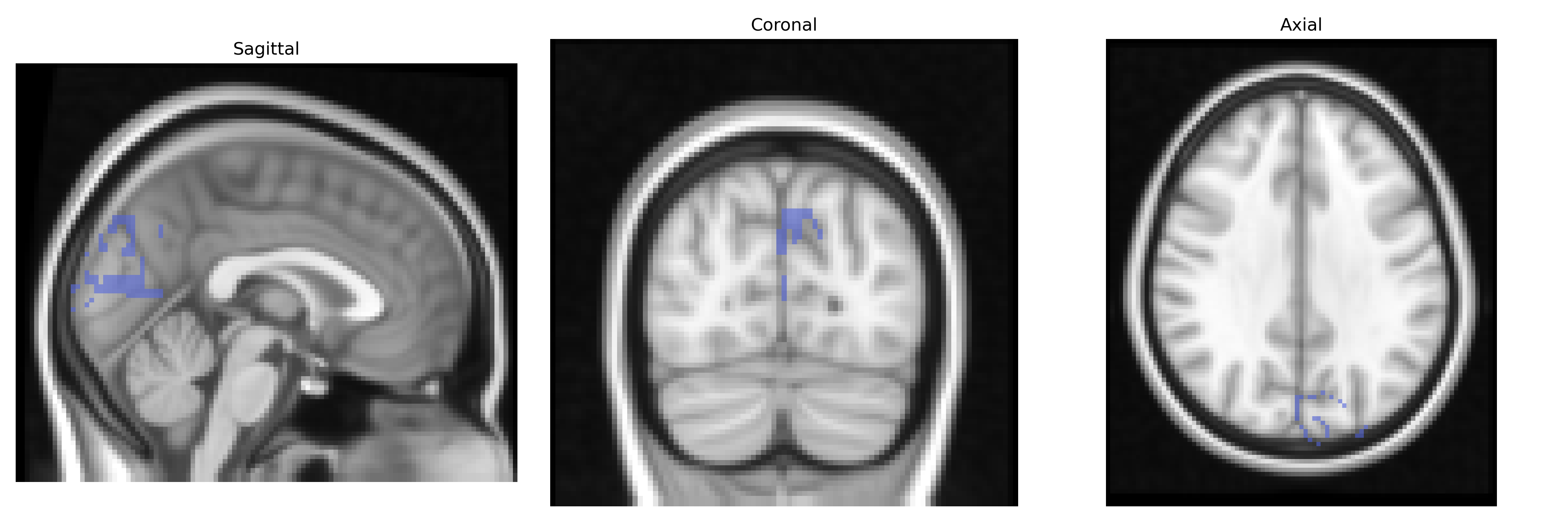
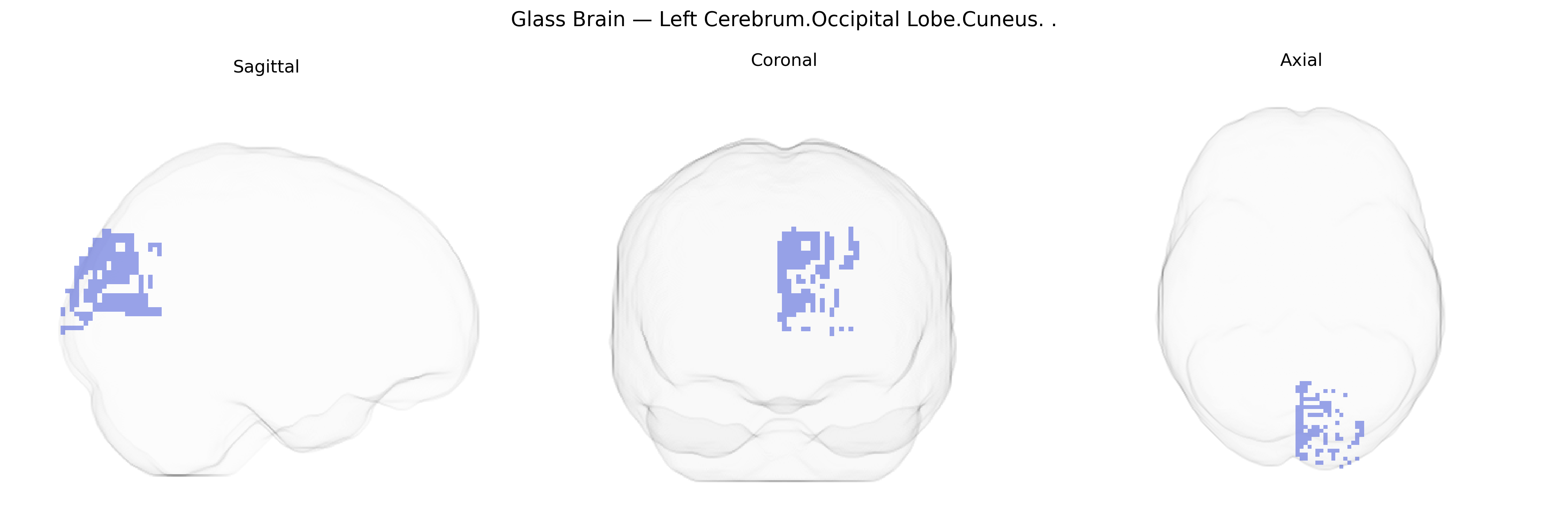
The bilateral Left Cerebrum.Occipital Lobe.Cuneus, as defined in the Talairach 2 mm Atlas, corresponds to the cuneus gyrus of the occipital lobe, a wedge-shaped region located on the medial surface of the cerebral hemisphere between the parieto-occipital sulcus and the calcarine sulcus. It is part of the primary and associative visual cortex, participating in early visual processing, especially of central visual field information, and contributes to functions such as visual attention, spatial orientation, and visuomotor integration. The cuneus receives prominent input from the lateral geniculate nucleus via the optic radiations and is interconnected with other occipital and parietal visual areas, supporting higher-order visual perception and integration of visual information with attention and eye-movement control.
Cuneus
The bilateral left cerebrum occipital lobe cuneus (Talairach 2 mm atlas label) is part of primary and associative visual cortex, and genetic associations involving this region largely emerge from imaging genetics and GWAS of brain structure and function rather than region-specific candidate studies. Large-scale neuroimaging GWAS (e.g., ENIGMA, UK Biobank) have identified multiple common variants influencing cortical thickness and surface area in occipital regions that encompass the cuneus, including loci near genes involved in neurodevelopment and synaptic function such as DACT1, PAX6, TBR1, and variants in or near the APOE region that show effects on occipital cortical metrics in aging and Alzheimer’s disease. Polygenic scores for schizophrenia, major depressive disorder, and bipolar disorder have been associated with altered occipital/cuneus volume or activation during visual and cognitive tasks, suggesting pleiotropic genetic effects on visual processing and higher-order integration. GWAS of visual acuity, myopia, and eye/retina traits have implicated genes (e.g., RAI1, GJD2, BCL11A) that, through developmental pathways, also modulate occipital cortical structure and visual network connectivity, indirectly linking cuneus anatomy and function to these traits. Additionally, genetic variants associated with migraine and visual aura (such as in CACNA1A and other ion-channel genes) have been related to altered excitability in occipital regions including the cuneus in imaging and electrophysiological studies, while risk alleles for autism spectrum disorder and ADHD show modest associations with atypical activation or connectivity in cuneus-centered visual networks. Overall, genetic influences on this region appear highly polygenic, shared across neurodevelopmental, psychiatric, and visual traits, and are typically detected as part of broader occipital or visual-network GWAS rather than as cuneus-specific findings.
Overview generated by GPT-4o (2026).
Region ID: 473
Hemisphere: bilateral
Atlas: Talairach labels 2mm
Full Quality Version: Download MP4
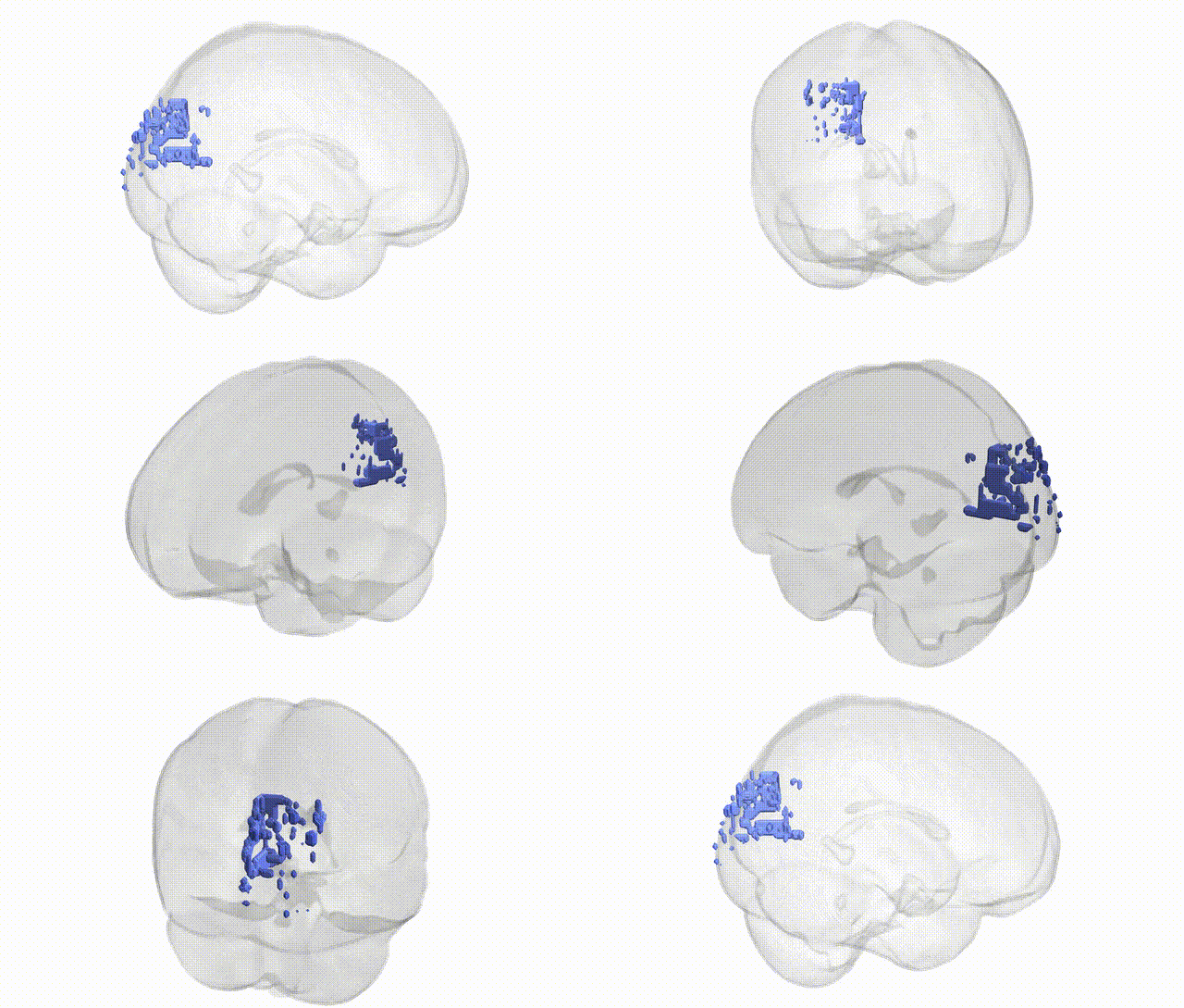
Full Quality Version: Download MP4
Wali Sidiqyar*, Gaurav Rudravaram*, Elyssa M. McMaster, Trent M. Schwartz, Adam M. Saunders, Kurt G. Schilling, Bennett A. Landman "Introducing SPINS: A Shared Public Visualization Library of Neuroanatomical Structures." Medical Imaging with Deep Learning- short paper
This resource is licensed under CC0 1.0 Universal (Public Domain).