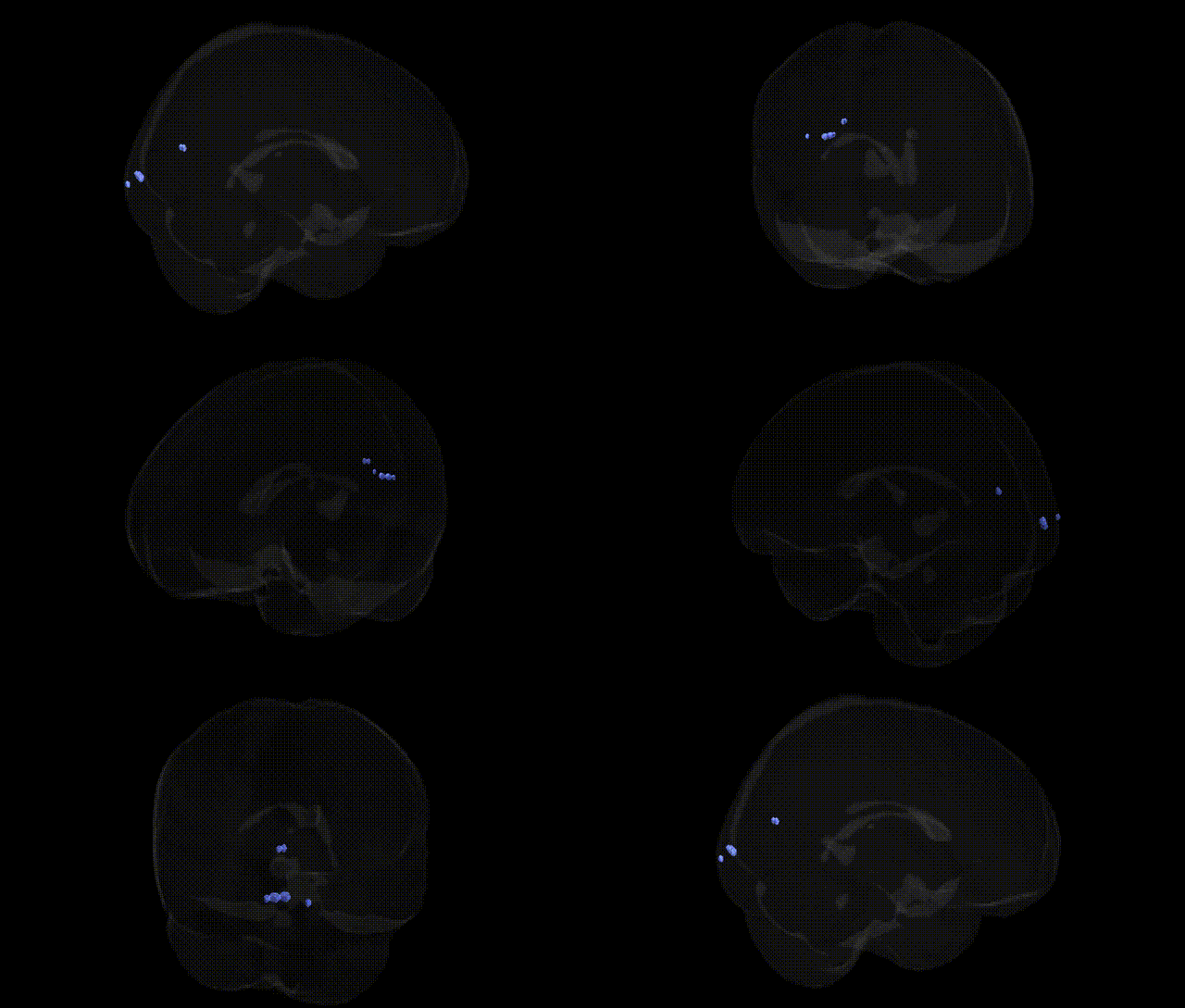
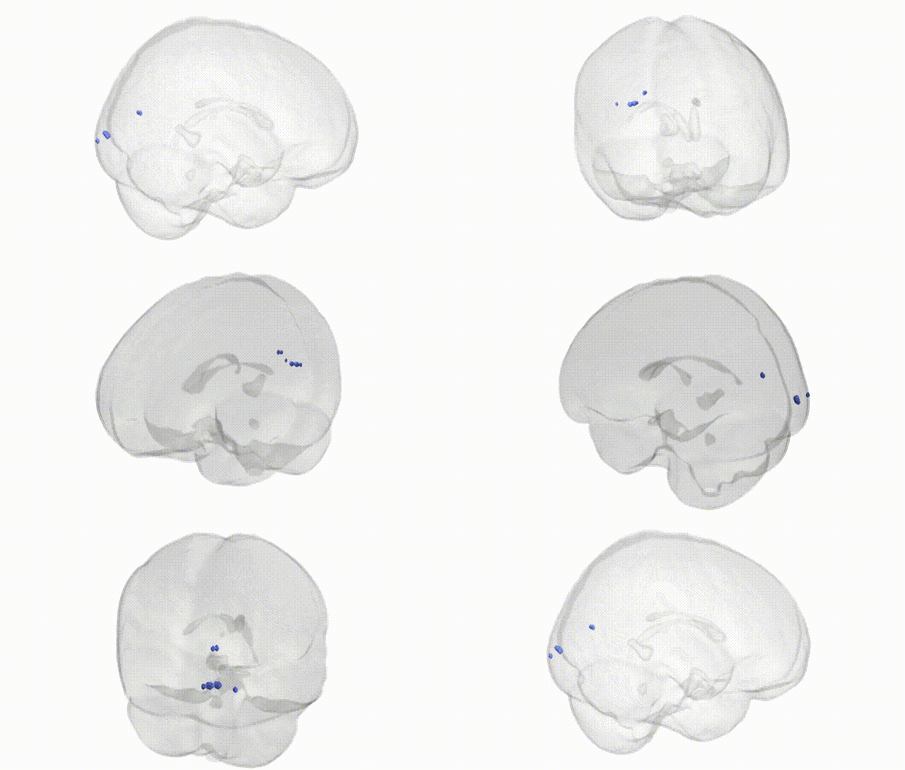
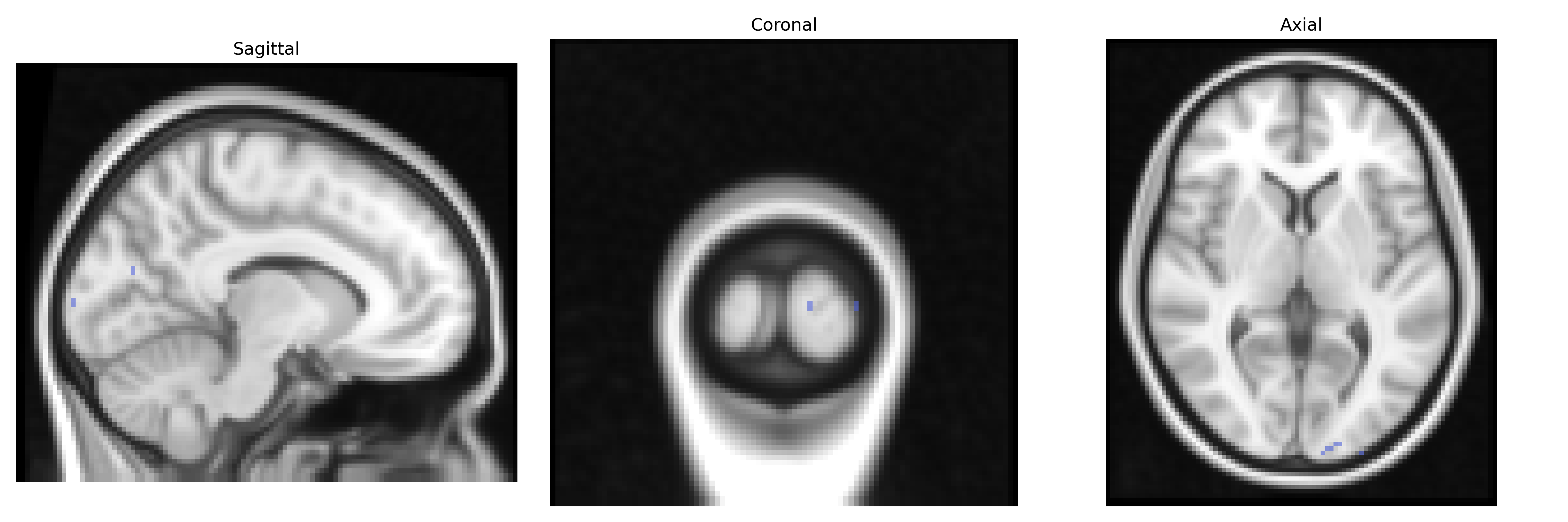
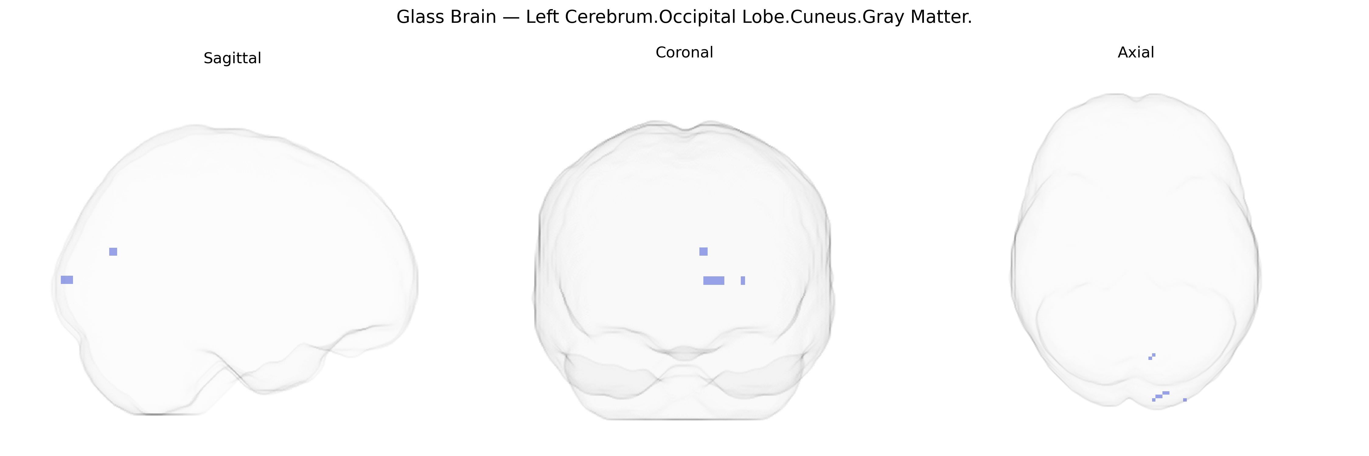
The bilateral Left Cerebrum.Occipital Lobe.Cuneus.Gray Matter corresponds to cortical tissue in the cuneus, a wedge-shaped region on the medial surface of the occipital lobe, bordered inferiorly by the calcarine sulcus and superiorly by the parieto-occipital sulcus. This region contains early visual cortical areas (including parts of V1 and V2) and is primarily involved in processing basic visual features such as orientation, spatial frequency, and contrast, as well as contributing to visual attention and integration of visual information across the visual field. The gray matter here comprises neuronal cell bodies, dendrites, and local circuitry that support the initial stages of conscious visual perception and the relay of processed visual signals to higher-order association areas. Cuneus
The bilateral Left Cerebrum.Occipital Lobe.Cuneus.Gray Matter, as defined in the Talairach labels 2mm Atlas, corresponds to a primary visual and higher-order visual integration region that appears in multiple imaging genetics and GWAS-based neuroimaging studies focused on cortical thickness, surface area, and gray matter volume. Large-scale consortia such as ENIGMA and UK Biobank have reported robust heritability of cuneus morphology and identified associations with variants near genes involved in neurodevelopment, synaptic function, and axonal guidance (e.g., loci near PAX6, TCF4, and neuroplasticity-related genes), though specific variants often differ by cohort and analytic approach. GWAS of brain structure frequently implicate cuneus measures in polygenic architectures shared with cognitive traits (general intelligence, processing speed, visual memory), educational attainment, and risk for neuropsychiatric disorders, including schizophrenia and major depressive disorder, where risk alleles or polygenic risk scores correlate with altered cuneus thickness or volume. Imaging–genetics studies in attention-deficit/hyperactivity disorder, autism spectrum disorder, and bipolar disorder also report cuneus structural or functional alterations that track with disorder-related genetic burden, and ophthalmologic/visual pathway GWAS sometimes show downstream structural differences in cuneus gray matter tied to genetic variation affecting retinal and occipital processing. Overall, the region is consistently treated as a genetically influenced node in visual and cognitive networks, with its gray matter measures serving as intermediate phenotypes linking polygenic variation to both normal variation in visual-cognitive performance and vulnerability to psychiatric and neurodevelopmental conditions.
Overview generated by GPT-4o (2026).
Region ID: 533
Hemisphere: bilateral
Atlas: Talairach labels 2mm
Full Quality Version: Download MP4
Full Quality Version: Download MP4
Wali Sidiqyar*, Gaurav Rudravaram*, Elyssa M. McMaster, Trent M. Schwartz, Adam M. Saunders, Kurt G. Schilling, Bennett A. Landman "Introducing SPINS: A Shared Public Visualization Library of Neuroanatomical Structures." Medical Imaging with Deep Learning- short paper
This resource is licensed under CC0 1.0 Universal (Public Domain).