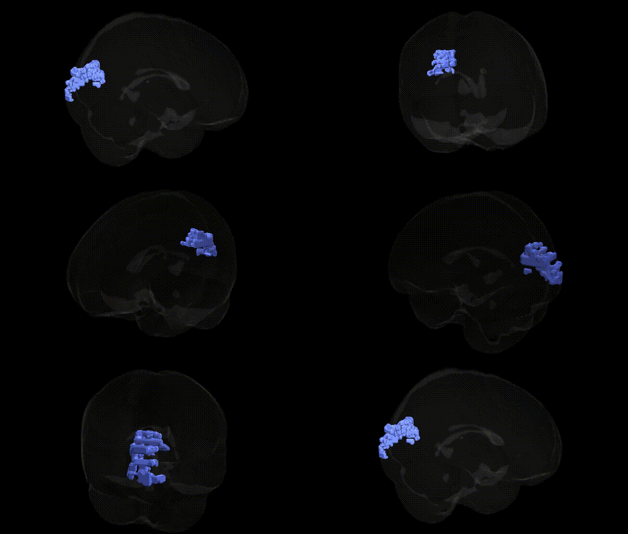
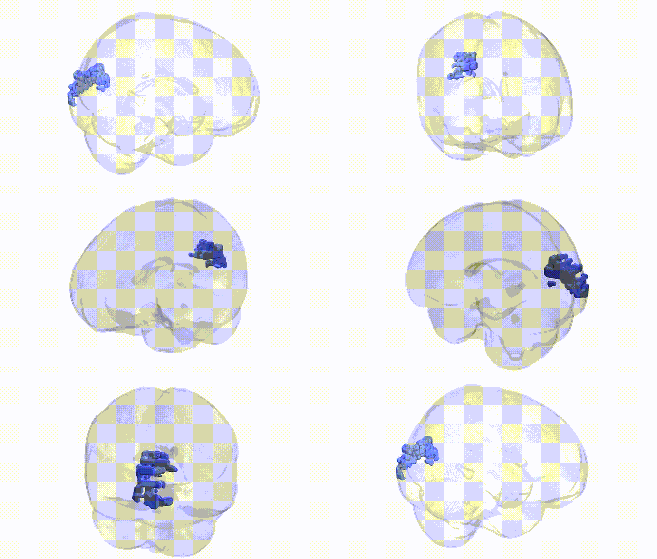
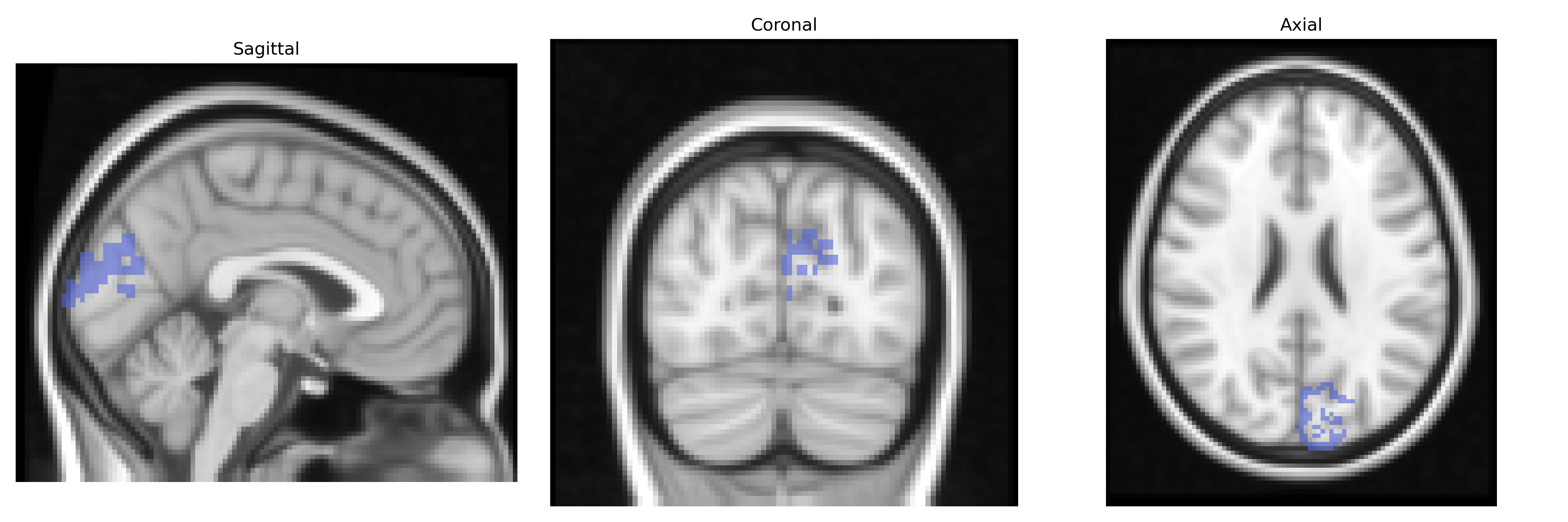
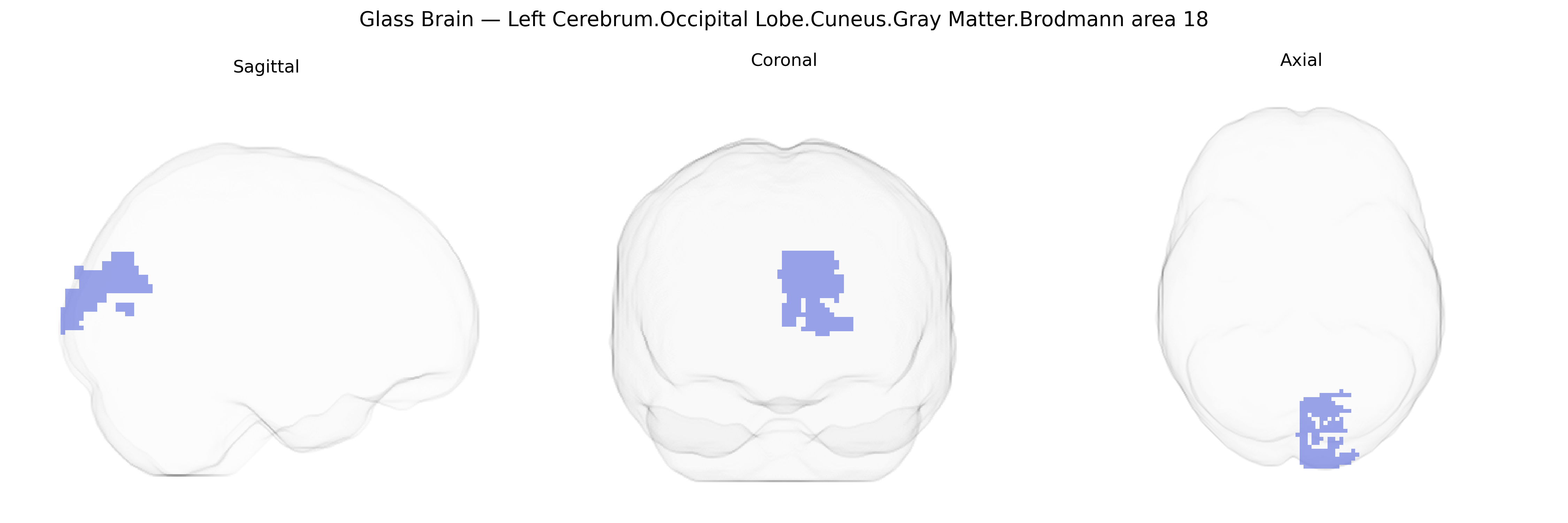
The bilateral Left Cerebrum.Occipital Lobe.Cuneus.Gray Matter.Brodmann area 18 corresponds to secondary visual cortex (V2) located in the cuneus region of the occipital lobe, immediately anterior to primary visual cortex (Brodmann area 17). This region receives dense feedforward input from area 17 and additional input from other visual areas, and is involved in the processing of more complex features of visual stimuli, including orientation, spatial frequency, contours, and binocular disparity, contributing to form, depth, and motion perception. Neuronal populations in Brodmann area 18 show modular organization (e.g., stripes in primates) and participate in early stages of visual integration that support higher-order visual recognition and visuospatial functions. There is no direct Wikipedia article specifically for “Brodmann area 18 in the cuneus,” but it is encompassed within Brodmann area 18.
The bilateral left cerebrum occipital lobe cuneus gray matter Brodmann area 18 (secondary visual cortex, V2) has been implicated in genetic studies primarily through its role in visual processing, cortical thickness, and functional activation patterns rather than via region-specific single-gene effects. GWAS of brain imaging phenotypes (e.g., ENIGMA, UK Biobank) have identified multiple loci affecting occipital and cuneus cortical thickness and surface area, including variants near genes involved in neurodevelopment and synaptic function (such as MIR137, HMGA2, and genes within Wnt and axon-guidance pathways), though these findings typically refer to broader occipital or visual cortex measures rather than Brodmann area 18 alone. Polygenic risk for schizophrenia, bipolar disorder, and major depression has been associated with structural and functional alterations in the cuneus and BA18, including reduced gray matter volume or altered activation to visual and emotional stimuli, suggesting that common psychiatric risk alleles exert downstream effects on this region. Genetic studies of migraine (especially visual aura), dyslexia and reading traits, autism spectrum disorder, and attention-deficit/hyperactivity disorder have reported associations between risk variants and atypical occipital/cuneus activation or morphology, consistent with the region’s role in visual attention and higher-order visual processing, though specific variants are usually linked to distributed networks rather than uniquely to BA18. Overall, genetic influences on BA18 appear to be highly polygenic and embedded within global neurodevelopmental architectures, with the region serving as one node through which common variants affecting cortical development, visual perception, and psychiatric liability manifest in brain imaging phenotypes.
Overview generated by GPT-4o (2026).
Region ID: 474
Hemisphere: bilateral
Atlas: Talairach labels 2mm
Full Quality Version: Download MP4
Full Quality Version: Download MP4
Wali Sidiqyar*, Gaurav Rudravaram*, Elyssa M. McMaster, Trent M. Schwartz, Adam M. Saunders, Kurt G. Schilling, Bennett A. Landman "Introducing SPINS: A Shared Public Visualization Library of Neuroanatomical Structures." Medical Imaging with Deep Learning- short paper
This resource is licensed under CC0 1.0 Universal (Public Domain).