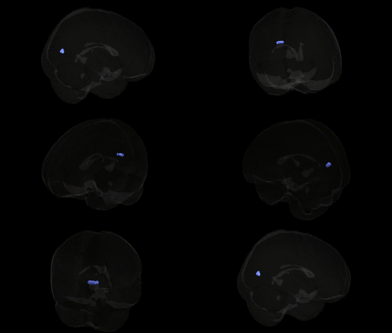
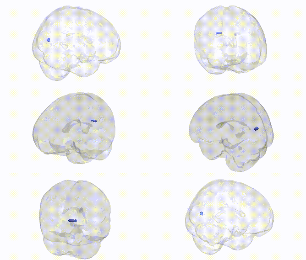
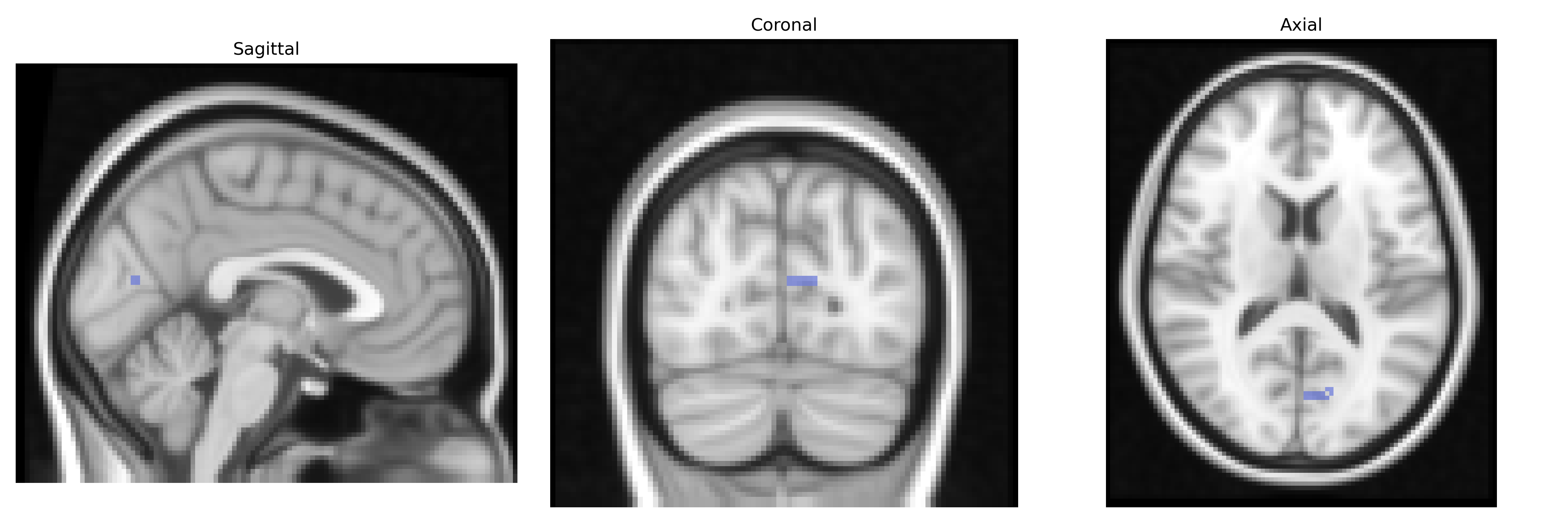
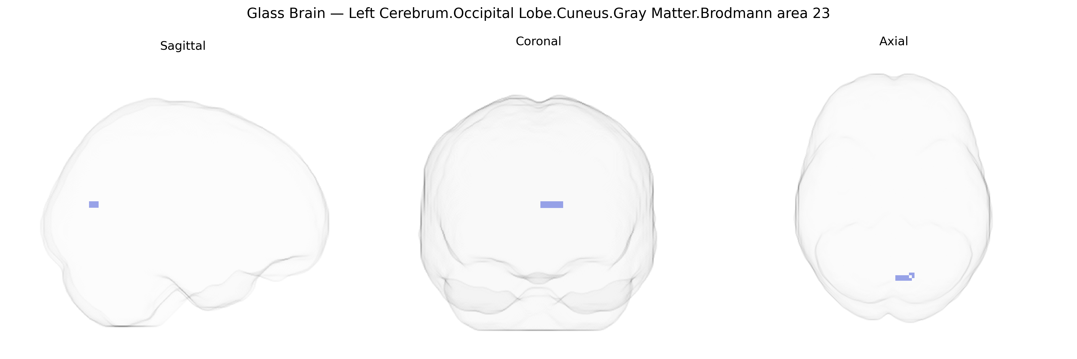
Bilateral Left Cerebrum.Occipital Lobe.Cuneus.Gray Matter.Brodmann area 23 refers to gray matter in the cuneus of the occipital lobe, localized in the left cerebral hemisphere but represented bilaterally in the Talairach 2 mm Atlas due to the symmetry of this region. Brodmann area 23 is classically defined as part of the ventral posterior cingulate cortex, a component of the limbic system involved in emotional processing, internal mentation, and default-mode network activity, but atlas-based label sets sometimes extend or map it to adjacent cortical territories such as the cuneus for coordinate-based parcellation. Functionally, the cuneus is primarily associated with early visual processing, visuospatial integration, and aspects of visual attention, and its interaction with limbic-associated areas like BA23 supports integration of visual information with contextual, mnemonic, and emotional states. There is no direct link for this compound label; a related and more general article is: Cuneus.
The left cuneus gray matter in the occipital lobe, corresponding to Brodmann area 23 in the Talairach 2 mm atlas (though BA23 is classically posterior cingulate, some atlases extend or mislabel into adjacent medial occipital territory), has been implicated in several imaging–genetics and GWAS findings, primarily through studies of cortical thickness, surface area, and functional activation rather than region-specific single-gene effects. Large neuroimaging GWAS consortia such as ENIGMA and UK Biobank have identified polygenic influences on occipital and cuneus cortical measures, with significant associations involving variants in loci related to neurodevelopment, synaptic function, and axon guidance (e.g., genes near 14q23.1 such as KIAA0586 and DAAM1, and other widespread cortical-structure loci), though these findings usually treat the cuneus as part of broader occipital or medial visual cortex rather than BA23 specifically. Genetic risk for psychiatric and neurodevelopmental disorders—including schizophrenia, bipolar disorder, major depression, and autism spectrum disorder—has been linked to altered structure or activity of the cuneus/medial occipital regions via polygenic risk scores, connecting distributed risk alleles (e.g., in CACNA1C, GRIN2A, complement and immune-related genes) to abnormalities in visual processing, self-referential and default-mode network function. Additionally, imaging–genetics studies of traits such as intelligence, educational attainment, and cognitive performance have associated polygenic scores with variation in cuneus thickness and activation during visual, attentional, and memory tasks, while GWAS of PTSD, anxiety, and pain sensitivity have implicated networks that include this region. Overall, genetic associations involving the left cuneus/BA23 region are highly polygenic and network-based, with no single variant uniquely tied to this specific Talairach-defined parcel, but consistent evidence that common and rare neuropsychiatric risk alleles influence its structure and function as part of broader visual–default mode circuitry.
Overview generated by GPT-4o (2026).
Region ID: 706
Hemisphere: bilateral
Atlas: Talairach labels 2mm
Full Quality Version: Download MP4
Full Quality Version: Download MP4
Wali Sidiqyar*, Gaurav Rudravaram*, Elyssa M. McMaster, Trent M. Schwartz, Adam M. Saunders, Kurt G. Schilling, Bennett A. Landman "Introducing SPINS: A Shared Public Visualization Library of Neuroanatomical Structures." Medical Imaging with Deep Learning- short paper
This resource is licensed under CC0 1.0 Universal (Public Domain).