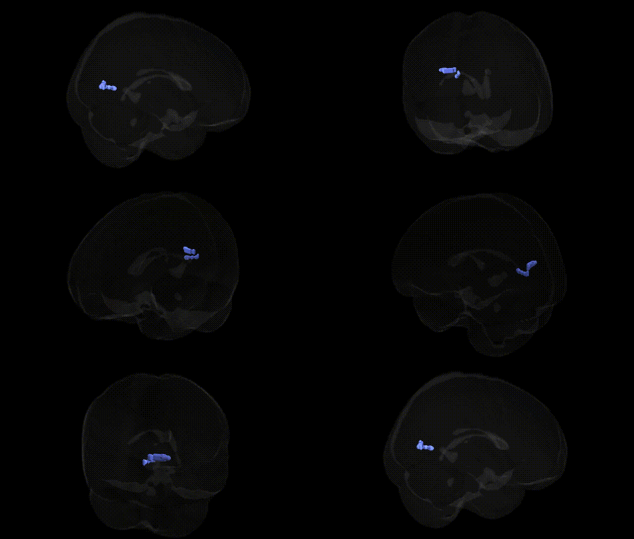
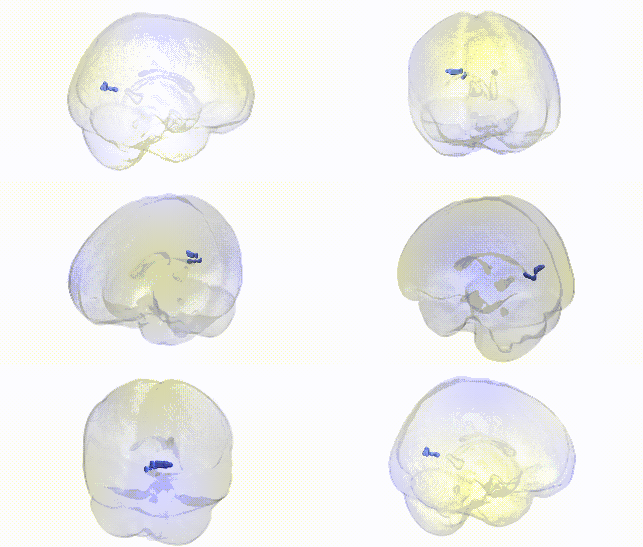
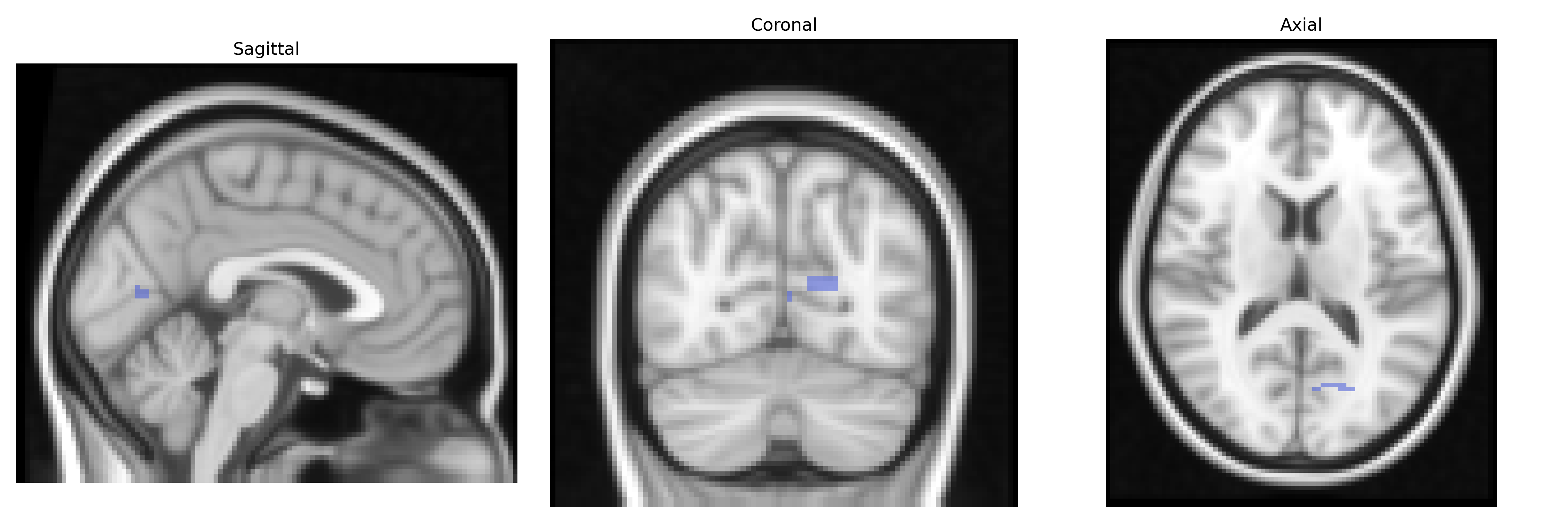
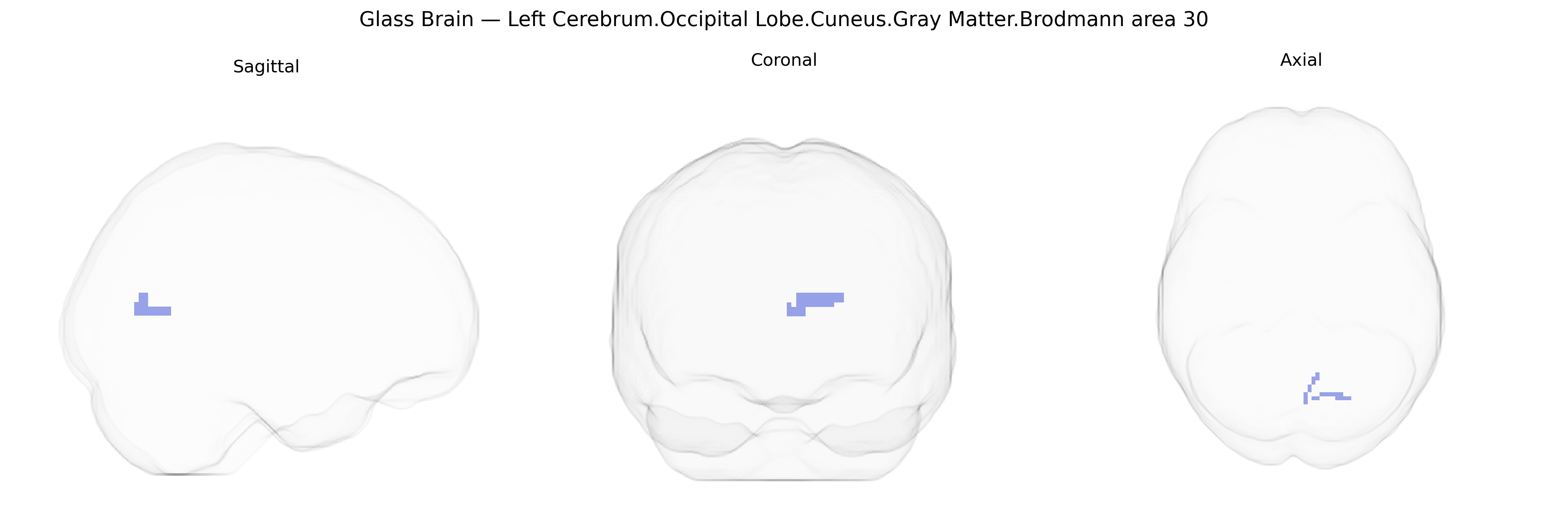
The bilateral Left Cerebrum.Occipital Lobe.Cuneus.Gray Matter.Brodmann area 30 corresponds to a portion of the retrosplenial cortex situated in the medial occipital region, bordering the posterior cingulate and parahippocampal areas. Brodmann area 30 is associated with visuospatial processing, scene recognition, and memory-related integration of visual information, and it plays a role in navigation and contextual associations. Although classified here within the cuneus of the occipital lobe, BA30 functionally belongs to the limbic network, contributing to episodic memory, environmental orientation, and the linkage of visual inputs with internal representations. There is no direct Wikipedia article for Brodmann area 30; a related structure and functional context can be found in Retrosplenial cortex.
The bilateral Left Cerebrum Occipital Lobe Cuneus Gray Matter Brodmann area 30 (Talairach 2 mm Atlas) is a retrosplenial/occipital region implicated in visual–spatial processing, memory, and self-referential cognition, and although relatively few studies target BA30 specifically, multiple GWAS and imaging genetics studies implicate overlapping genetic influences on its structure and function. Common variants in genes involved in synaptic plasticity and neurodevelopment—such as BDNF (e.g., Val66Met), APOE (especially ε4), and COMT—have been associated with gray matter volume, cortical thickness, or functional activation in retrosplenial and adjacent cuneus/precuneus areas, often in the context of episodic memory, visuospatial navigation, and default mode network activity. Large-scale neuroimaging GWAS (e.g., ENIGMA and UK Biobank) show polygenic influences on occipital and cuneus volumes, with significant loci near genes related to neuronal development, axon guidance, and myelination (such as HMGA2, DLG2, and others), and these structural metrics partially mediate genetic risk for cognitive traits including intelligence, educational attainment, and processing speed. BA30-adjacent cuneus and retrosplenial cortex have been repeatedly linked, via imaging genetic and GWAS-derived polygenic risk scores, to Alzheimer’s disease, schizophrenia, and major depressive disorder, where disease risk alleles and polygenic burden correlate with altered activation or connectivity within the posterior medial network, including BA30, during memory and self-related tasks. Additional GWAS and candidate gene work in post-traumatic stress disorder and anxiety disorders has connected variants in stress-regulation genes (FKBP5, CRHR1) and glutamatergic signaling genes to altered retrosplenial/cuneus responses to trauma cues, supporting a role for BA30-region circuitry in fear memory and contextual processing. Overall, genetic associations for BA30 are largely inferred from studies of overlapping retrosplenial, posterior cingulate, and cuneus territories rather than BA30-specific GWAS, with convergent evidence that polygenic variation affecting synaptic function, neurodevelopment, and neurodegeneration shapes the structure and function of this region and contributes to risk for memory-related and psychiatric disorders.
Overview generated by GPT-4o (2026).
Region ID: 660
Hemisphere: bilateral
Atlas: Talairach labels 2mm
Full Quality Version: Download MP4
Full Quality Version: Download MP4
Wali Sidiqyar*, Gaurav Rudravaram*, Elyssa M. McMaster, Trent M. Schwartz, Adam M. Saunders, Kurt G. Schilling, Bennett A. Landman "Introducing SPINS: A Shared Public Visualization Library of Neuroanatomical Structures." Medical Imaging with Deep Learning- short paper
This resource is licensed under CC0 1.0 Universal (Public Domain).