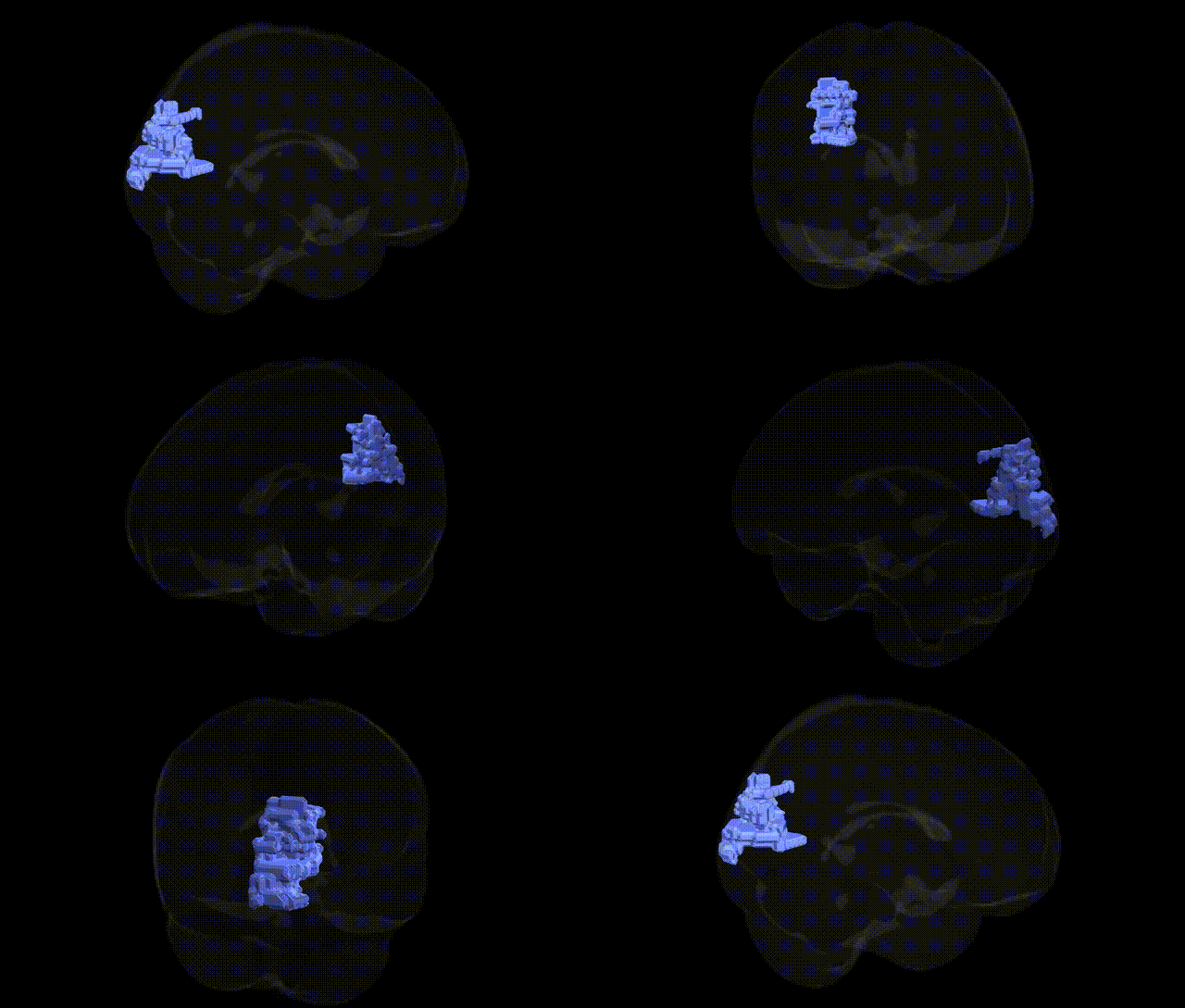
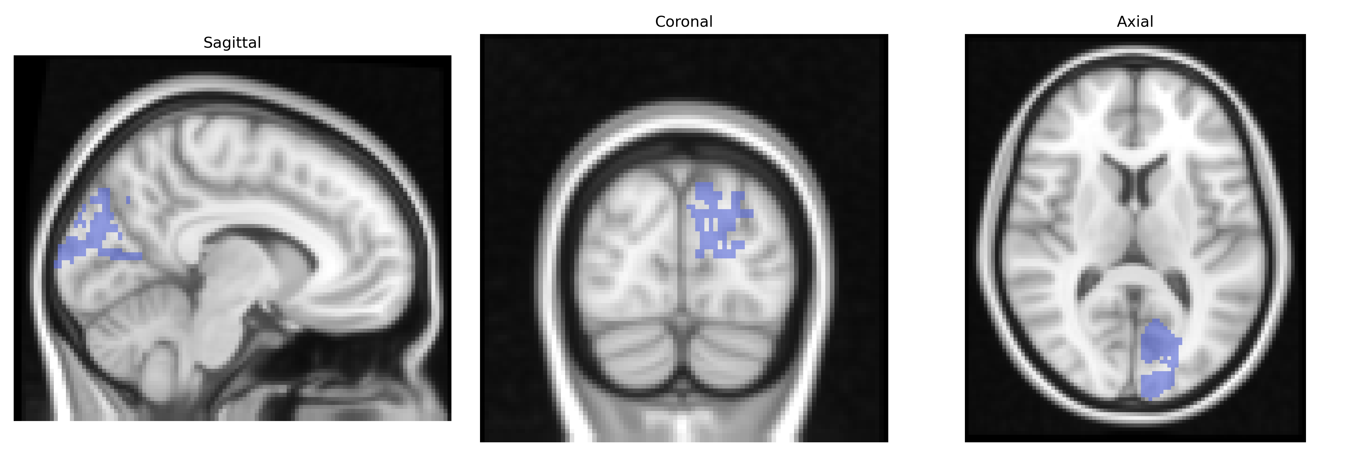
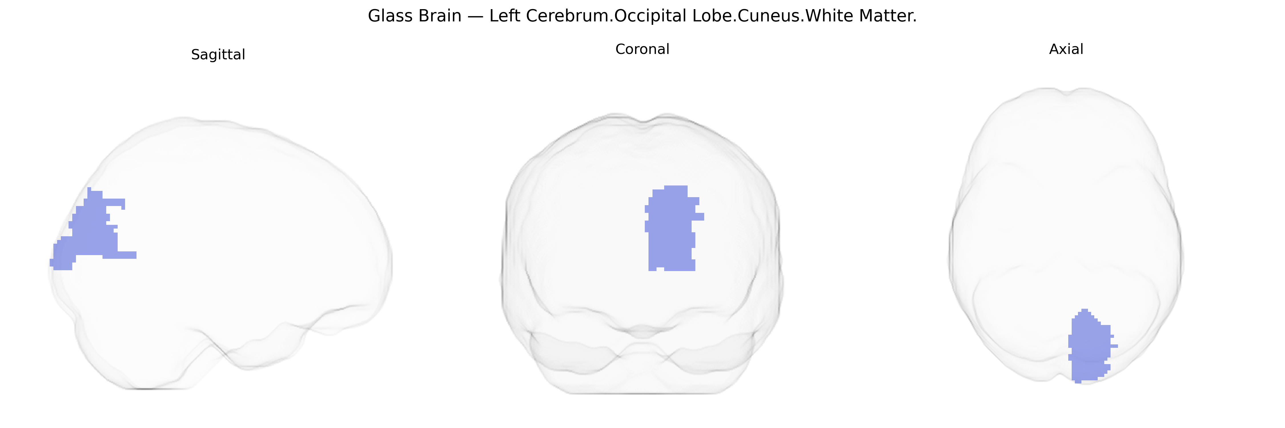
The bilateral Left Cerebrum.Occipital Lobe.Cuneus.White Matter corresponds to the subcortical fiber pathways underlying the cuneus region of the occipital lobe, a dorsal medial portion of visual cortex located between the parieto-occipital sulcus and the calcarine sulcus. Although the white matter itself contains no neuronal cell bodies, it comprises intra- and interhemispheric axonal projections that relay visual information between primary and higher-order visual areas, and toward associative and attentional networks. Functionally, this region supports the processing of visual inputs related to the central visual field and contributes to integration of visual signals necessary for perception and visuospatial analysis. There is no direct Wikipedia article for the cuneus white matter; a related cortical structure is the Cuneus.
The bilateral left cerebrum occipital lobe cuneus white matter, as defined in the Talairach 2mm atlas, is part of the visual processing network whose structural and functional characteristics show heritable variation and multiple genetic associations, although few studies target this exact labeled parcel. Genome-wide association studies (GWAS) of brain imaging phenotypes (e.g., ENIGMA and UK Biobank) have identified variants near genes such as EMX2, PAX6, TESC, and genes in Wnt and axon guidance pathways that influence occipital and cuneus cortical thickness, surface area, and white matter microstructure (including fractional anisotropy and mean diffusivity in occipital tracts). Polygenic risk scores for disorders like schizophrenia, major depressive disorder, bipolar disorder, and autism spectrum disorder show associations with cuneus/occipital white matter integrity and cortical metrics, suggesting a genetic contribution to altered visual and default-mode–related processing in these conditions. Variants near NRXN1, CNTNAP2, CACNA1C, and synaptic/neuronal adhesion genes have been linked to white matter abnormalities involving occipital and cuneus regions in psychosis and developmental disorders, while GWAS of migraine, visual hallucinations in Parkinson’s disease, and visual attention traits have also implicated occipital/cuneus networks, though typically at a broader regional level. Overall, heritability estimates for occipital/cuneus structural measures are moderate to high, and genetic influences appear to act through neurodevelopmental, myelination, and synaptic plasticity pathways that broadly affect visual and associative networks rather than this specific Talairach parcel alone.
Overview generated by GPT-4o (2026).
Region ID: 485
Hemisphere: bilateral
Atlas: Talairach labels 2mm
Full Quality Version: Download MP4
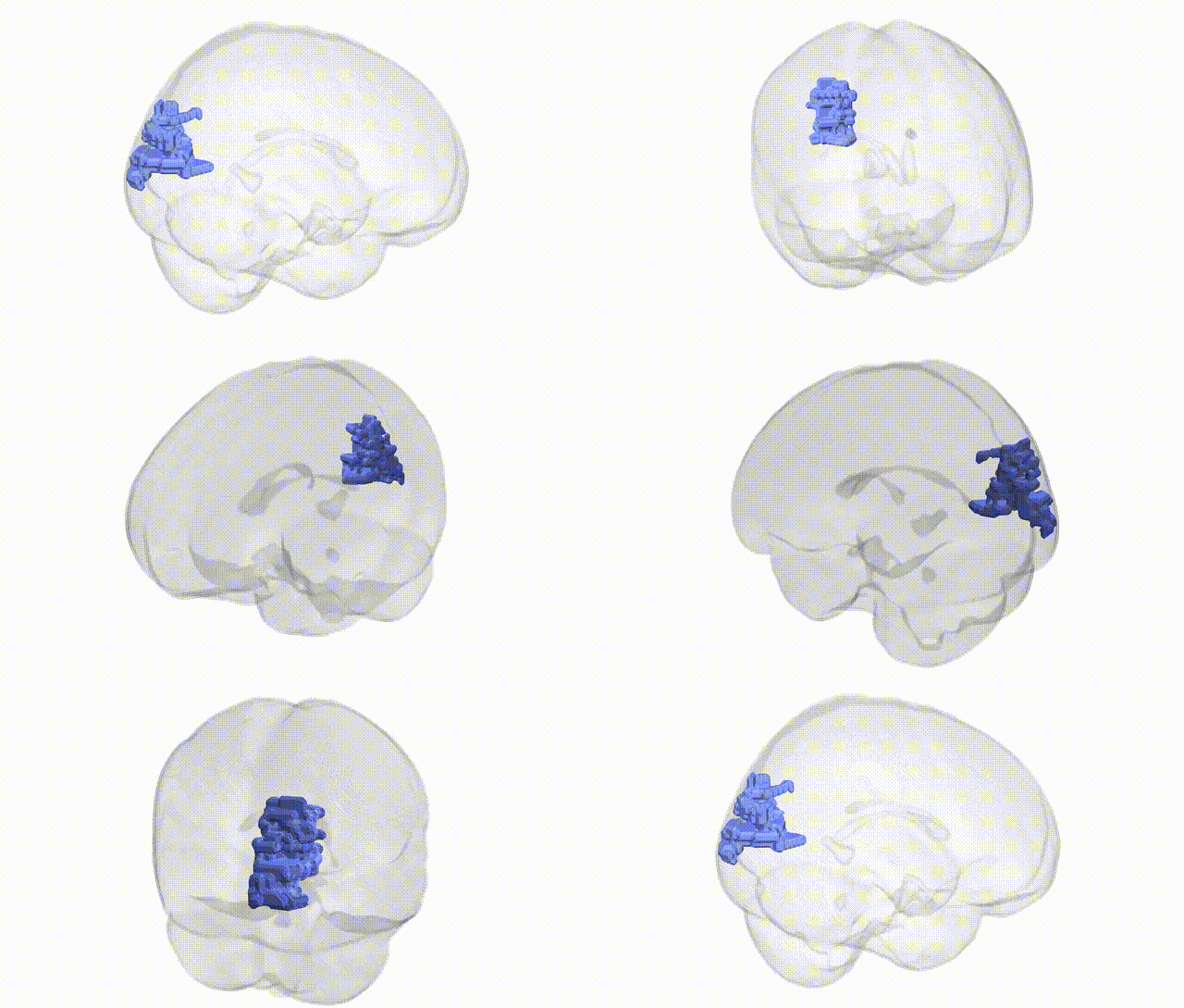
Full Quality Version: Download MP4
Wali Sidiqyar*, Gaurav Rudravaram*, Elyssa M. McMaster, Trent M. Schwartz, Adam M. Saunders, Kurt G. Schilling, Bennett A. Landman "Introducing SPINS: A Shared Public Visualization Library of Neuroanatomical Structures." Medical Imaging with Deep Learning- short paper
This resource is licensed under CC0 1.0 Universal (Public Domain).