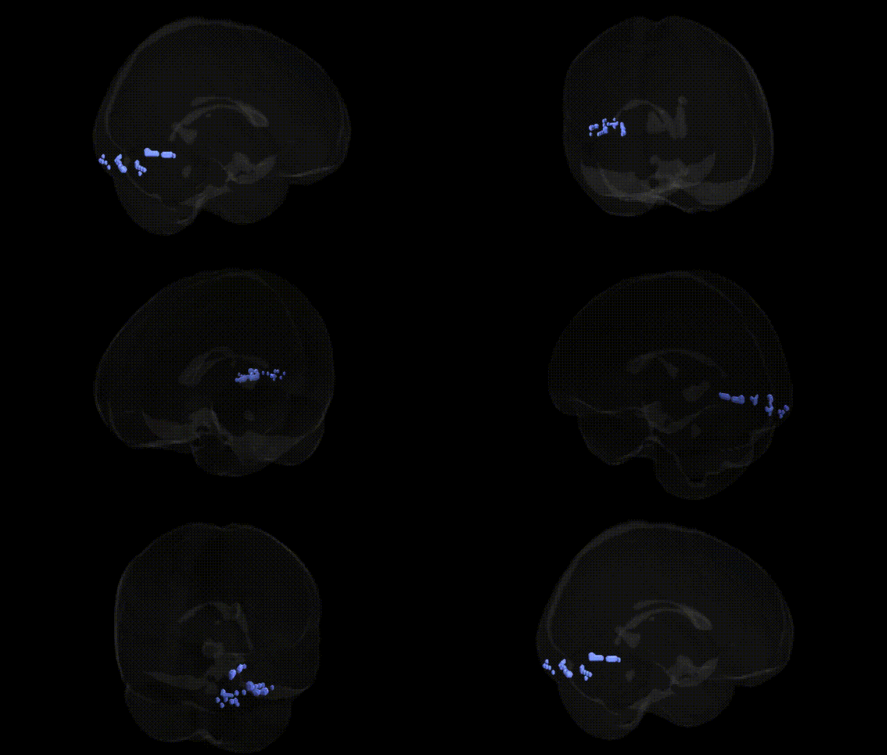
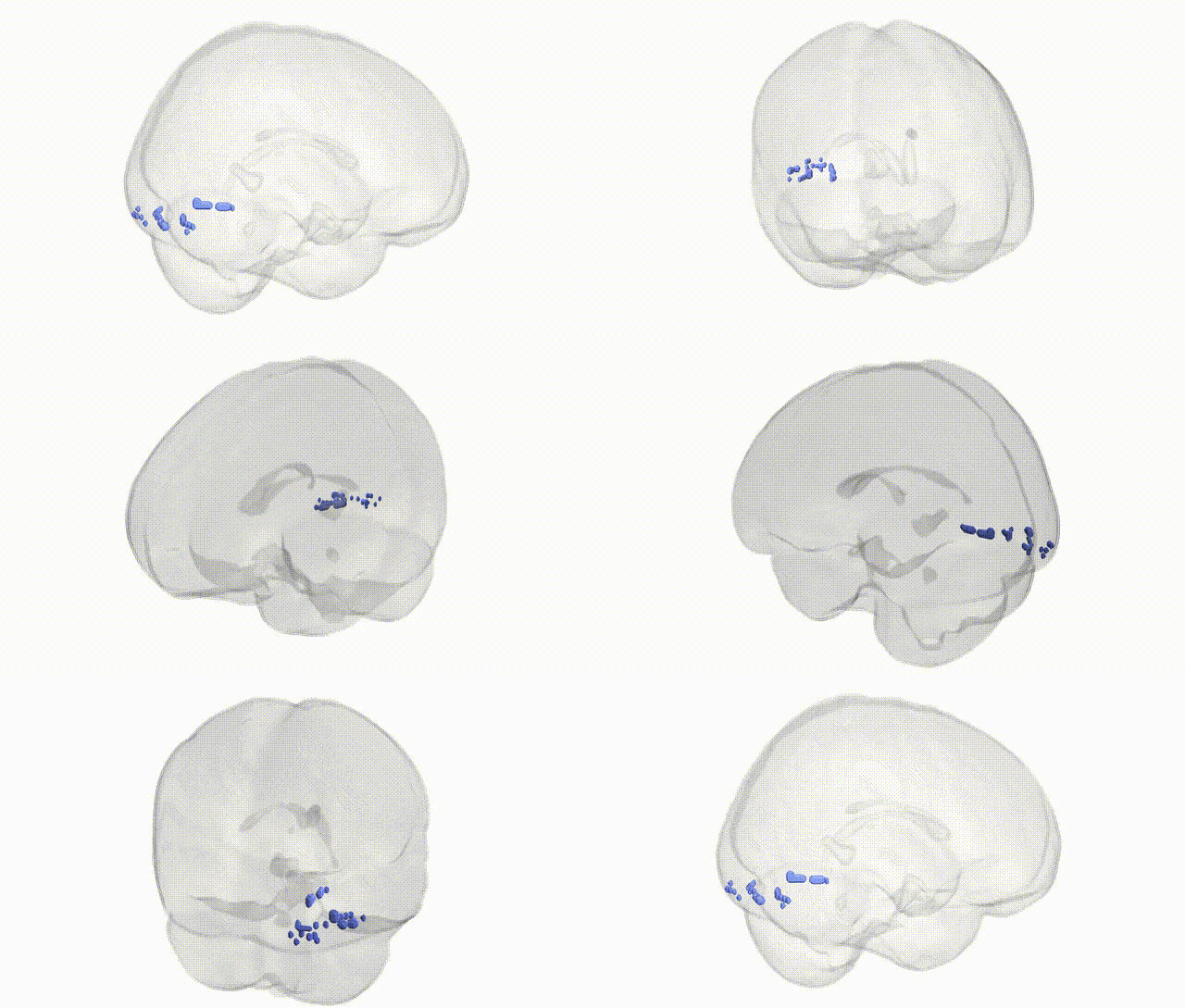
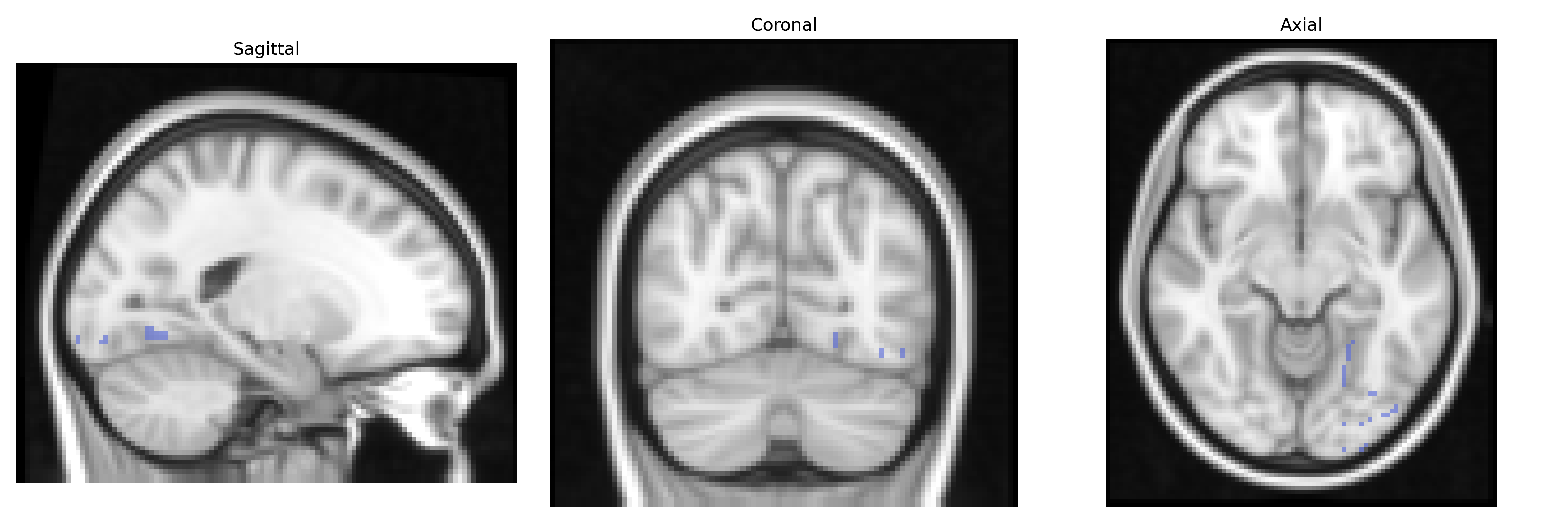
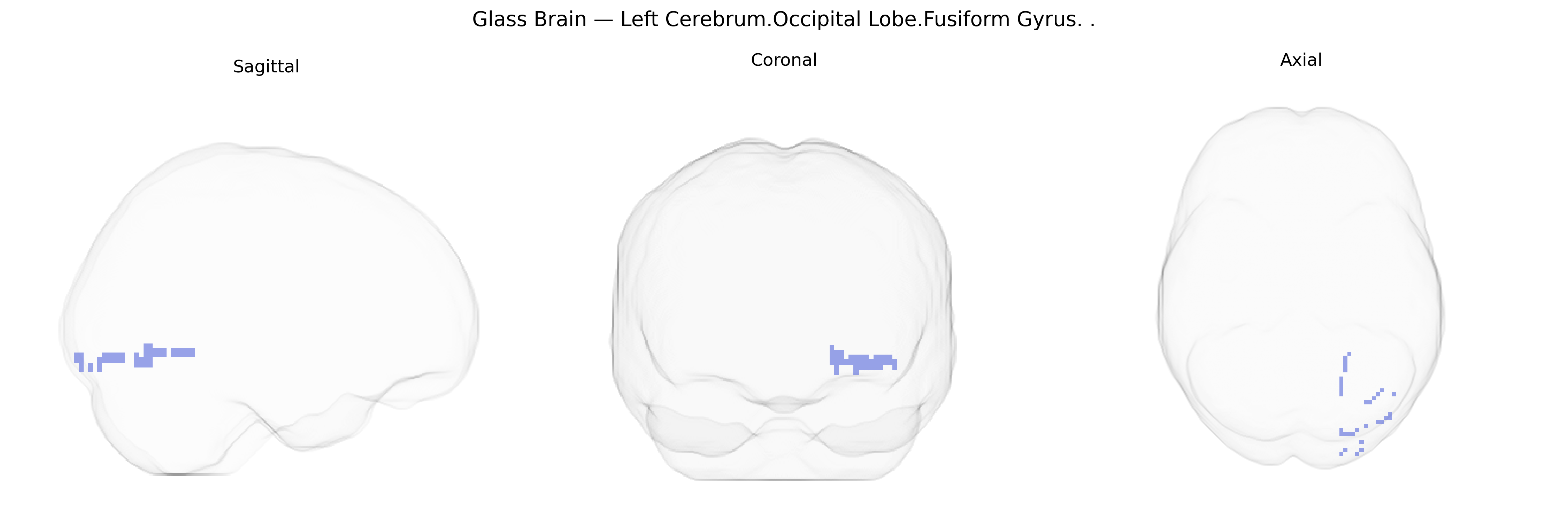
The bilateral Left Cerebrum.Occipital Lobe.Fusiform Gyrus corresponds to cortex within the fusiform gyrus located in the occipitotemporal portion of the left cerebral hemisphere, extending along the ventral surface of the temporal and occipital lobes. This region is implicated in high-level visual processing, including object recognition and category-specific perception such as faces, words, and within-class discriminations, and forms part of the ventral “what” visual stream. It has extensive connectivity with visual association areas, the inferior temporal cortex, and limbic structures, supporting integration of visual features with memory and semantic information. Functionally, it includes portions of the visual word form area and fusiform face area, although these are typically defined more narrowly within the broader fusiform gyrus. Fusiform gyrus
The bilateral fusiform gyrus of the occipital lobe, including the Left Cerebrum.Occipital Lobe.Fusiform Gyrus as defined in the Talairach 2 mm atlas, has been implicated in genetic associations primarily through imaging genetics and GWAS of brain structure and function rather than region-specific disease loci. Large-scale neuroimaging GWAS (e.g., ENIGMA, UK Biobank) have identified common variants influencing fusiform cortical thickness and surface area, notably within genes related to neurodevelopment and synaptic function such as HMGA2, microtubule-related loci, and broad polygenic influences overlapping with general brain size and cortical morphology loci. Functionally, fusiform gyrus activation patterns (especially in face and word recognition networks) show heritability and polygenic modulation, with genetic correlations reported between fusiform structure/function and traits like cognitive ability, educational attainment, and neurodevelopmental conditions. Autism spectrum disorder and dyslexia studies have repeatedly implicated atypical fusiform morphology or activation, and rare or common variants in genes involved in cortical development (e.g., CNTNAP2, FOXP2-related pathways, synaptic and chromatin-remodeling genes) have been linked to altered fusiform function, though not uniquely or exclusively to this region. Genetic risk for schizophrenia and major depressive disorder shows distributed effects on temporal–occipital cortices, including fusiform regions, via polygenic risk scores that correlate with subtle volumetric or functional differences, but specific GWAS loci are generally interpreted as affecting broad networks rather than the fusiform gyrus alone. Overall, current genetic evidence supports the fusiform gyrus as a heritable, polygenically influenced node within visual, language, and social-cognition networks, with its alterations in disorders such as autism, dyslexia, and schizophrenia reflecting widespread genetic influences on cortical development and connectivity rather than clearly defined, region-specific genetic variants.
Overview generated by GPT-4o (2026).
Region ID: 202
Hemisphere: bilateral
Atlas: Talairach labels 2mm
Full Quality Version: Download MP4
Full Quality Version: Download MP4
Wali Sidiqyar*, Gaurav Rudravaram*, Elyssa M. McMaster, Trent M. Schwartz, Adam M. Saunders, Kurt G. Schilling, Bennett A. Landman "Introducing SPINS: A Shared Public Visualization Library of Neuroanatomical Structures." Medical Imaging with Deep Learning- short paper
This resource is licensed under CC0 1.0 Universal (Public Domain).