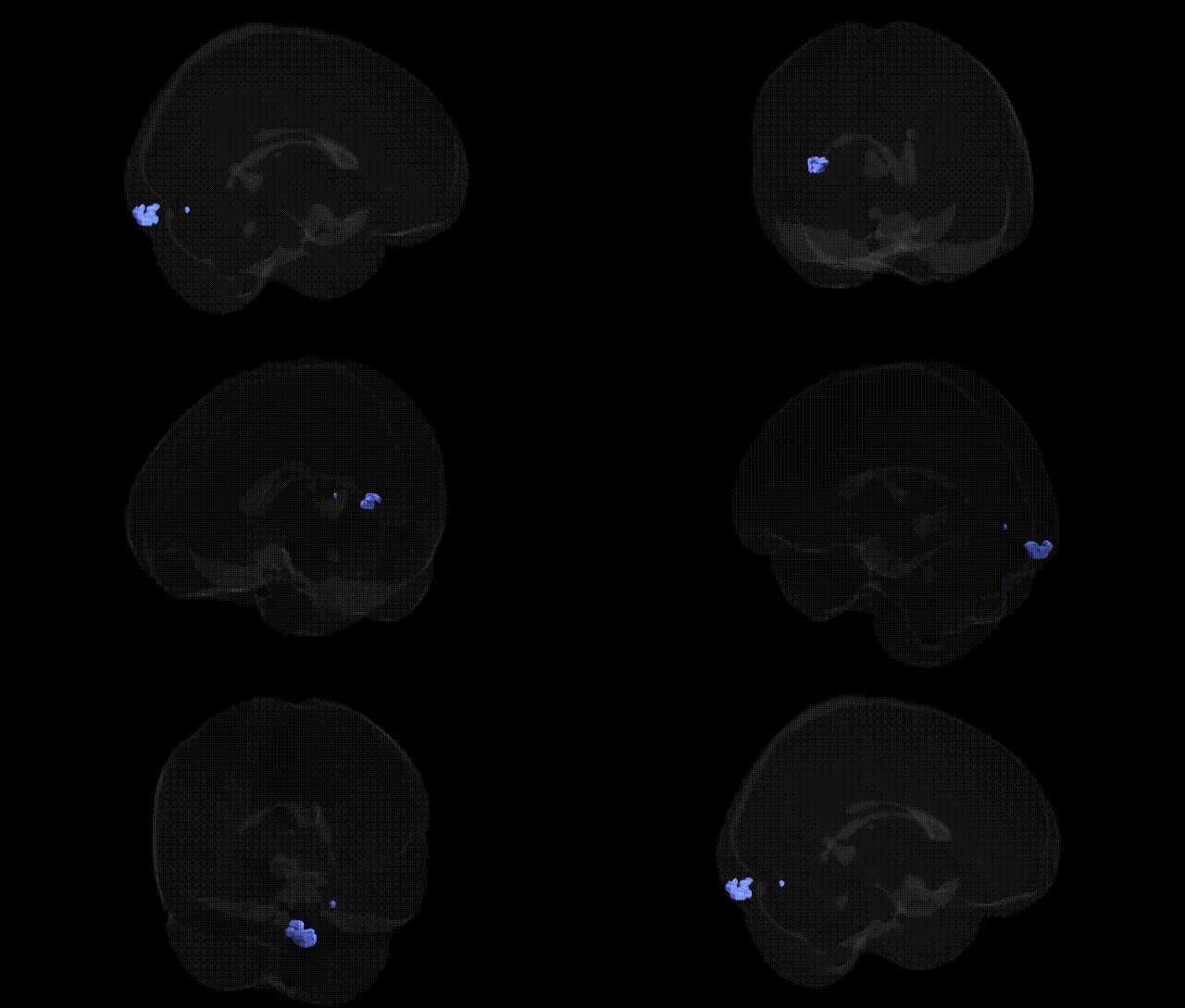
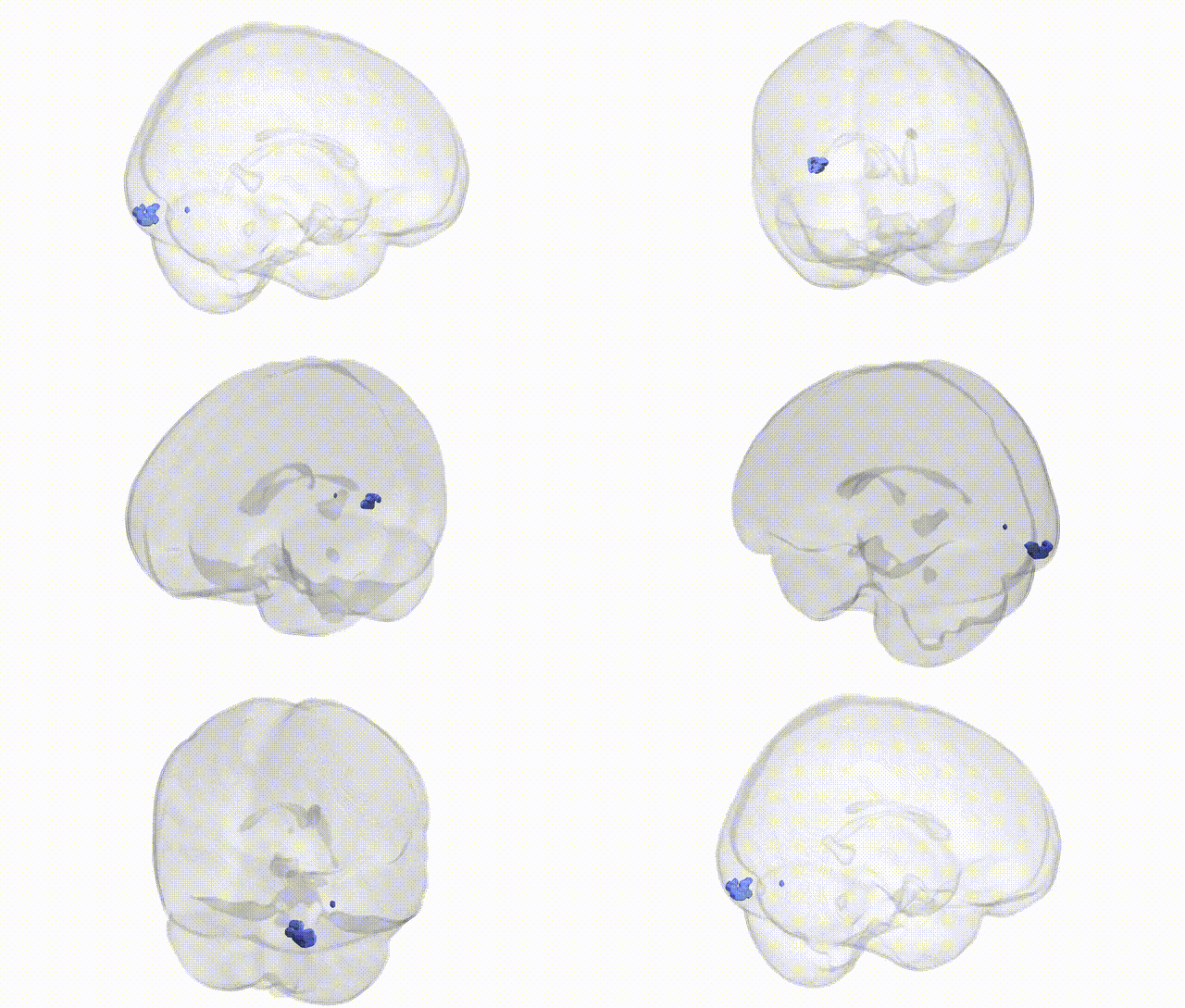
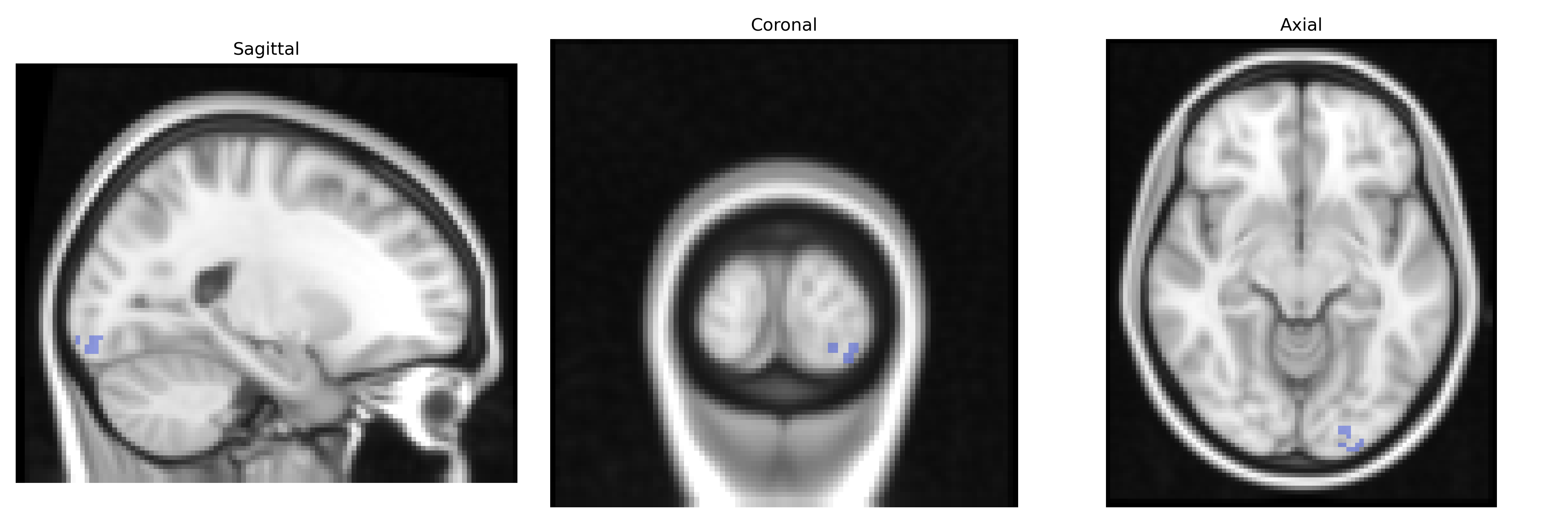
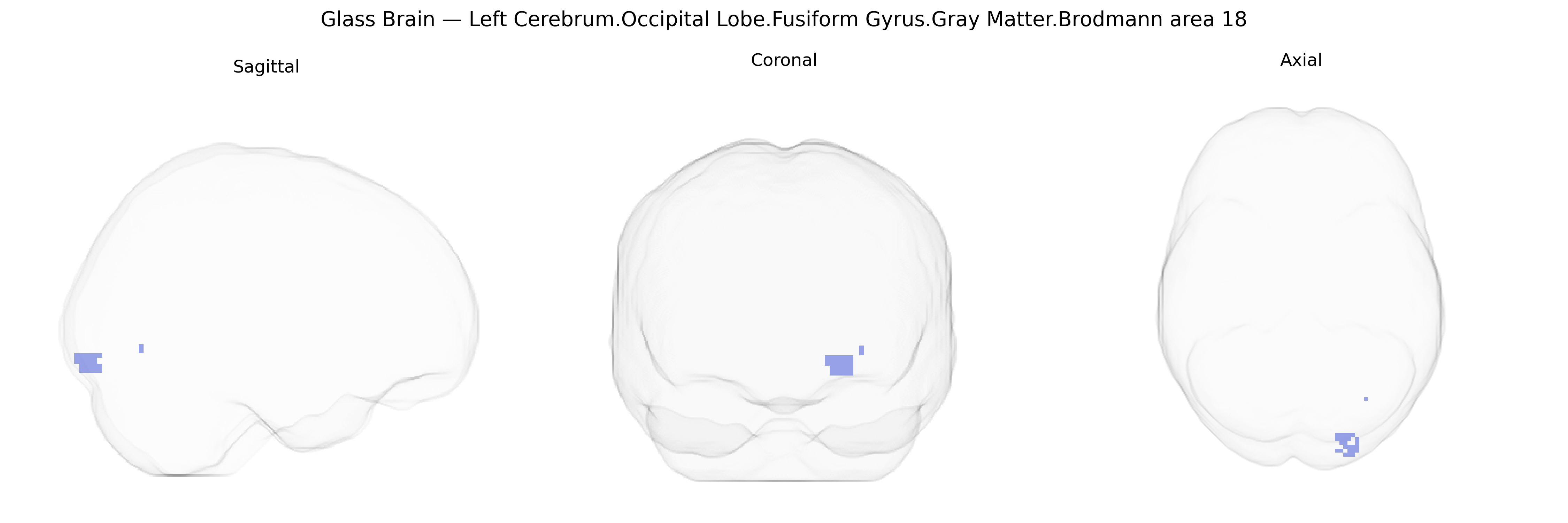
The bilateral Left Cerebrum.Occipital Lobe.Fusiform Gyrus.Gray Matter.Brodmann area 18 corresponds to secondary visual cortex (V2) located along the ventral aspect of the occipital lobe within the fusiform gyrus, adjacent to primary visual cortex (BA17). This cytoarchitectonic region participates in early-stage processing of visual information, including orientation, spatial frequency, and more complex pattern analysis, and serves as a relay between primary visual cortex and higher-order visual association areas in the temporal and parietal lobes. Activity in this region contributes to the integration of visual features that support object recognition, form perception, and aspects of visual attention. There is no direct Wikipedia article for this specific fusiform Brodmann area 18 region; a closely related structure is Brodmann area 18.
The bilateral left occipital fusiform gyrus gray matter in Brodmann area 18 (Talairach 2 mm Atlas) lies at the intersection of visual association cortex and ventral occipitotemporal regions implicated in higher-order visual processing, and genetic associations involving this area typically emerge from large-scale neuroimaging genetics and GWAS of brain structure, function, and related cognitive or clinical phenotypes. ENIGMA and UK Biobank MRI–GWAS have identified heritable variation in occipital and fusiform cortical thickness and surface area linked to loci near genes involved in neurodevelopment and synaptic function (e.g., variants in or near MIR137, APOE, and other neurodevelopmental or neurodegeneration-related genes), though most findings are reported at lobar or regional rather than fine-grained Brodmann-area resolution. Functional imaging genetics has associated BA18/occipital fusiform activation patterns with polymorphisms in dopaminergic and serotonergic genes (such as COMT and 5-HTTLPR) during visual attention, emotional face processing, and reading-related tasks, indicating genotype-dependent modulation of visual and socio-emotional processing in this region. In dyslexia and other reading disorders, GWAS have implicated loci near KIAA0319, DCDC2, and other neurodevelopmental genes, and imaging–genetic studies have linked these variants to structural and functional differences in occipitotemporal and fusiform cortices that partially overlap BA18-related networks. Schizophrenia, autism spectrum disorder, and major depression GWAS highlight polygenic influences on occipital and fusiform structure and connectivity—often via synaptic, glutamatergic, and immune-related genes—corresponding with reported abnormalities in early visual processing and face recognition that recruit BA18. However, due to the relatively coarse spatial resolution of most GWAS-based parcellations, genetic associations are typically attributed to broader occipital or ventral visual stream territories rather than specifically and exclusively to the bilateral left fusiform BA18 region.
Overview generated by GPT-4o (2026).
Region ID: 203
Hemisphere: bilateral
Atlas: Talairach labels 2mm
Full Quality Version: Download MP4
Full Quality Version: Download MP4
Wali Sidiqyar*, Gaurav Rudravaram*, Elyssa M. McMaster, Trent M. Schwartz, Adam M. Saunders, Kurt G. Schilling, Bennett A. Landman "Introducing SPINS: A Shared Public Visualization Library of Neuroanatomical Structures." Medical Imaging with Deep Learning- short paper
This resource is licensed under CC0 1.0 Universal (Public Domain).