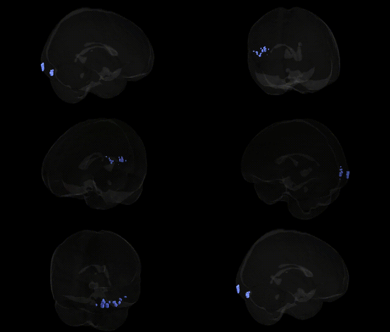
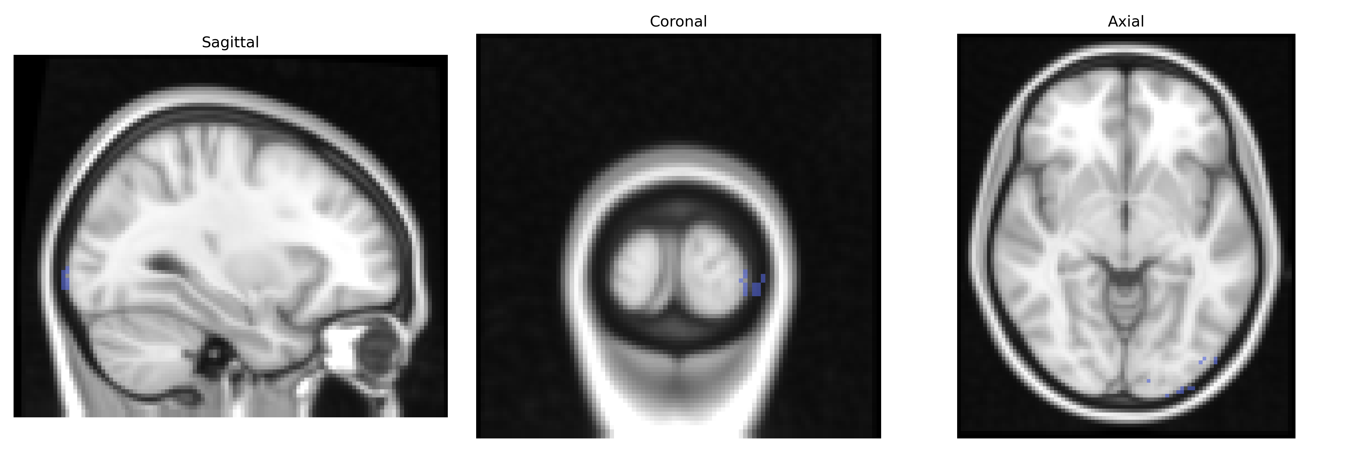
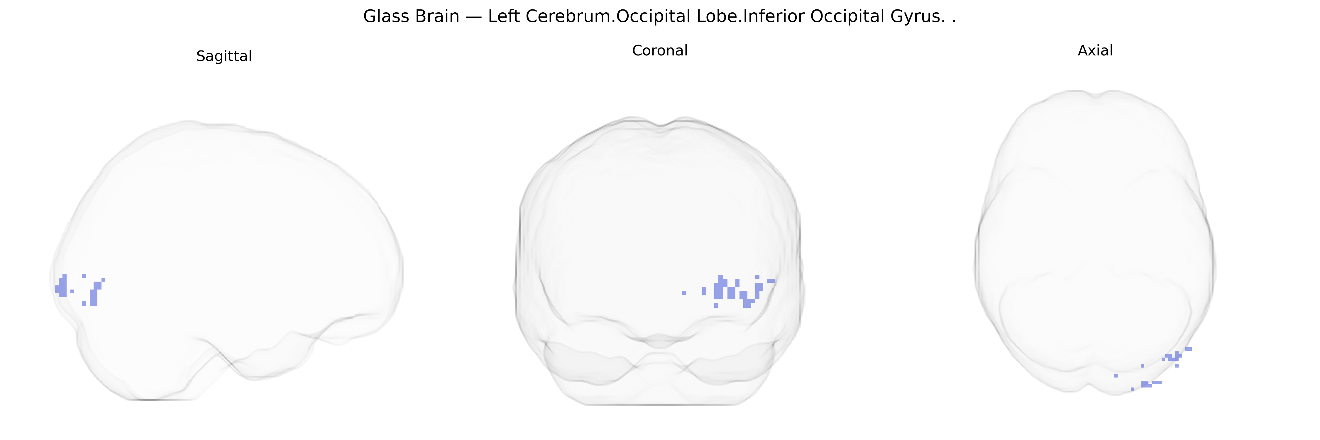
The bilateral Left Cerebrum.Occipital Lobe.Inferior Occipital Gyrus, as defined in the Talairach 2 mm atlas, refers to the inferior portions of the occipital cortex in the left hemisphere, extending ventrally along the occipital pole and posterior temporal border. This region is part of the ventral visual stream and is associated with early to intermediate stages of visual processing, including analysis of complex shapes, contours, and object features, and contributes to the integration of visual information that supports object recognition. It lies adjacent to and interacts with neighboring visual association areas, such as the lateral occipital complex and portions of the fusiform and lingual gyri. There is no direct Wikipedia article for the “Inferior Occipital Gyrus”; a closely related and overlapping visual processing area is the Lateral occipital cortex.
The bilateral Left Cerebrum Occipital Lobe Inferior Occipital Gyrus (Talairach 2 mm Atlas) lies within ventral visual association cortex, particularly the lateral occipital/ventral occipito-temporal territory, and genetic associations typically emerge from imaging genetics and GWAS of brain structure and function rather than region-specific candidate studies. GWAS of occipital cortical thickness and surface area have identified loci near genes involved in neurodevelopment and synaptic function, such as MEF2C, PAX6, TESC, FOXO3, and variants in the 15q14 and 6q22 regions, which broadly influence visual and associative cortex morphology and have downstream links to cognitive performance, educational attainment, and general intelligence. Functional MRI–GWAS work on face and object recognition implicates polygenic influences on ventral occipital activation, with contributions from genes related to synaptic plasticity (BDNF), glutamatergic signaling (GRIN2B), and white-matter development (CNTNAP2), although findings are typically reported at the level of lateral occipital or fusiform regions rather than the inferior occipital gyrus alone. Structural and functional variation in this territory has also been tied to neurodevelopmental and psychiatric disorders in genetic studies: polygenic risk scores for schizophrenia and autism spectrum disorder show associations with altered occipital and ventral visual cortex thickness or connectivity, and rare CNVs (for example at 16p11.2 and 22q11.2) consistently affect posterior cortical development, including occipital association areas. In addition, GWAS of migraine and visual aura, as well as occipital lobe epilepsy, highlight genes involved in vascular regulation and cortical excitability (e.g., CACNA1A, ATP1A2, TRPM8), which may modulate susceptibility of occipital cortex, including the inferior occipital gyrus, to spreading depolarization and hyperexcitability. Overall, genetic influences on this region reflect general determinants of cortical development and visual processing rather than highly specific loci unique to the inferior occipital gyrus.
Overview generated by GPT-4o (2026).
Region ID: 251
Hemisphere: bilateral
Atlas: Talairach labels 2mm
Full Quality Version: Download MP4
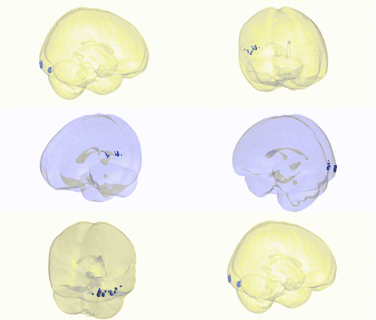
Full Quality Version: Download MP4
Wali Sidiqyar*, Gaurav Rudravaram*, Elyssa M. McMaster, Trent M. Schwartz, Adam M. Saunders, Kurt G. Schilling, Bennett A. Landman "Introducing SPINS: A Shared Public Visualization Library of Neuroanatomical Structures." Medical Imaging with Deep Learning- short paper
This resource is licensed under CC0 1.0 Universal (Public Domain).