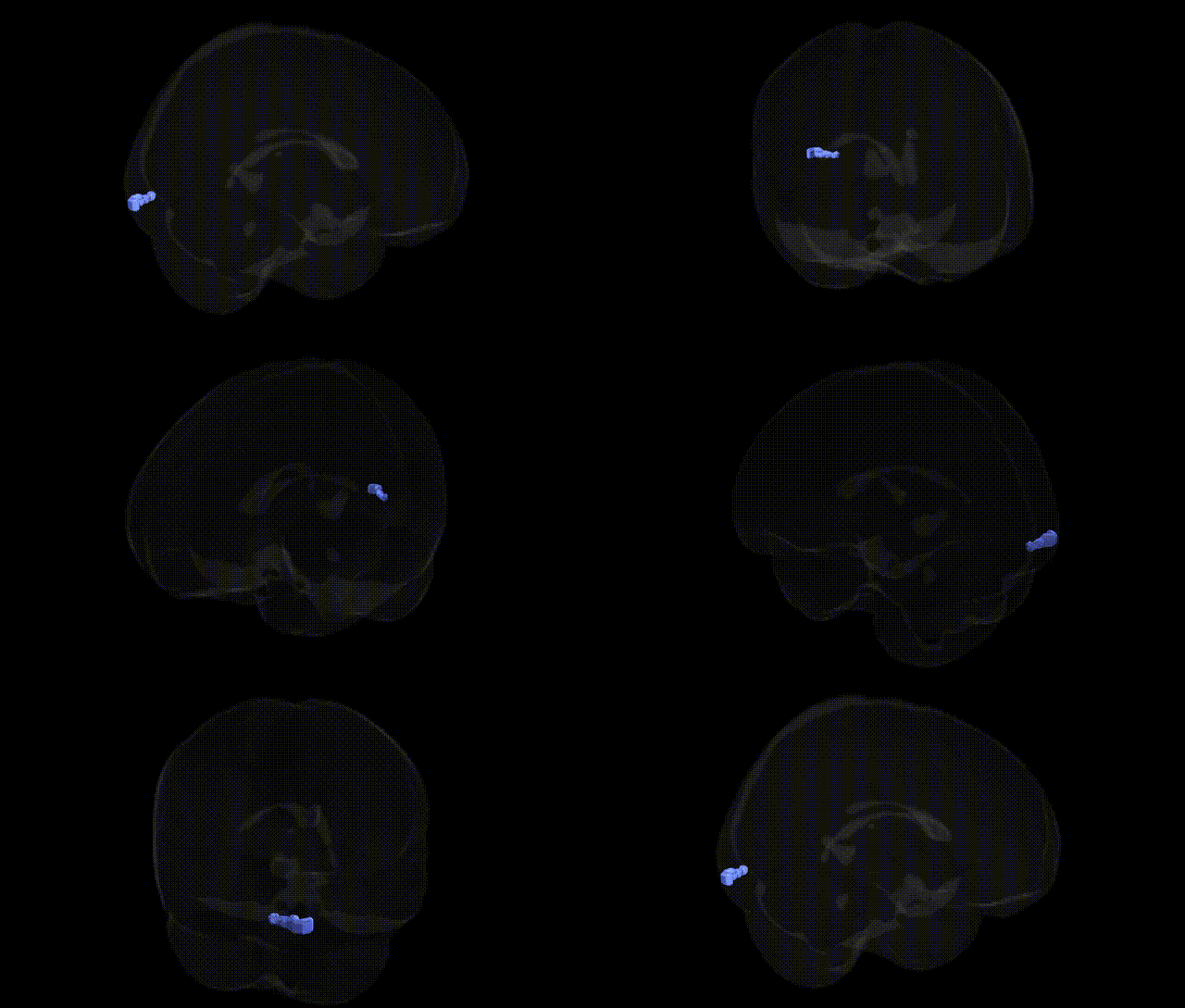
The bilateral Left Cerebrum.Occipital Lobe.Inferior Occipital Gyrus.Gray Matter.Brodmann area 17 corresponds to primary visual cortex (V1), the main cortical recipient of visual input from the lateral geniculate nucleus of the thalamus. Situated along the banks of the calcarine fissure in the occipital lobe, this cytoarchitectonic region (BA17) is characterized by a prominent granular layer (layer IV) specialized for processing retinotopically organized visual information, with distinct upper and lower visual field representations mapped across its cortical surface. Neuronal populations within BA17 are tuned to basic visual features such as orientation, spatial frequency, contrast, and motion, forming the initial cortical stage of visual perception that feeds forward to extrastriate visual areas (e.g., V2, V3). The inferior occipital portion of BA17 contributes particularly to processing visual input from specific sectors of the contralateral visual field and serves as a critical hub for conscious visual experience and visually guided behavior.
Primary visual cortex
The bilateral inferior occipital gyrus gray matter within Brodmann area 17 (primary visual cortex) has been implicated in genetic studies of cortical structure, visual function, and neuropsychiatric risk, though most associations target V1 as a whole rather than this subregion specifically. Large-scale MRI GWAS, such as those using UK Biobank and ENIGMA data, have identified multiple common variants influencing occipital cortical thickness and surface area, including loci near genes involved in neurodevelopment, synaptic signaling, and axon guidance (for example, variants near PAX6, TBR1, and other corticogenesis-related genes), with some of these occipital structural traits showing genetic correlations with general cognitive ability, educational attainment, and risk for psychiatric disorders. Regional heritability analyses consistently demonstrate substantial genetic contributions to volume and thickness in primary visual cortex, and twin studies indicate strong heritability of V1 surface area and organization, supporting a genetic basis for individual differences in visual processing. GWAS of visual acuity, refractive error (myopia), and other ophthalmologic traits have linked genes such as GJD2, RASGRF1, and ZIC2 to both ocular parameters and occipital activation patterns, suggesting a genetically mediated pathway from eye structure to cortical visual representation. Neuroimaging genetics in schizophrenia, major depression, autism spectrum disorder, and bipolar disorder occasionally report disorder- or risk-allele–associated alterations in occipital and BA17 gray matter (for example, reduced occipital cortical thickness or volume in carriers of certain schizophrenia polygenic risk profiles), though these effects are generally less robust and more diffuse than those observed in frontal or temporal regions. In summary, the inferior occipital gyrus portion of BA17 participates in a broader, highly heritable occipital network whose structure and function are modulated by variants in neurodevelopmental and synaptic genes and whose morphology shows polygenic overlap with visual traits, cognition, and neuropsychiatric risk, but no single gene or disorder has been uniquely and consistently tied to this specific Talairach-defined subregion.
Overview generated by GPT-4o (2026).
Region ID: 303
Hemisphere: bilateral
Atlas: Talairach labels 2mm
Full Quality Version: Download MP4
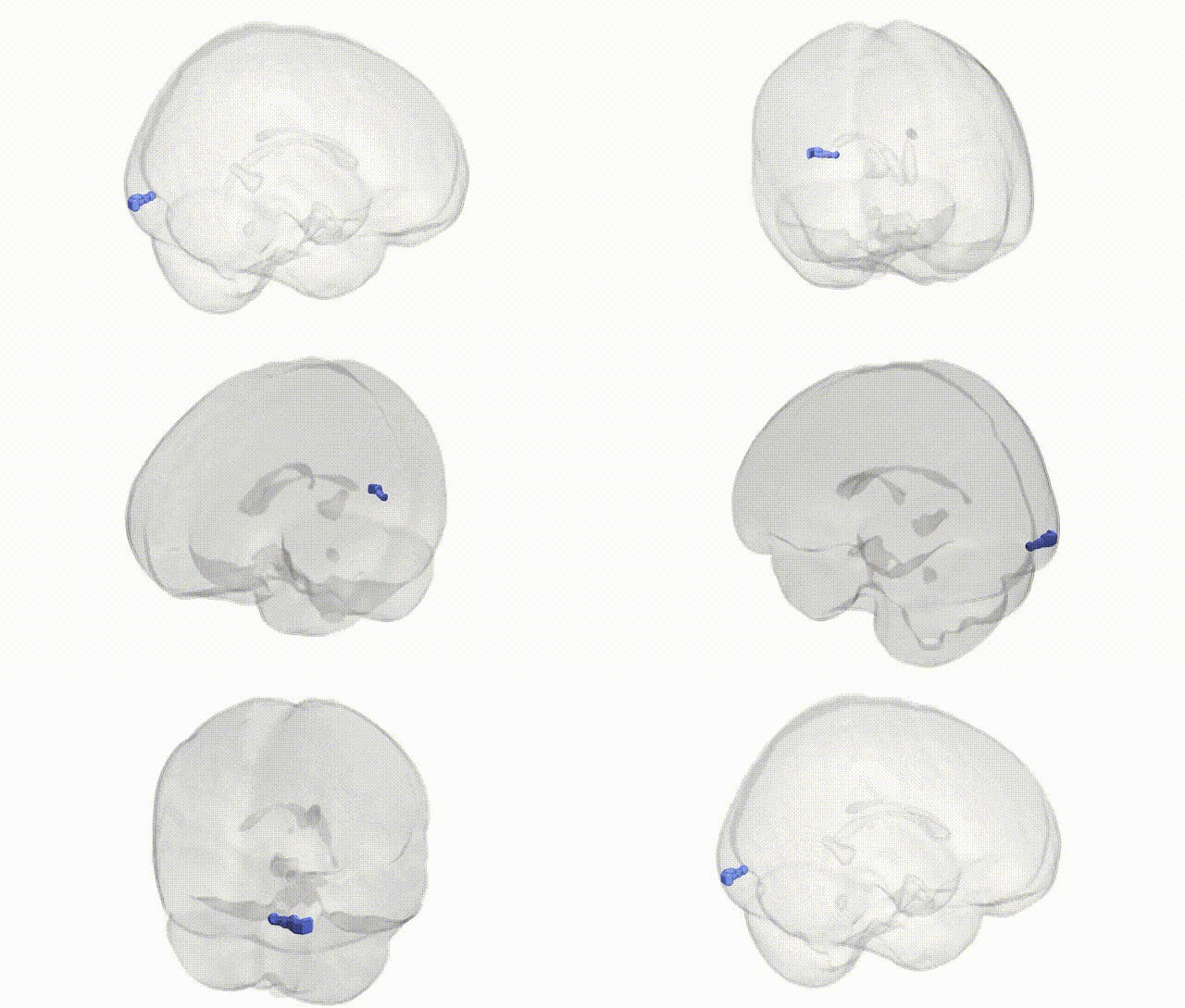
Full Quality Version: Download MP4
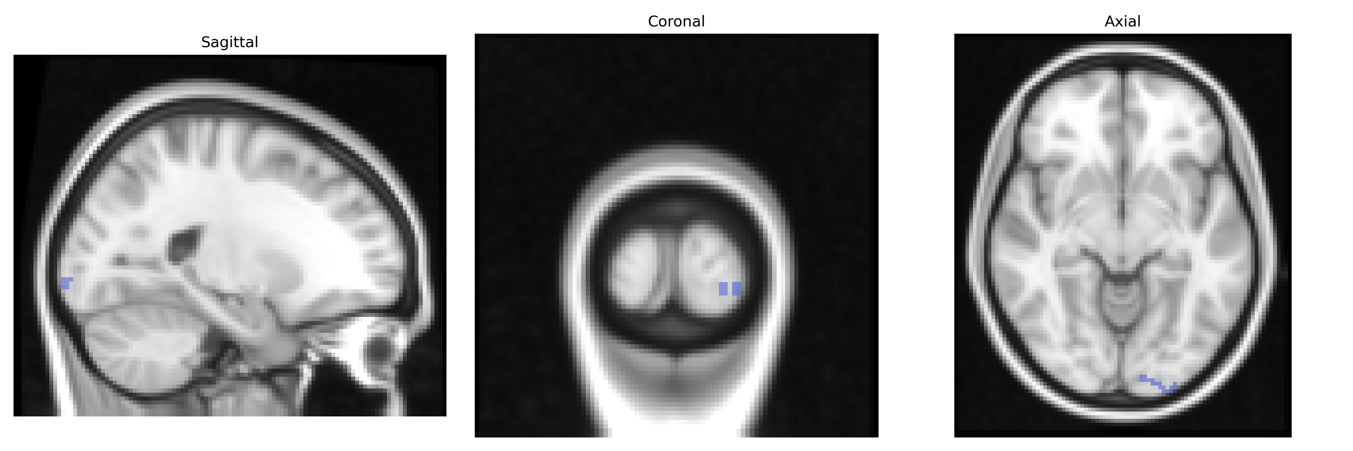
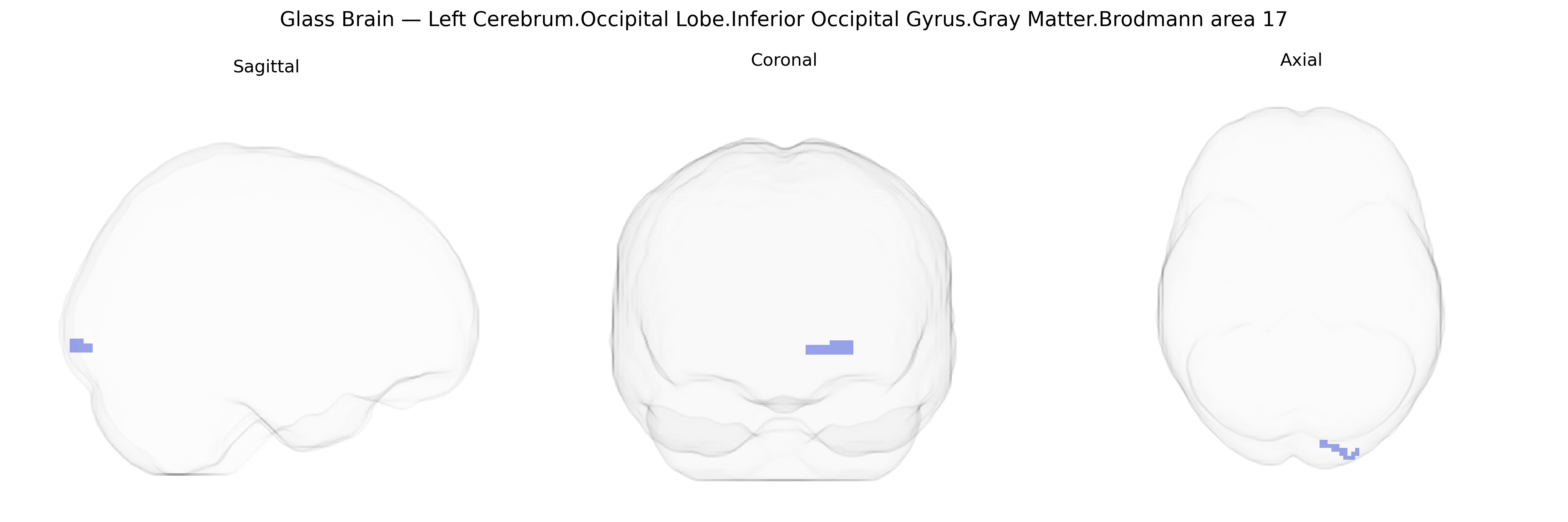
Wali Sidiqyar*, Gaurav Rudravaram*, Elyssa M. McMaster, Trent M. Schwartz, Adam M. Saunders, Kurt G. Schilling, Bennett A. Landman "Introducing SPINS: A Shared Public Visualization Library of Neuroanatomical Structures." Medical Imaging with Deep Learning- short paper
This resource is licensed under CC0 1.0 Universal (Public Domain).