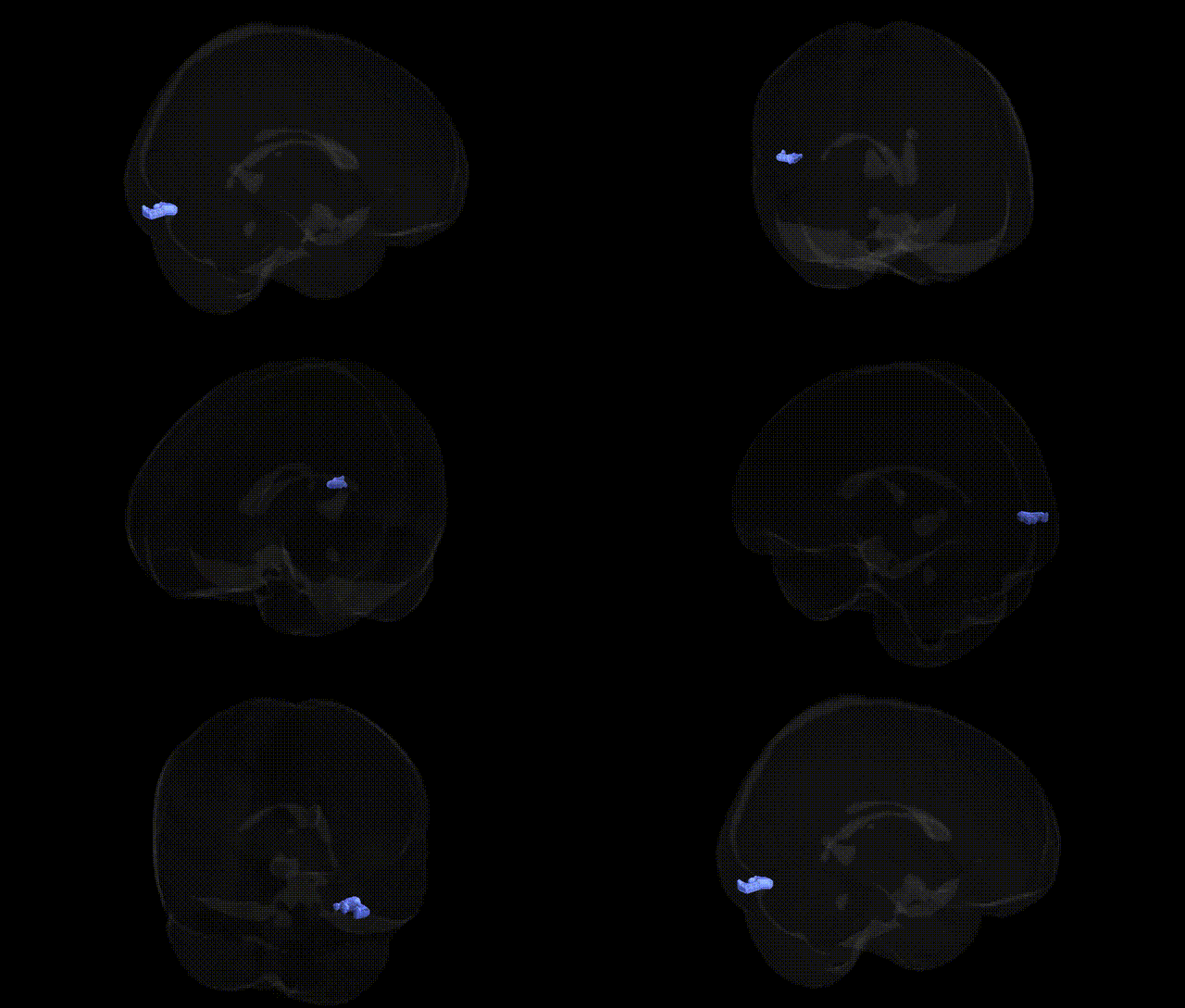
The bilateral Left Cerebrum.Occipital Lobe.Inferior Occipital Gyrus.Gray Matter.Brodmann area 19 corresponds to a higher-order visual association cortex located in the inferior portion of the occipital lobe, lateral and ventral to primary visual cortex (BA17) and surrounding BA18. This region participates in complex visual processing, including integration of visual features, object and form recognition, and the association of visual input with stored representations, contributing to the ventral “what” visual pathway. Histologically, BA19 is characterized by a heterotypical granular neocortex with well-developed granular layers and dense cortico-cortical connections to neighboring occipital, temporal, and parietal association areas. There is no direct Wikipedia article for this exact Talairach label; a closely related structure is Brodmann area 19.
Brodmann area 19 of the inferior occipital gyrus (bilateral left cerebrum, occipital lobe, gray matter) is a key component of the extrastriate visual cortex, and genetic associations involving this region primarily emerge from neuroimaging GWAS and studies of neuropsychiatric and neurodevelopmental disorders with visual processing components. Large-scale imaging genetics consortia (e.g., ENIGMA, UK Biobank) have identified common variants near genes involved in neurodevelopment, synaptic function, and axon guidance (such as MEF2C, DCC, PAX6, and genes in Wnt and MAPK pathways) that are associated with occipital cortical thickness, surface area, and gray-matter volume, which include BA19 territory. Polygenic risk scores for schizophrenia, bipolar disorder, and major depression have been linked to altered occipital cortex structure and activation during visual tasks, implicating broad neurodevelopmental risk architecture rather than single loci specific to BA19. Genetic variants associated with reading and dyslexia (e.g., near DCDC2, KIAA0319) and with autism spectrum disorder have been related to functional and structural differences in posterior visual regions involved in higher-order visual integration, overlapping BA19. In addition, GWAS of visual acuity, refractive error, and migraine have reported associations with cortical measures in the occipital lobe, suggesting shared genetic influences on peripheral and central visual systems, although these findings generally implicate distributed networks rather than isolating Brodmann area 19 as a uniquely affected locus.
Overview generated by GPT-4o (2026).
Region ID: 414
Hemisphere: bilateral
Atlas: Talairach labels 2mm
Full Quality Version: Download MP4
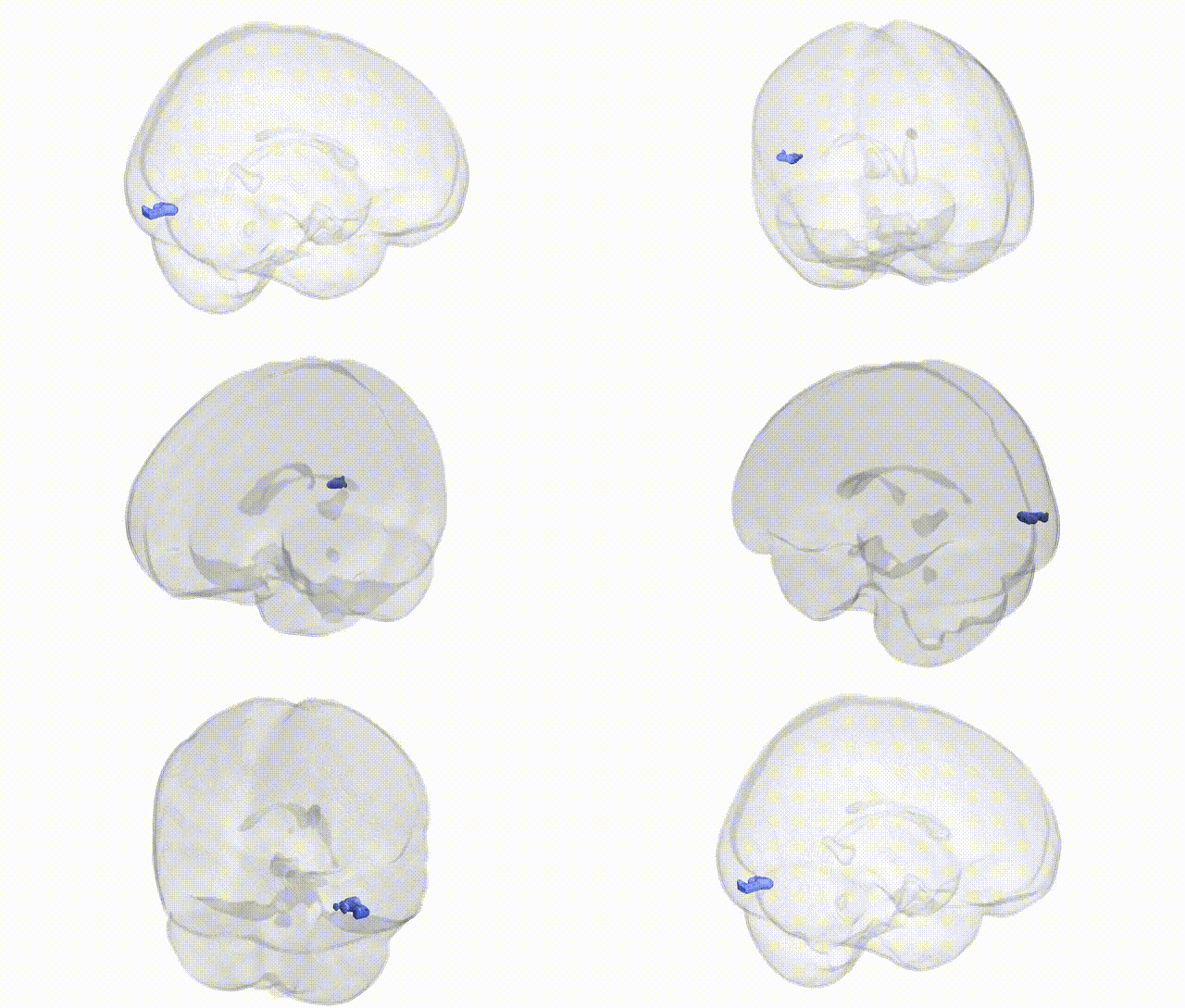
Full Quality Version: Download MP4
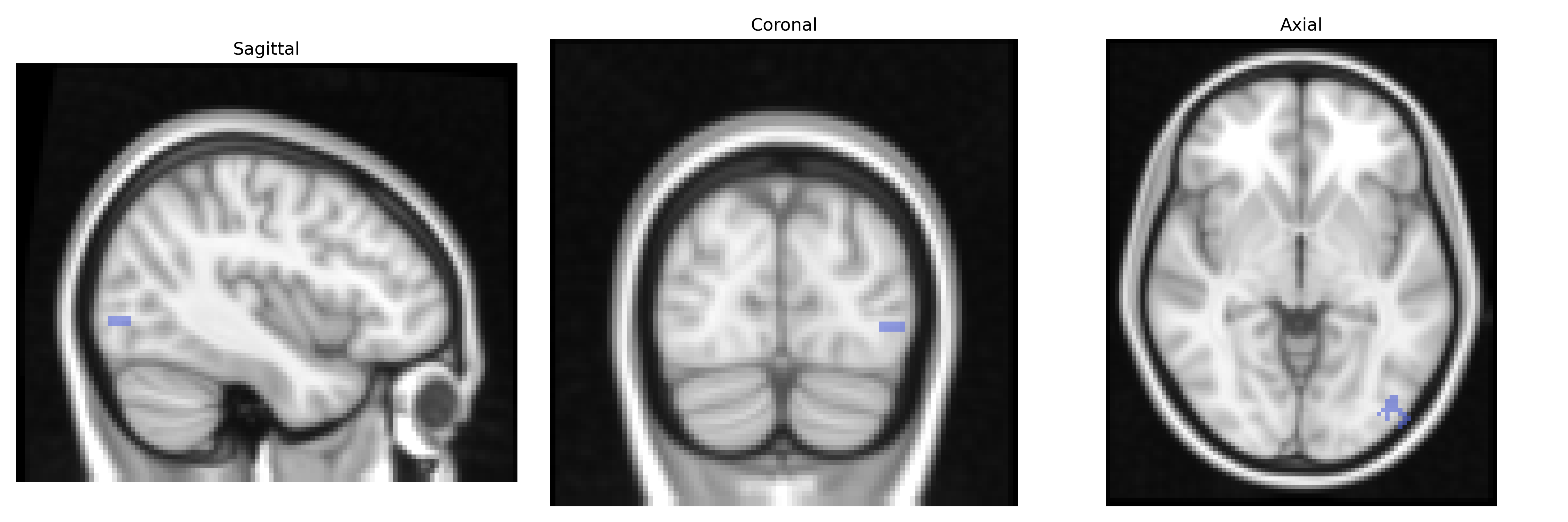
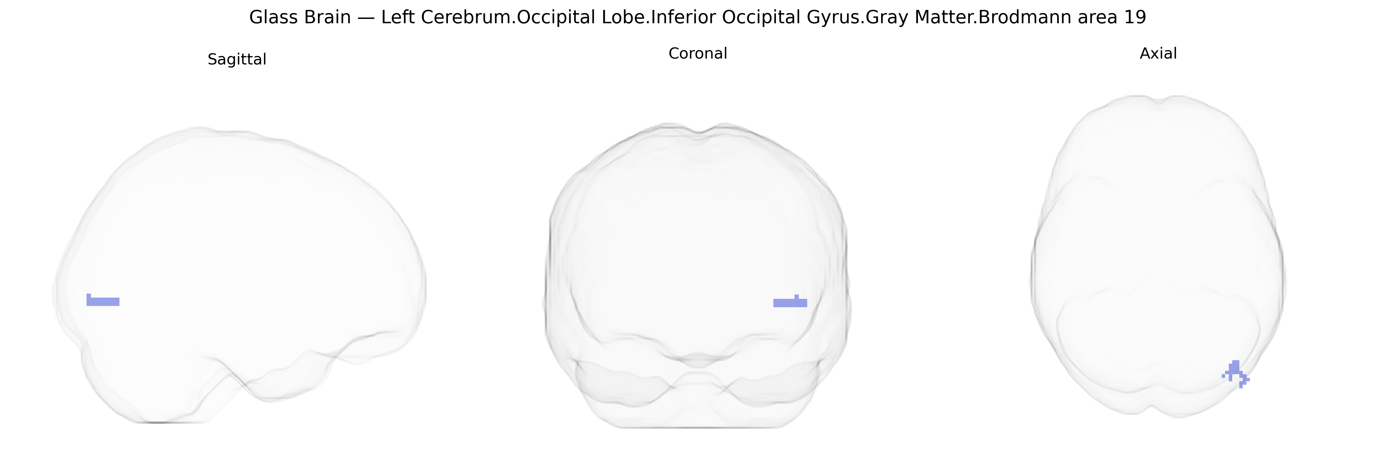
Wali Sidiqyar*, Gaurav Rudravaram*, Elyssa M. McMaster, Trent M. Schwartz, Adam M. Saunders, Kurt G. Schilling, Bennett A. Landman "Introducing SPINS: A Shared Public Visualization Library of Neuroanatomical Structures." Medical Imaging with Deep Learning- short paper
This resource is licensed under CC0 1.0 Universal (Public Domain).