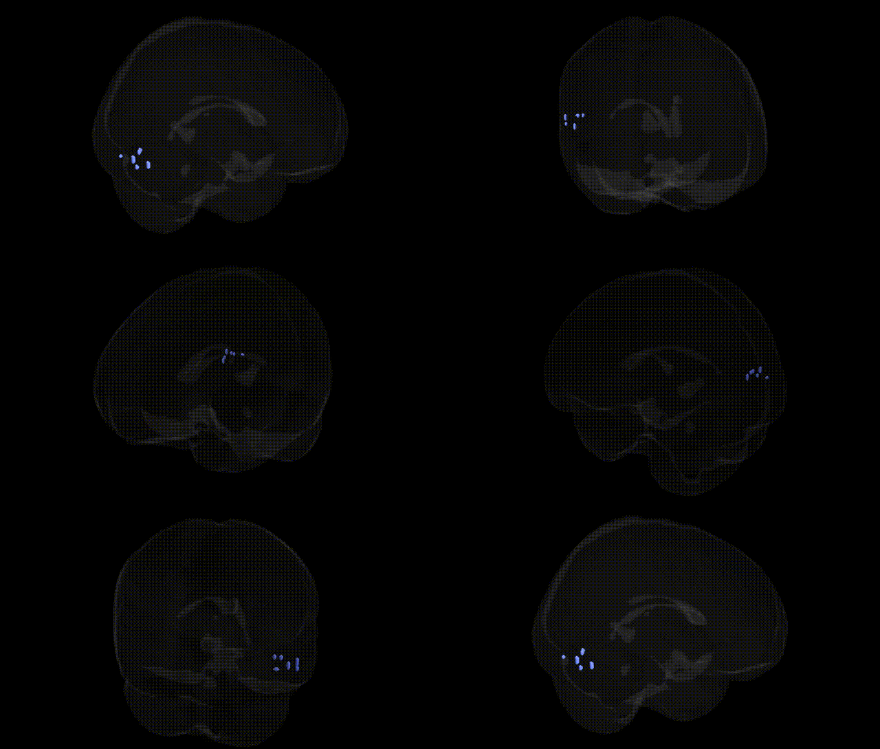
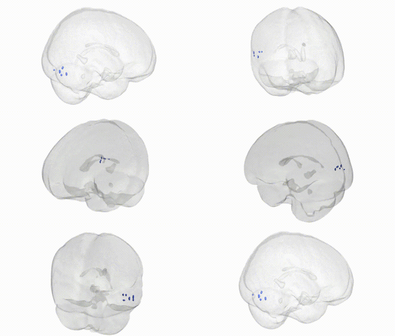
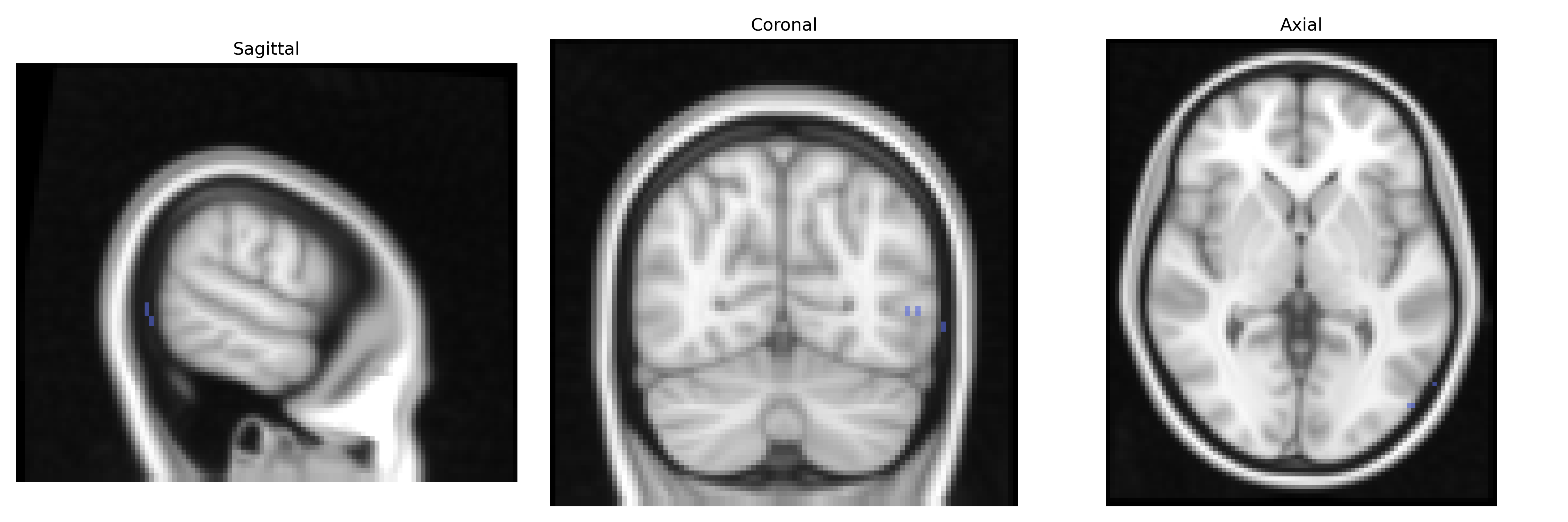
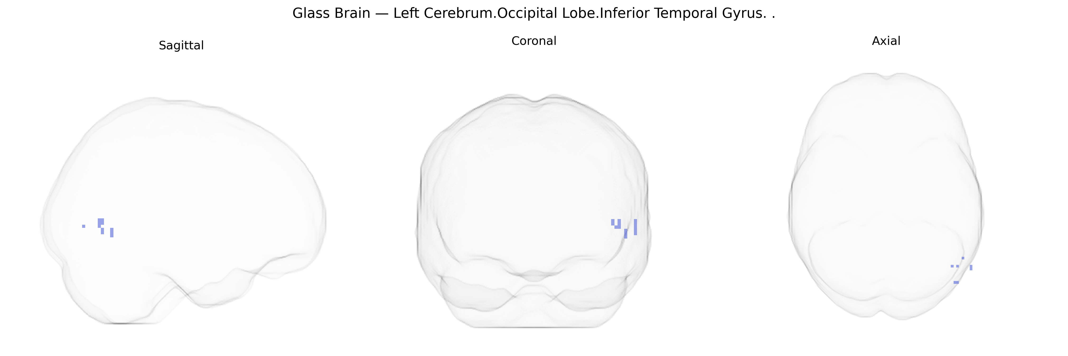
The bilateral Left Cerebrum.Occipital Lobe.Inferior Temporal Gyrus corresponds to the inferior portion of the temporal cortex extending into the occipitotemporal region on the left hemisphere, a key part of the ventral visual processing stream (“what pathway”). This region is involved in high-level visual object recognition, including complex form and pattern processing, and contributes to the perception of faces, scenes, and written words through integration of visual inputs from occipital areas. It also participates in multimodal association functions, linking visual representations with semantic and memory-related information stored in temporal lobe structures. There is no direct link for this composite Talairach label; a closely related structure is the inferior temporal gyrus: Inferior temporal gyrus.
The bilateral left cerebrum occipital lobe inferior temporal gyrus (ITG), as defined in the Talairach 2 mm atlas, participates in high-level visual processing, object recognition, and aspects of semantic memory, and genetic associations involving this region largely emerge from imaging-genetics and GWAS of cortical structure and function. Large-scale cortical thickness and surface-area GWAS (e.g., ENIGMA, UK Biobank) have implicated common variants in and near genes such as CENPW, KIAA0586, MIR548F3, and DAAM1 with occipital and temporal cortical morphology, including inferior temporal regions, suggesting polygenic influences on its development and maintenance. In functional imaging genetics, ITG activation during visual, face, and language-related tasks has been associated with allelic variation in genes affecting synaptic plasticity and neurotransmission, including BDNF (notably Val66Met), COMT, and dopaminergic and glutamatergic pathway genes, although these effects are typically small and task-specific. Neurodevelopmental and psychiatric GWAS—particularly for autism spectrum disorder, schizophrenia, and dyslexia—have repeatedly identified risk loci that, in downstream imaging work, show altered structure or functional connectivity involving inferior temporal and adjacent occipitotemporal regions, linking variants in synaptic, cell-adhesion, and chromatin-remodeling genes (e.g., CNTNAP2, NRXN1, CACNA1C, and others) to atypical visual and language-related processing supported by this gyrus. In Alzheimer’s disease and related dementias, risk variants such as APOE ε4 and loci near CLU, PICALM, and BIN1 have been associated with accelerated cortical thinning and hypometabolism in temporal and occipital association cortices, including inferior temporal areas, reinforcing the role of lipid transport, endocytosis, and amyloid-related pathways in the vulnerability of this region. Overall, GWAS and imaging-genetic findings indicate that the inferior temporal gyrus is influenced by a widely distributed polygenic architecture rather than by region-specific loci, with many associated variants acting through broad neurodevelopmental, synaptic, and neurodegenerative mechanisms that manifest in ITG structure, connectivity, and task-related activation.
Overview generated by GPT-4o (2026).
Region ID: 419
Hemisphere: bilateral
Atlas: Talairach labels 2mm
Full Quality Version: Download MP4
Full Quality Version: Download MP4
Wali Sidiqyar*, Gaurav Rudravaram*, Elyssa M. McMaster, Trent M. Schwartz, Adam M. Saunders, Kurt G. Schilling, Bennett A. Landman "Introducing SPINS: A Shared Public Visualization Library of Neuroanatomical Structures." Medical Imaging with Deep Learning- short paper
This resource is licensed under CC0 1.0 Universal (Public Domain).