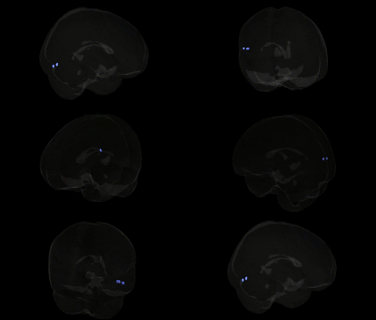
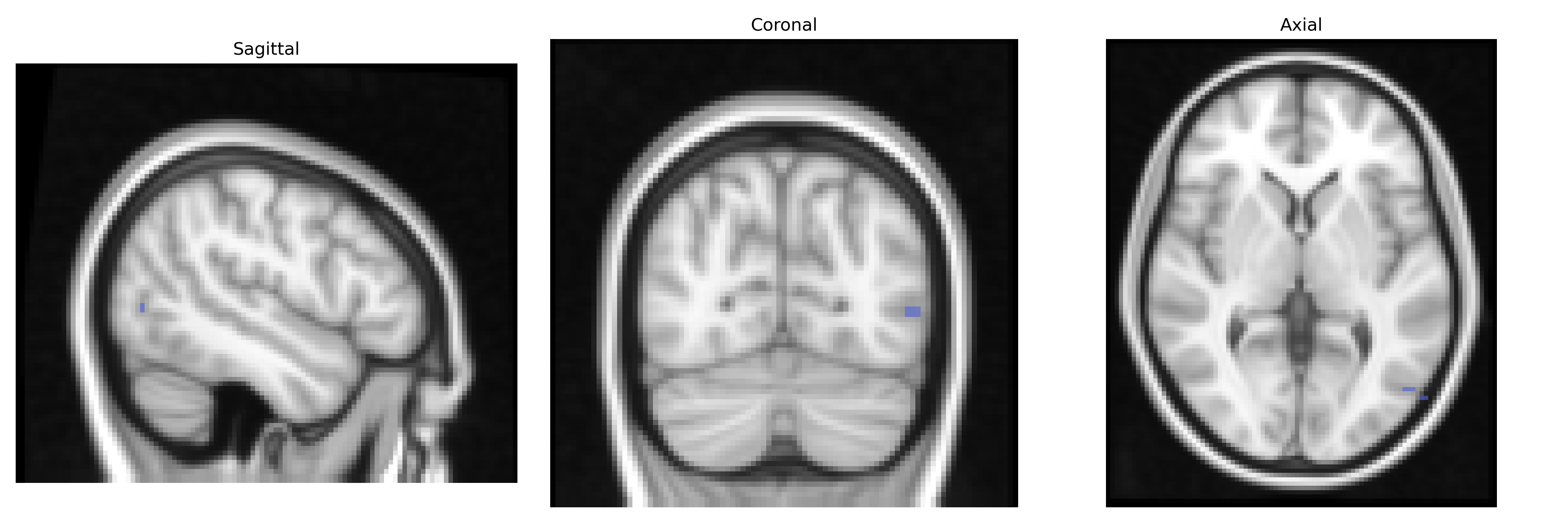
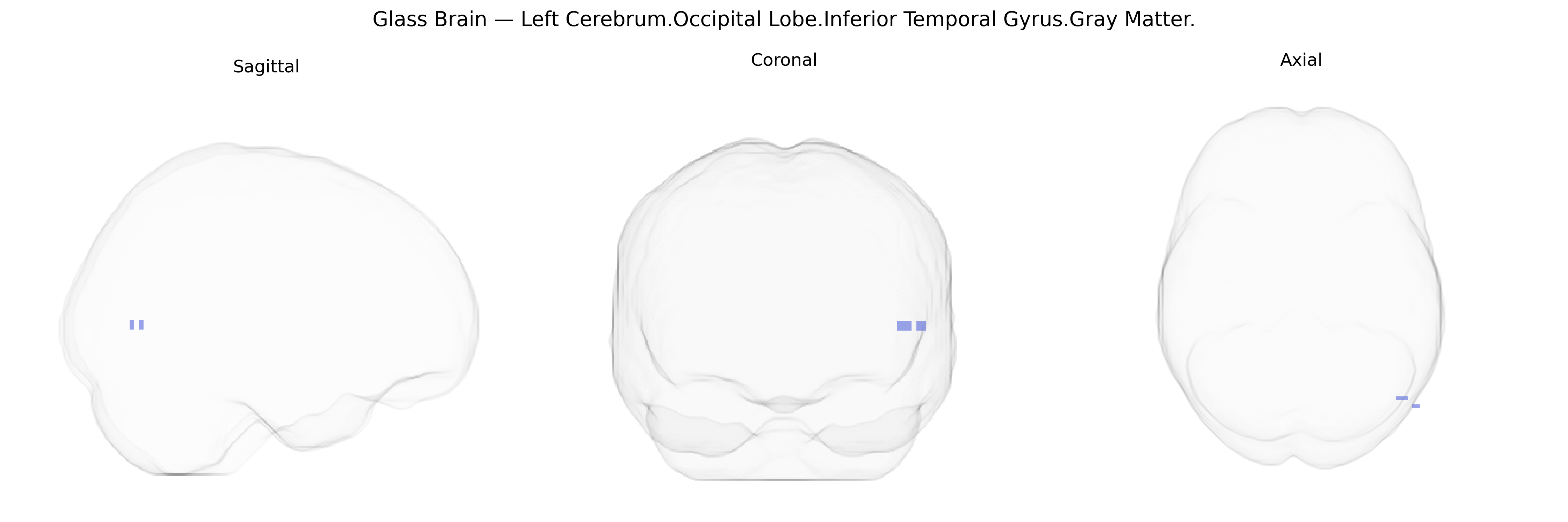
The bilateral Left Cerebrum.Occipital Lobe.Inferior Temporal Gyrus.Gray Matter corresponds to cortical tissue located along the ventral aspect of the temporal lobe near its junction with the occipital lobe, forming part of the lateral occipital–temporal cortex. This region participates in high-level visual processing, including object recognition and aspects of visual memory, integrating complex form, color, and texture information from primary and associative visual areas. Functionally, it contributes to the ventral “what” pathway and is involved in category-specific processing such as faces, objects, and possibly word forms, depending on the precise subregion. Its gray matter consists of layered neocortex with dense reciprocal connections to adjacent temporal and occipital areas, as well as to limbic and frontal regions implicated in memory and semantic processing. There is no direct link for this combined occipital–inferior temporal designation; a closely related structure is the Inferior temporal gyrus.
The bilateral Left Cerebrum Occipital Lobe Inferior Temporal Gyrus gray matter region (as defined in the Talairach 2 mm Atlas, overlapping ventral occipitotemporal cortex including lateral fusiform and inferior temporal areas) is implicated by imaging genetics and GWAS primarily through its roles in higher-order visual processing, object recognition, and reading-related functions. Large neuroimaging GWAS (e.g., ENIGMA, UK Biobank) have identified polygenic influences on cortical thickness and surface area in inferior temporal/ventral occipitotemporal regions, with enrichment for neurodevelopmental and synaptic genes, though few locus-specific findings target this exact Talairach parcel; associated loci often include variants near genes involved in axon guidance (e.g., ROBO, SLIT families), chromatin and transcriptional regulation (e.g., KIAA0319, DCDC2, FOXP2 in reading/language-related studies), and general brain size/cortical morphology genes (e.g., HMGA2, IGF1 pathways). Functional and structural variation in overlapping inferior temporal and fusiform territories has been linked genetically to developmental dyslexia and reading disability, where risk alleles in KIAA0319, DCDC2, and related loci show associations with altered activation or gray matter in left ventral occipitotemporal “visual word form” regions. GWAS and candidate-gene studies in autism spectrum disorder and schizophrenia report group differences and polygenic score associations in inferior temporal/fusiform gray matter, consistent with broader neurodevelopmental and synaptic genetic architectures (e.g., CNTNAP2, NRXN, SHANK genes), although these typically implicate networks rather than a single gyrus. Additional imaging-genetic work connects inferior temporal/occipitotemporal morphology and activation to genetic risk for face-processing variation, prosopagnosia, and social cognition traits, again via distributed polygenic influences rather than region-specific genes. Overall, genetic associations for this Talairach-defined region arise from large-scale brain-structure GWAS and disorder/trait imaging genetics, highlighting a polygenic, neurodevelopmental and synaptic basis for variability in its gray matter rather than discrete, uniquely mapped loci.
Overview generated by GPT-4o (2026).
Region ID: 548
Hemisphere: bilateral
Atlas: Talairach labels 2mm
Full Quality Version: Download MP4
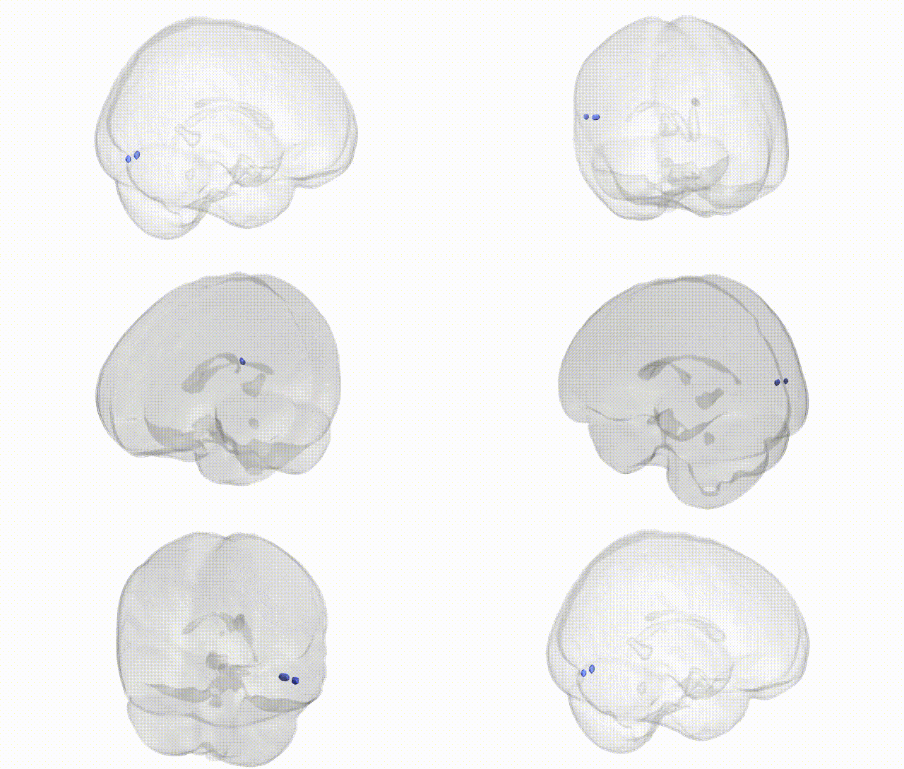
Full Quality Version: Download MP4
Wali Sidiqyar*, Gaurav Rudravaram*, Elyssa M. McMaster, Trent M. Schwartz, Adam M. Saunders, Kurt G. Schilling, Bennett A. Landman "Introducing SPINS: A Shared Public Visualization Library of Neuroanatomical Structures." Medical Imaging with Deep Learning- short paper
This resource is licensed under CC0 1.0 Universal (Public Domain).