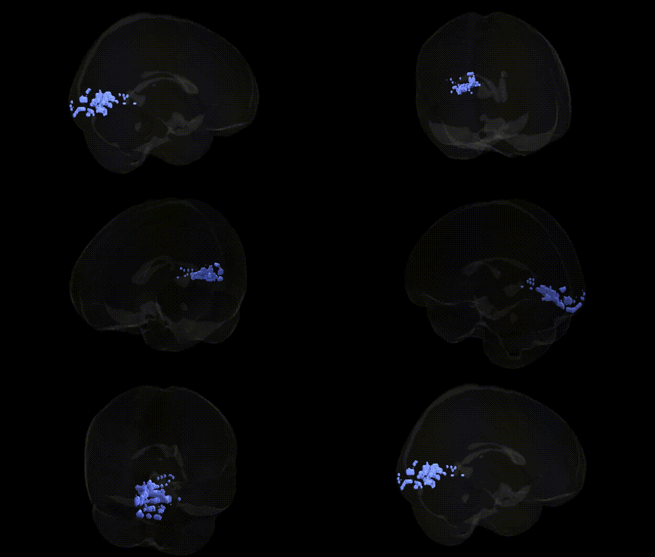
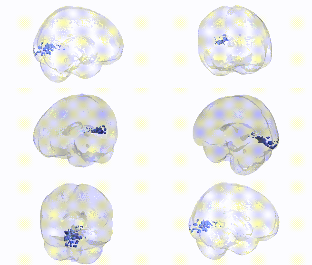
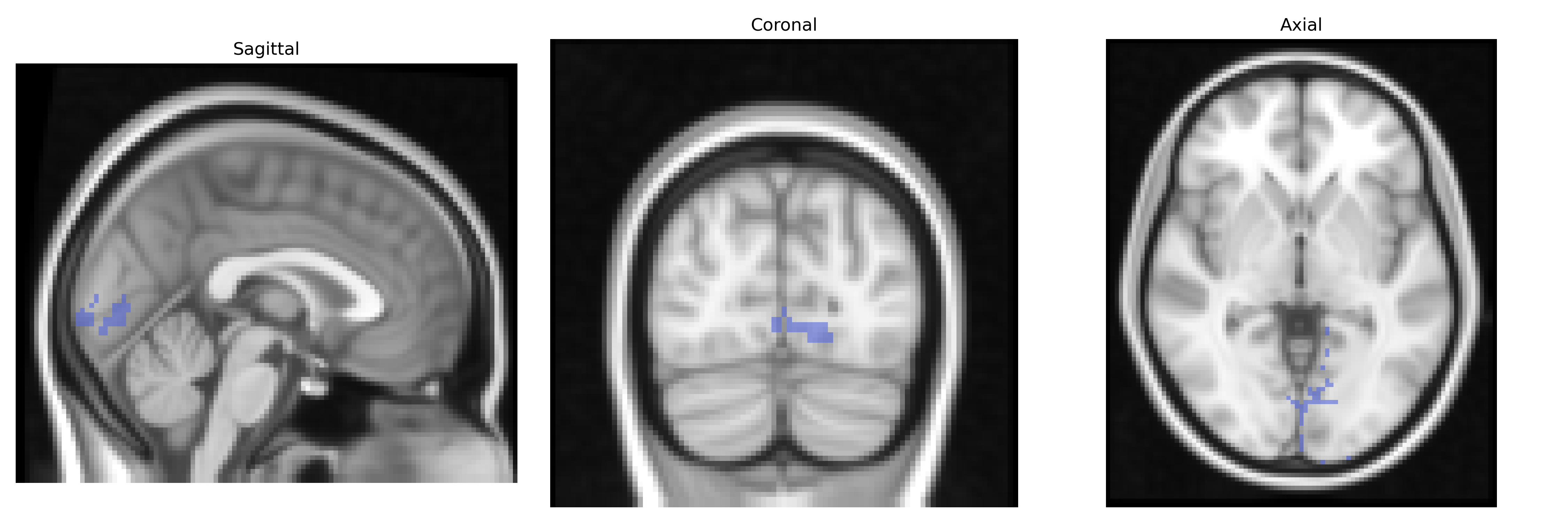
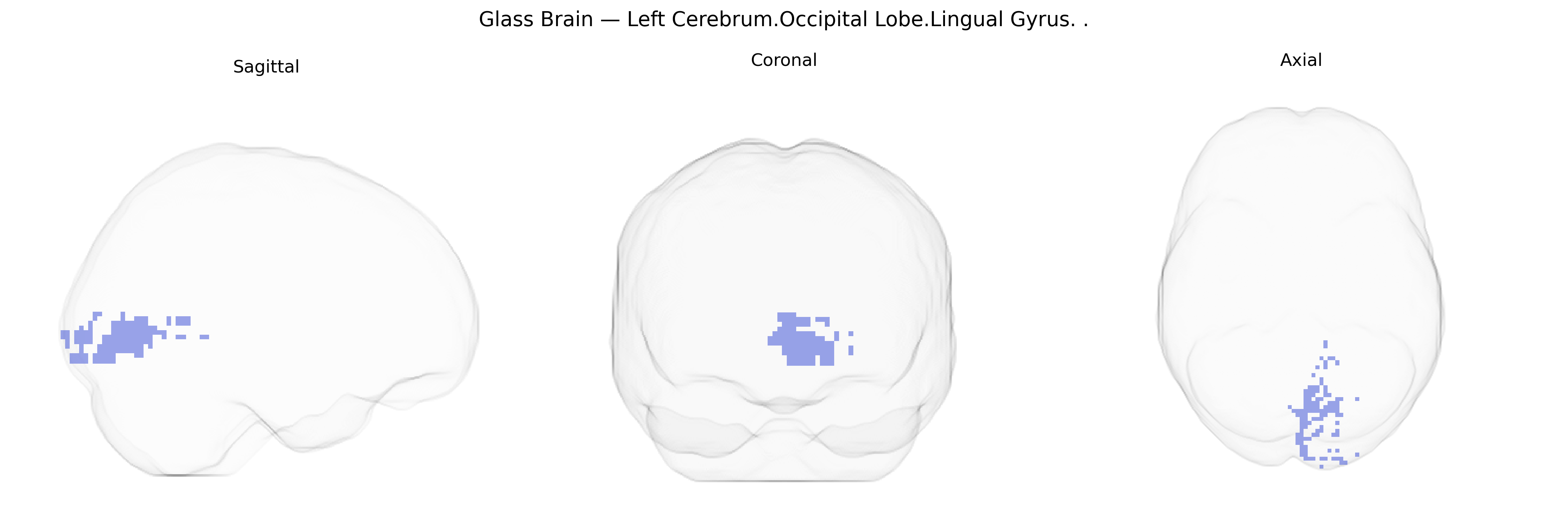
The bilateral Left Cerebrum.Occipital Lobe.Lingual Gyrus is a medial occipital cortical region located on the ventral surface of the occipital lobe, extending from the calcarine sulcus toward the posterior fusiform and parahippocampal gyri. It is primarily involved in early and intermediate stages of visual processing, including analysis of complex visual patterns, letter and word forms, and aspects of color and scene perception. The lingual gyrus contributes to visual memory and is often implicated in tasks involving reading, visual recognition, and processing of symbolic stimuli. Functionally, it forms part of the ventral visual stream and interacts with adjacent occipitotemporal areas to support higher-order object and face recognition, as well as visuospatial integration.
Lingual gyrus
The lingual gyrus, a medial-occipital region involved in basic and higher-order visual processing (including visual word form recognition and aspects of memory), has been implicated in several genetic and GWAS-based associations, although often as part of broader occipital or visual cortical networks rather than in isolation. Imaging genetics studies have linked common variants in genes influencing synaptic plasticity and neurodevelopment—such as BDNF (e.g., Val66Met), COMT, and DISC1—to altered activation or structural measures in occipital and lingual regions during visual and memory tasks. Large-scale brain morphology GWAS (e.g., ENIGMA and UK Biobank studies) have identified multiple loci associated with occipital cortical thickness and surface area, including regions overlapping the lingual gyrus, with variants in genes related to neuronal migration, axonal guidance, and calcium signaling among implicated pathways. Functional and structural changes in the lingual gyrus have been repeatedly observed in genetically influenced neuropsychiatric and neurodevelopmental conditions—including schizophrenia, bipolar disorder, major depressive disorder, autism spectrum disorder, and dyslexia—with risk variants in loci such as CACNA1C, ZNF804A, and NRG1 linked to abnormal occipital activation patterns or connectivity in visual networks. Moreover, GWAS of cognitive traits and educational attainment have associated polygenic scores with differences in occipital and lingual gyrus structure and function, suggesting a distributed genetic influence on visual-cognitive integration. While no single gene or variant is uniquely specific to the bilateral lingual gyrus in the Talairach 2 mm atlas, convergent evidence from imaging genetics and brain-structure GWAS supports a polygenic architecture in which multiple common variants affecting cortical development, synaptic signaling, and visual processing contribute to individual variation and disorder-related alterations in this region.
Overview generated by GPT-4o (2026).
Region ID: 238
Hemisphere: bilateral
Atlas: Talairach labels 2mm
Full Quality Version: Download MP4
Full Quality Version: Download MP4
Wali Sidiqyar*, Gaurav Rudravaram*, Elyssa M. McMaster, Trent M. Schwartz, Adam M. Saunders, Kurt G. Schilling, Bennett A. Landman "Introducing SPINS: A Shared Public Visualization Library of Neuroanatomical Structures." Medical Imaging with Deep Learning- short paper
This resource is licensed under CC0 1.0 Universal (Public Domain).