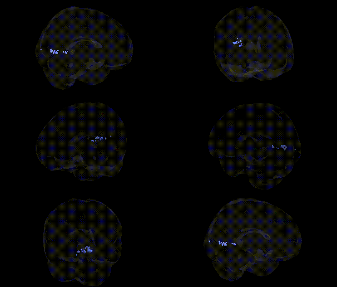
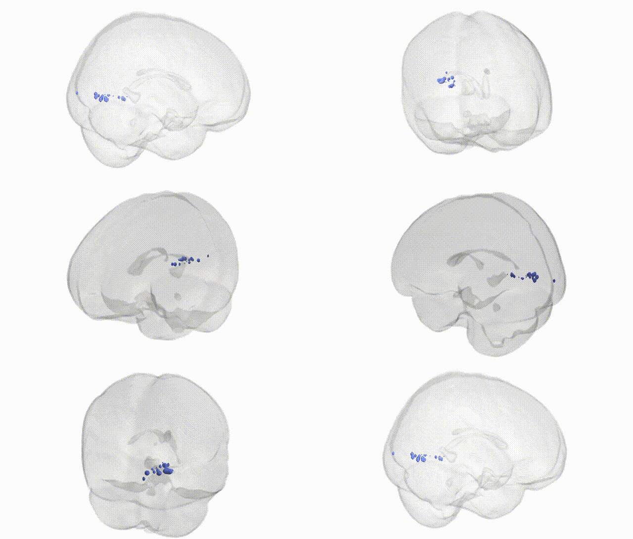
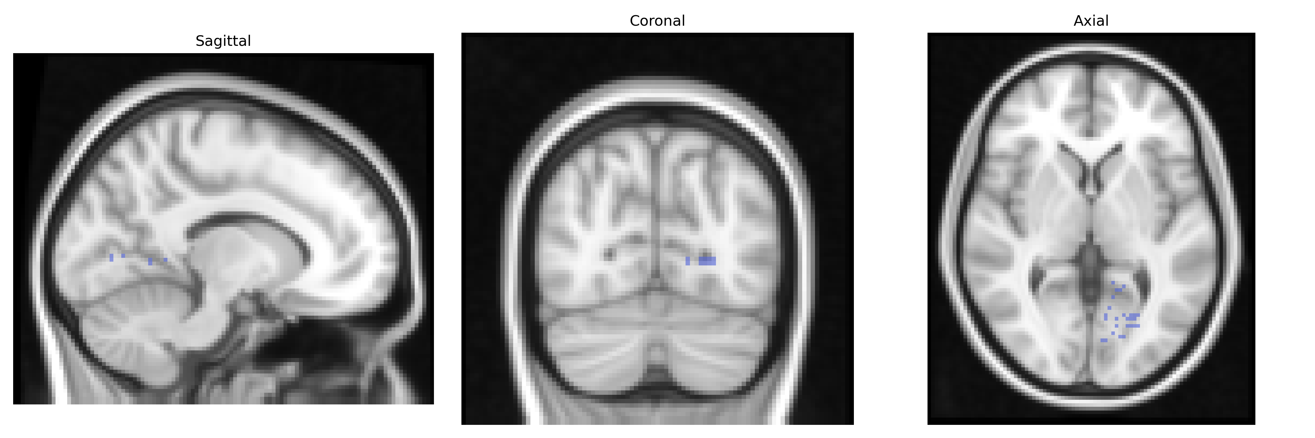
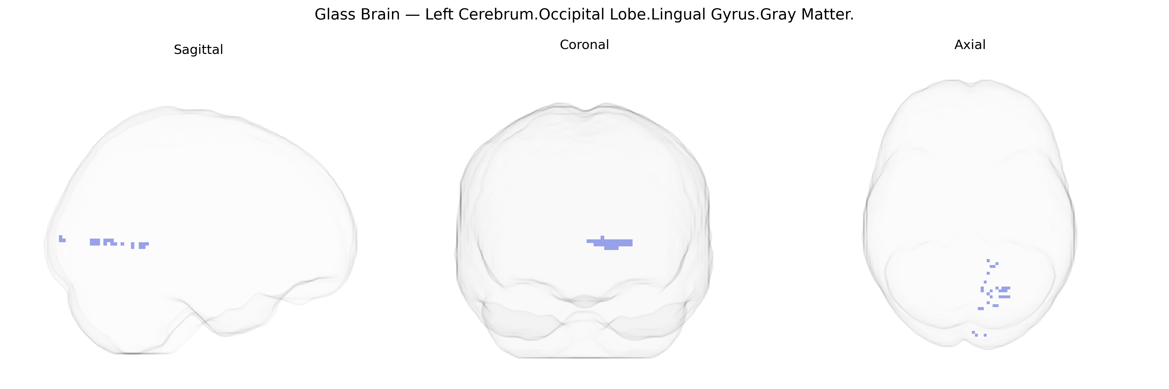
The bilateral Left Cerebrum.Occipital Lobe.Lingual Gyrus.Gray Matter corresponds to cortical gray matter within the lingual gyrus of the occipital lobe, a ventromedial region on the inferior aspect of the cerebral hemisphere extending from the posterior calcarine sulcus toward the temporal lobe. This region is heavily involved in early visual processing, including analysis of complex visual features, contrast, and orientation, and contributes to higher-order visual functions such as visual word form recognition and aspects of visual memory. Functionally, it participates in the ventral visual stream, integrating inputs from primary visual cortex (V1) and associational visual areas to support pattern recognition and visuospatial processing. Cytoarchitectonically, it comprises densely packed pyramidal neurons typical of neocortical gray matter, receiving thalamocortical projections from the lateral geniculate nucleus and projecting to other occipitotemporal association cortices. There is no direct link for the full Talairach label; a closely related structure is: Lingual gyrus.
The bilateral left occipital lingual gyrus gray matter (Talairach 2 mm Atlas) is a primary visual association region whose structure and function have been linked to numerous genetic influences, though often studied at coarser parcellations than this exact label. Twin and family studies show high heritability of occipital cortical thickness, surface area, and volume, with polygenic contributions involving general neurodevelopmental loci (e.g., variants near genes such as PAX6, EMX2, and other regulators of cortical patterning), and large-scale brain GWAS (UK Biobank and related cohorts) regularly identify SNPs and gene sets affecting occipital/lingual cortical morphology through pathways in axon guidance, synaptic plasticity, and myelination. Functionally, the lingual gyrus is central to visual processing, word and symbol recognition, and visuospatial memory, and imaging-genetic studies have associated its activation and connectivity patterns with genetic risk for neuropsychiatric and neurodevelopmental disorders, including schizophrenia, bipolar disorder, autism spectrum disorder, and dyslexia, as well as traits such as reading ability, visual memory, and susceptibility to visual hallucinations. GWAS of migraine, occipital epilepsy, and photic sensitivity implicate genes in glutamatergic signaling and cortical excitability that modulate occipital network function, sometimes with downstream effects on lingual gyrus activation. Neurodegenerative GWAS (e.g., Alzheimer’s disease loci such as APOE and CLU) and polygenic risk scores for cognitive decline have been associated with occipital atrophy patterns that include the lingual cortex, and imaging-genetic work on depression and anxiety has linked serotonergic and stress-related genes (e.g., SLC6A4, FKBP5) to altered occipital/lingual responses to emotional visual stimuli. Overall, genetic influences on this specific gray matter region reflect a distributed polygenic architecture shaping visual cortex development, cortical microstructure, and participation in broader cognitive and psychiatric risk networks rather than single, region-specific susceptibility genes.
Overview generated by GPT-4o (2026).
Region ID: 541
Hemisphere: bilateral
Atlas: Talairach labels 2mm
Full Quality Version: Download MP4
Full Quality Version: Download MP4
Wali Sidiqyar*, Gaurav Rudravaram*, Elyssa M. McMaster, Trent M. Schwartz, Adam M. Saunders, Kurt G. Schilling, Bennett A. Landman "Introducing SPINS: A Shared Public Visualization Library of Neuroanatomical Structures." Medical Imaging with Deep Learning- short paper
This resource is licensed under CC0 1.0 Universal (Public Domain).