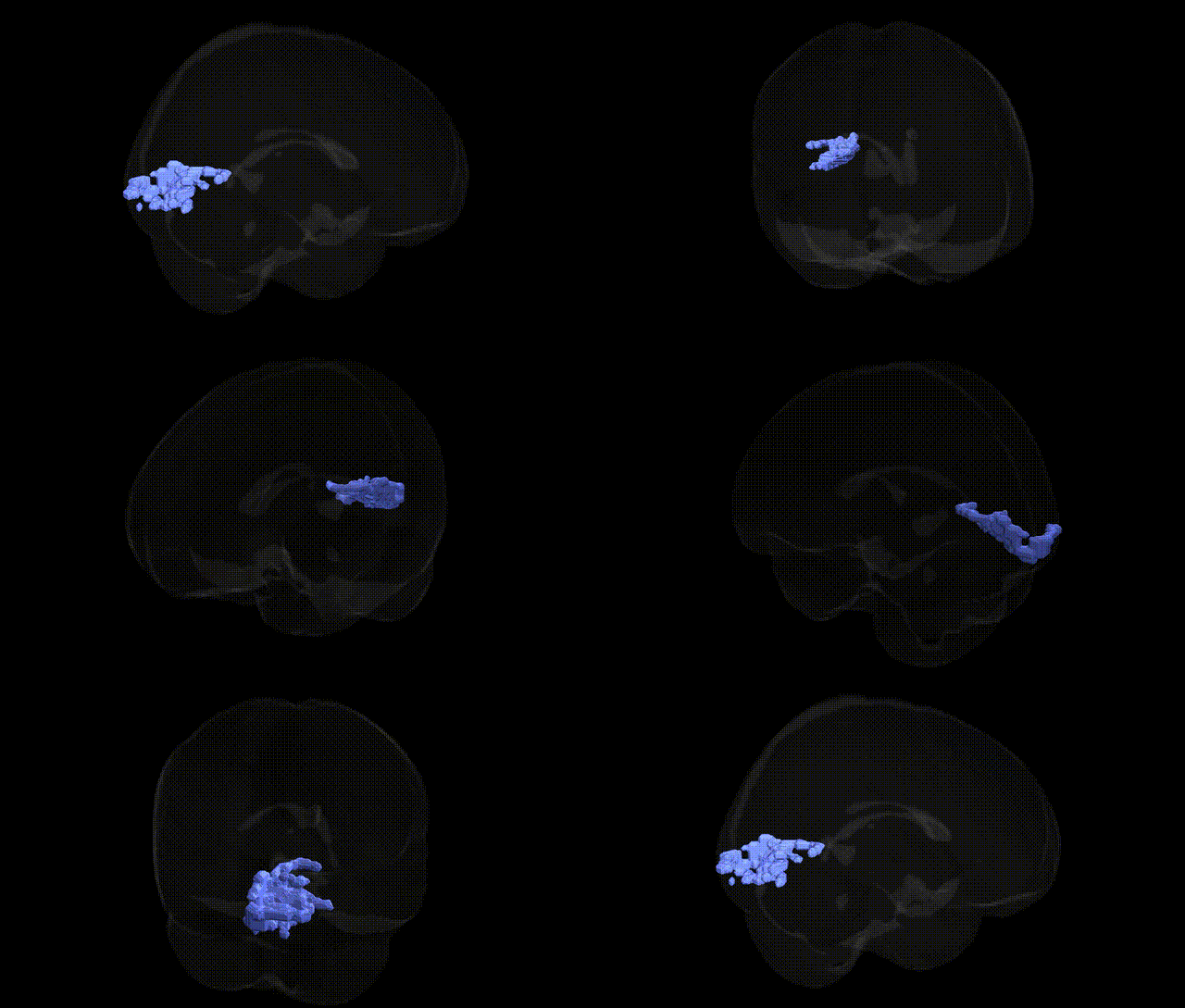
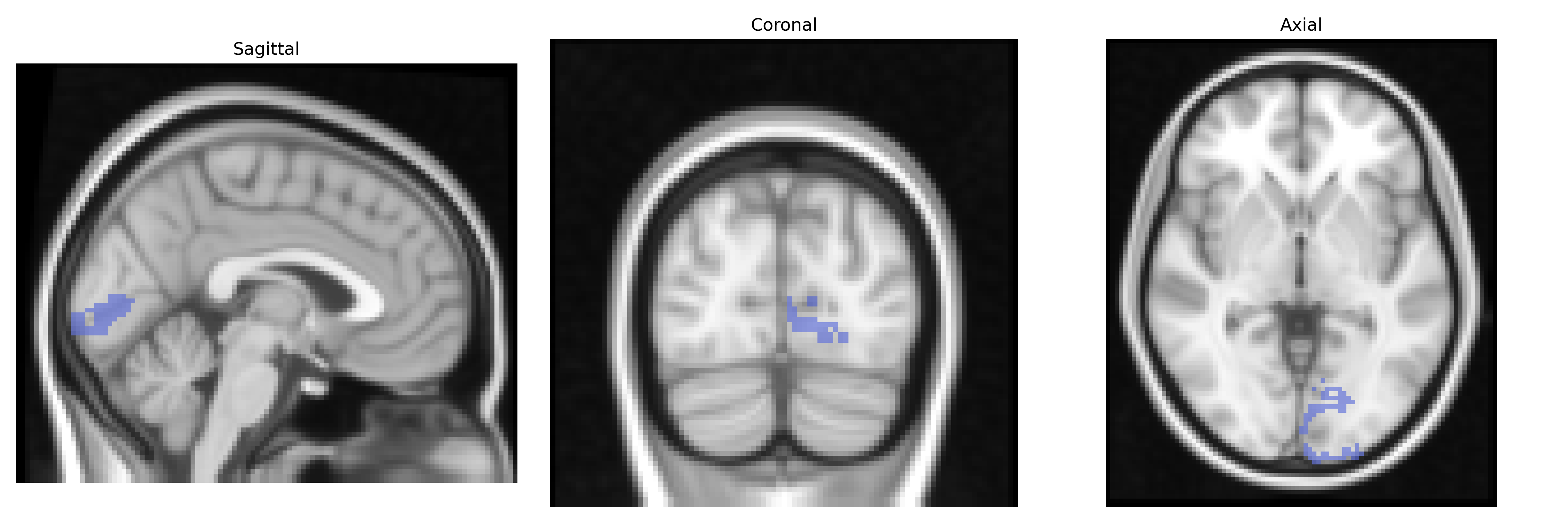
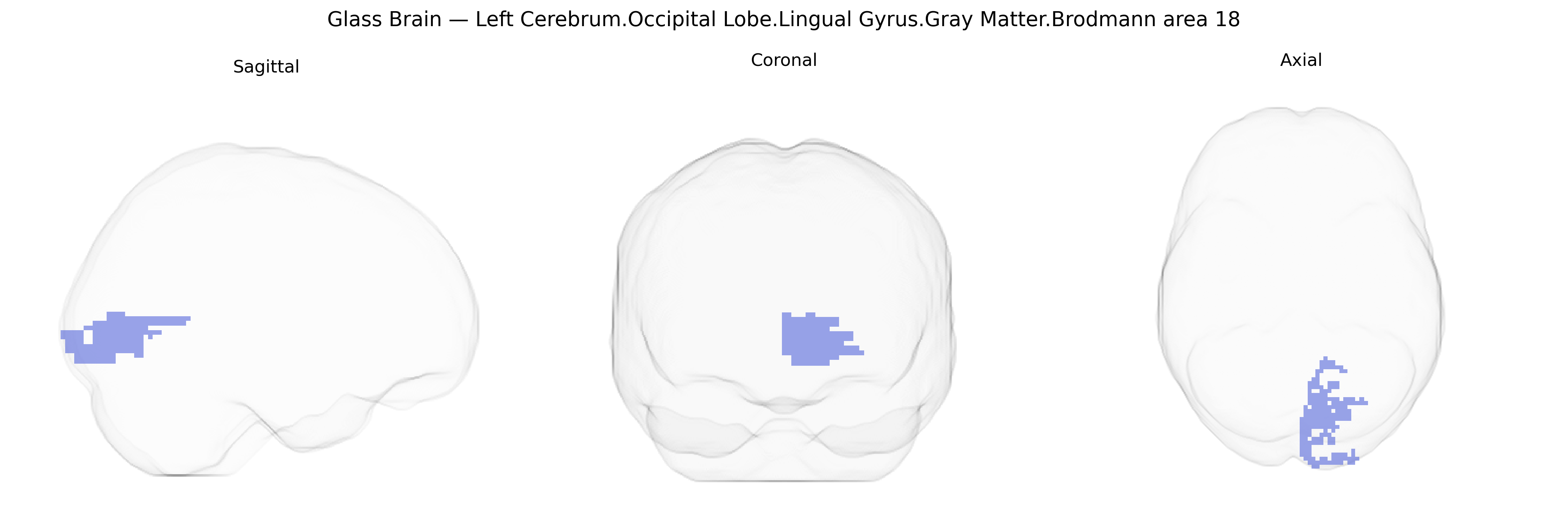
The bilateral Left Cerebrum.Occipital Lobe.Lingual Gyrus.Gray Matter.Brodmann area 18 corresponds to secondary visual cortex (V2) located along the lingual gyrus on the medial aspect of the occipital lobe, just anterior to the primary visual cortex (BA17). This region is cytoarchitectonically defined by its distinct laminar organization and functions primarily in early-stage visual processing, including analysis of orientation, spatial frequency, contours, and binocular disparity, as well as integration of information from the primary visual cortex for more complex pattern and form recognition. Activity in Brodmann area 18 contributes to construction of detailed visual representations that support higher-order visual tasks such as object recognition and visual perception of scenes. There is no direct link for this exact composite region; see the related area Brodmann area 18.
The bilateral left cerebrum occipital lobe lingual gyrus gray matter in Brodmann area 18 (secondary visual cortex) is implicated in genetic studies primarily through its role in visual processing, reading, and higher-order cognitive functions. GWAS and imaging-genetics analyses from large cohorts (e.g., ENIGMA, UK Biobank) have shown that gray matter volume and cortical thickness in occipital and lingual regions are heritable and influenced by common variants in pathways involved in synaptic development, axon guidance, and neurogenesis, including genes such as BDNF, HMGA2, and variants near MEF2C and WNT signaling components, although most findings are polygenic and regionally nonspecific. BA18/lingual gyrus structure and function have been associated with risk loci for neurodevelopmental and psychiatric disorders that alter visual and visuo-cognitive processing, including schizophrenia (e.g., variants in CACNA1C, GRIN2A, and genome-wide polygenic risk scores correlating with occipital cortex morphology), autism spectrum disorder (with widespread cortical patterning genes showing effects in occipital regions), and major depressive disorder (where polygenic risk is linked to occipital and lingual cortical thickness). Genetic variants affecting reading and language-related traits (such as dyslexia-associated loci near DCDC2, KIAA0319, and ROBO1) have been connected through neuroimaging to altered activation and structure in the left lingual and occipitotemporal regions, consistent with a role in orthographic and visual word processing. Additionally, GWAS of visual acuity, refractive error (e.g., variants near GJD2, RASGRF1), and migraine have reported associations with occipital cortex involvement, and polygenic influences on general intelligence and educational attainment show correlations with visual association cortex volumes, including BA18, underscoring that genetic effects on this region are largely pleiotropic and embedded within broad, distributed brain-genetic architectures rather than being specific to the Talairach-defined label.
Overview generated by GPT-4o (2026).
Region ID: 247
Hemisphere: bilateral
Atlas: Talairach labels 2mm
Full Quality Version: Download MP4
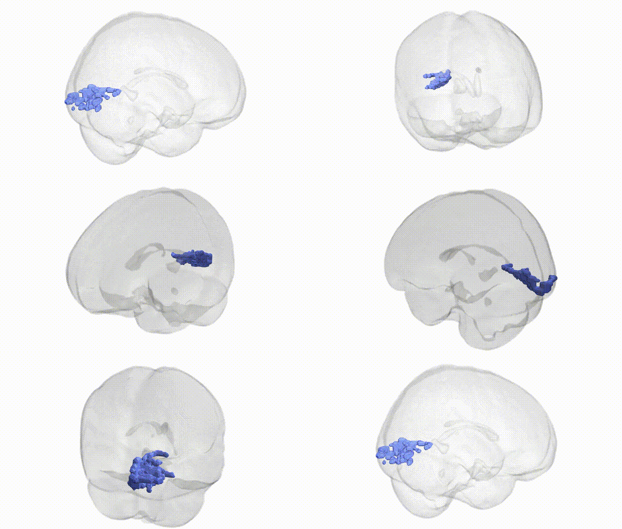
Full Quality Version: Download MP4
Wali Sidiqyar*, Gaurav Rudravaram*, Elyssa M. McMaster, Trent M. Schwartz, Adam M. Saunders, Kurt G. Schilling, Bennett A. Landman "Introducing SPINS: A Shared Public Visualization Library of Neuroanatomical Structures." Medical Imaging with Deep Learning- short paper
This resource is licensed under CC0 1.0 Universal (Public Domain).