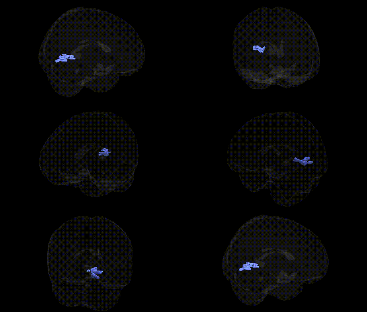
The bilateral Left Cerebrum.Occipital Lobe.Lingual Gyrus.Gray Matter.Brodmann area 19 corresponds to a portion of secondary visual cortex within the lingual gyrus of the occipital lobe, involved in higher-order visual processing such as complex form, object, and scene analysis. As part of the extrastriate cortex, BA19 receives input from primary visual cortex (BA17) and other visual association areas, contributing to integration of visual features, visuospatial perception, and aspects of visual memory. Neuronal populations in this region exhibit sensitivity to patterns, motion, and contextual visual information, supporting tasks like recognition of shapes and environmental layouts. There is no direct Wikipedia link for this exact composite Talairach label; a closely related structure is Brodmann area 19.
The bilateral Left Cerebrum Occipital Lobe Lingual Gyrus gray matter in Brodmann area 19 (BA19) is a higher-order visual association region whose genetic associations are largely inferred from imaging genetics and GWAS of brain structure and visual-related traits rather than BA19-specific gene mapping. Structural and functional variation in the lingual gyrus and BA19 has been linked to polygenic influences involving neurodevelopmental and synaptic genes identified in large MRI-based GWAS (e.g., UK Biobank), where common variants near genes such as MEF2C, DLG2, and other neuronal differentiation and synaptic plasticity genes contribute to occipital cortical thickness and surface area. BA19 is consistently implicated in visual processing and reading; imaging genetics studies in dyslexia and related learning disorders report associations between candidate genes involved in neuronal migration and axonal guidance (e.g., DCDC2, KIAA0319, ROBO1) and altered activation or morphology in occipitotemporal and occipital regions that encompass BA19, although these findings are often indirect and regionally broad. Psychiatric and neurodevelopmental GWAS (schizophrenia, autism spectrum disorder, ADHD) show polygenic risk effects on occipital cortex volume and connectivity, including BA19, reflecting distributed genetic architectures involving synaptic and immune-related pathways rather than BA19-specific loci. Migraine and visual aura GWAS identify multiple loci in ion channel and vascular regulation genes (e.g., CACNA1A-related pathways), and imaging studies frequently report structural and functional differences in occipital cortex and lingual gyrus, suggesting that genetic susceptibility to cortical excitability may manifest in BA19. Overall, genetic influences on this Talairach-defined BA19 region are highly polygenic and shared with broader visual and association cortex networks, with known associations emerging mainly from large-scale imaging GWAS of cortical morphology and from disorder GWAS whose polygenic risk impacts occipital visual regions rather than from genes uniquely mapped to this specific Brodmann area.
Overview generated by GPT-4o (2026).
Region ID: 417
Hemisphere: bilateral
Atlas: Talairach labels 2mm
Full Quality Version: Download MP4
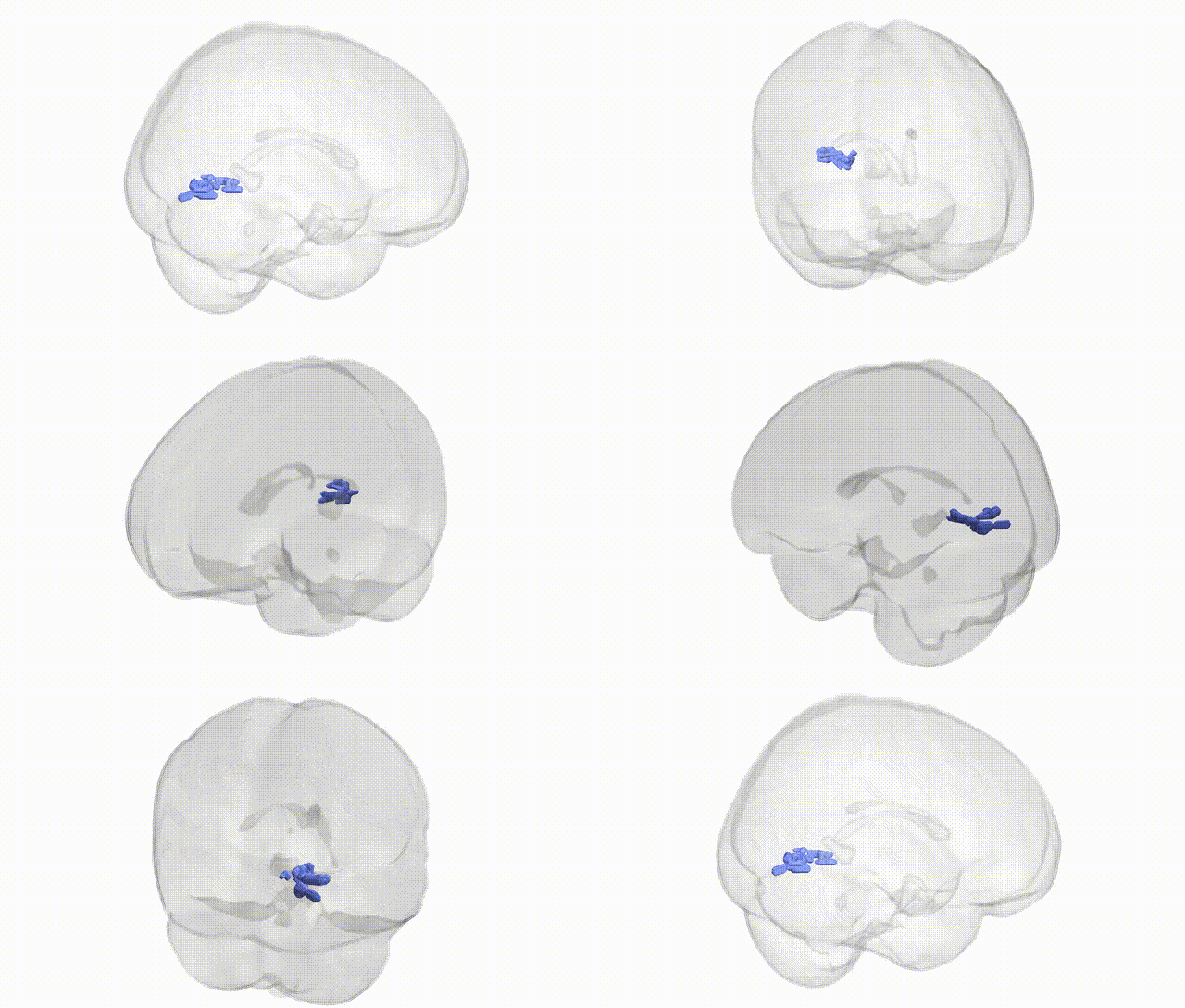
Full Quality Version: Download MP4
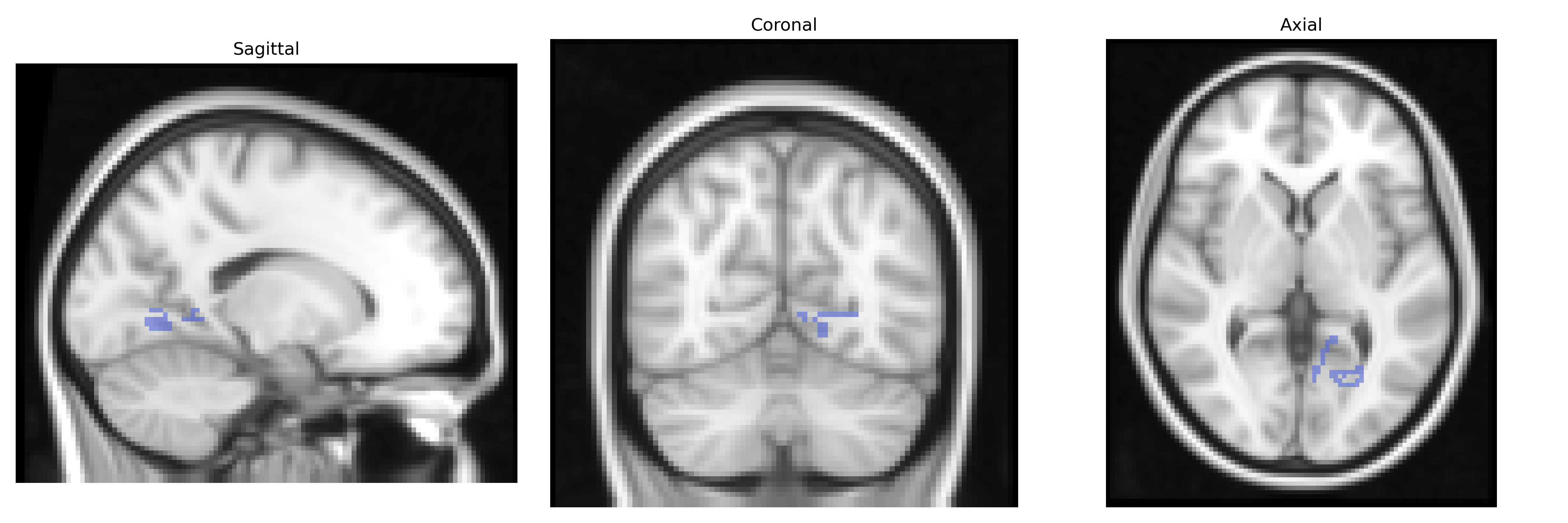
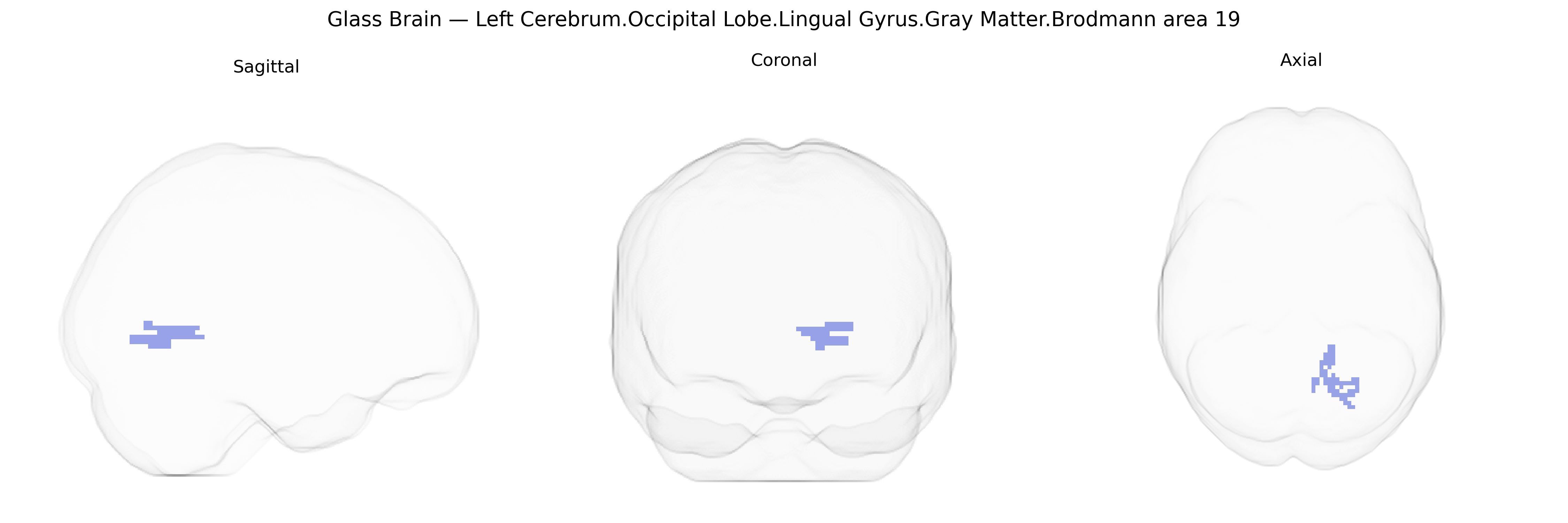
Wali Sidiqyar*, Gaurav Rudravaram*, Elyssa M. McMaster, Trent M. Schwartz, Adam M. Saunders, Kurt G. Schilling, Bennett A. Landman "Introducing SPINS: A Shared Public Visualization Library of Neuroanatomical Structures." Medical Imaging with Deep Learning- short paper
This resource is licensed under CC0 1.0 Universal (Public Domain).