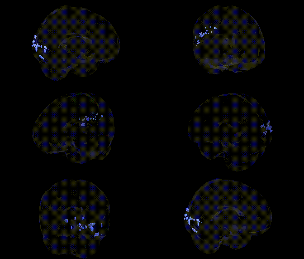
The bilateral Left Cerebrum.Occipital Lobe.Middle Occipital Gyrus corresponds to cortical tissue situated on the lateral aspect of the occipital lobe, extending along the middle portion of the occipital cortex between the superior and inferior occipital gyri. Functionally, this region is involved in intermediate-level visual processing, including analysis of object form, motion, and spatial relationships, and participates in integrating visual inputs with higher-order associative regions in parietal and temporal cortices. It contributes to the dorsal and ventral visual streams, supporting visuospatial perception and visual object recognition. There is no direct Wikipedia article for the middle occipital gyrus; a closely related structure is the Occipital lobe.
The bilateral Middle Occipital Gyrus (Talairach Left Cerebrum.Occipital Lobe.Middle Occipital Gyrus) is a key visual-association region implicated in several genetic and imaging-genetics findings, though few studies target it in isolation. GWAS of cortical thickness and surface area have identified associations between common variants in loci including 15q14, 14q23 (near KIAA0586, DACT1), and 17q21 (near MAPT) with occipital and visual-association cortex measures that encompass the middle occipital gyrus, suggesting polygenic influences on its morphology. Imaging-genetics work has linked variants in genes related to synaptic plasticity and neurodevelopment (e.g., BDNF Val66Met, NRG1, DISC1) and major psychiatric risk loci (e.g., CACNA1C, ZNF804A, and schizophrenia polygenic risk scores) to altered activation or structure in lateral occipital regions during visual processing, face and object recognition, and working memory, sometimes including middle occipital clusters. Occipital regions overlapping the middle occipital gyrus show genetically influenced vulnerability in disorders such as schizophrenia, bipolar disorder, major depression, and autism spectrum disorder, where case–control MRI and GWAS-meta analyses report heritable reductions in cortical thickness or volume. Genetic variation associated with dyslexia and other reading/visual word form traits (including KIAA0319, DCDC2 and other neurodevelopmental loci) has also been linked to functional differences in occipito-temporal circuits that extend into middle occipital territory. Overall, current evidence indicates that the middle occipital gyrus shares the broadly polygenic, pleiotropic architecture of visual-association cortex, with morphology and function influenced by neurodevelopmental, synaptic, and psychiatric risk genes, but precise, region-specific genetic associations remain limited and typically arise within larger occipital or visual network analyses rather than this Talairach label alone.
Overview generated by GPT-4o (2026).
Region ID: 310
Hemisphere: bilateral
Atlas: Talairach labels 2mm
Full Quality Version: Download MP4
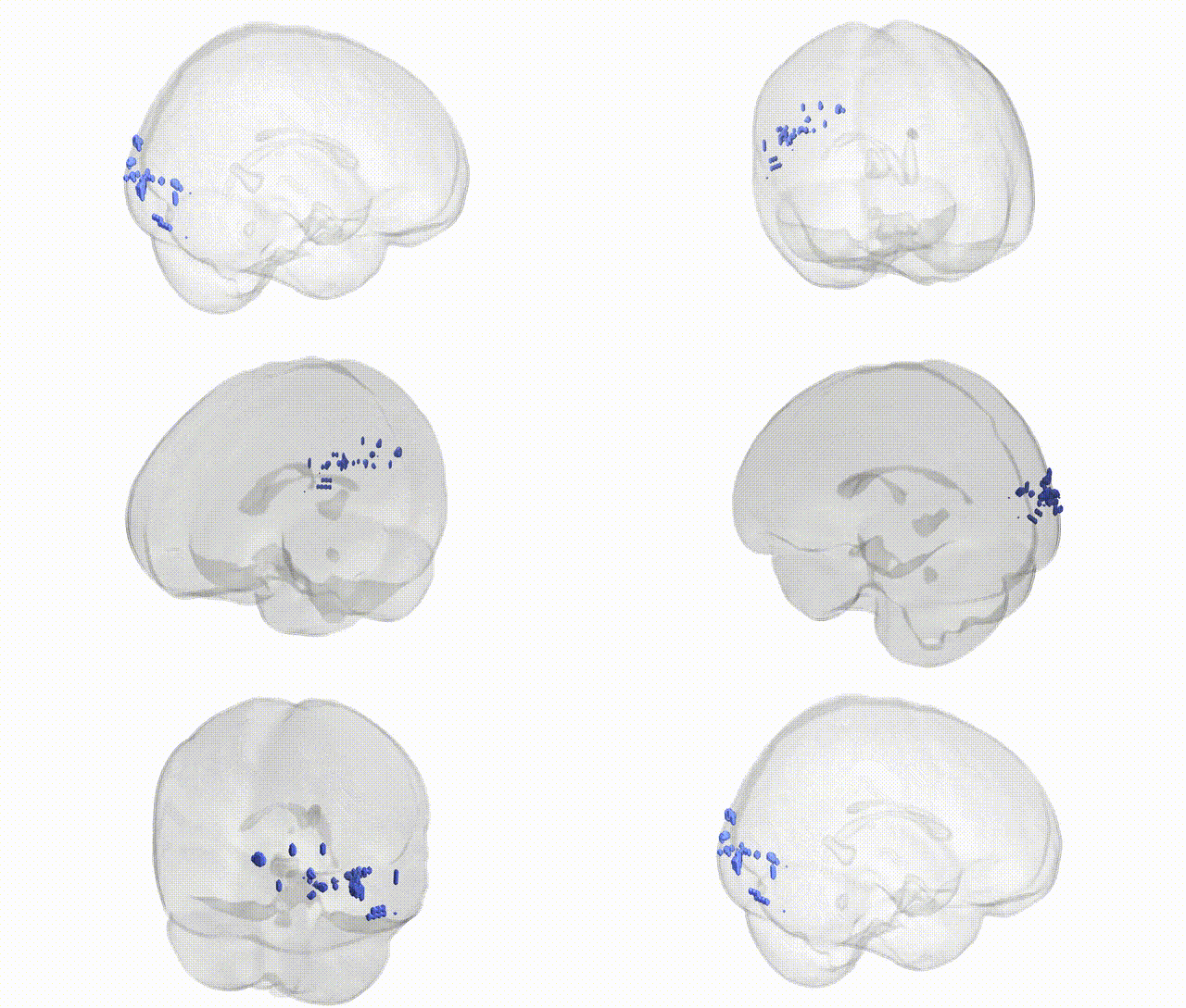
Full Quality Version: Download MP4
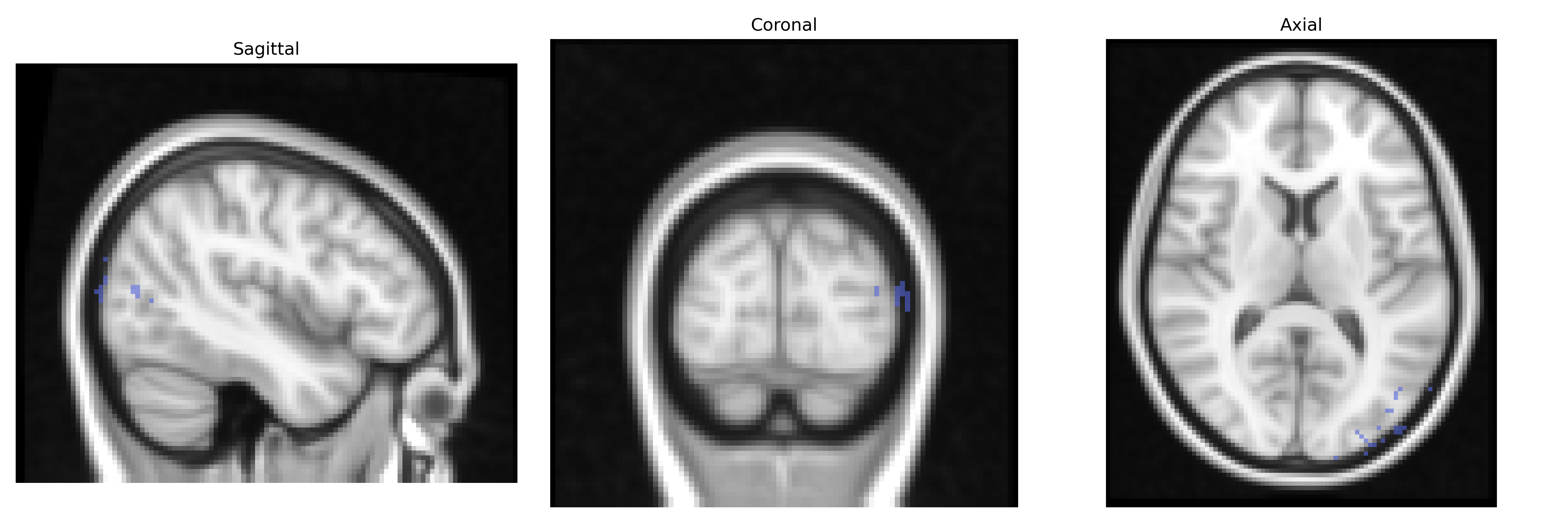
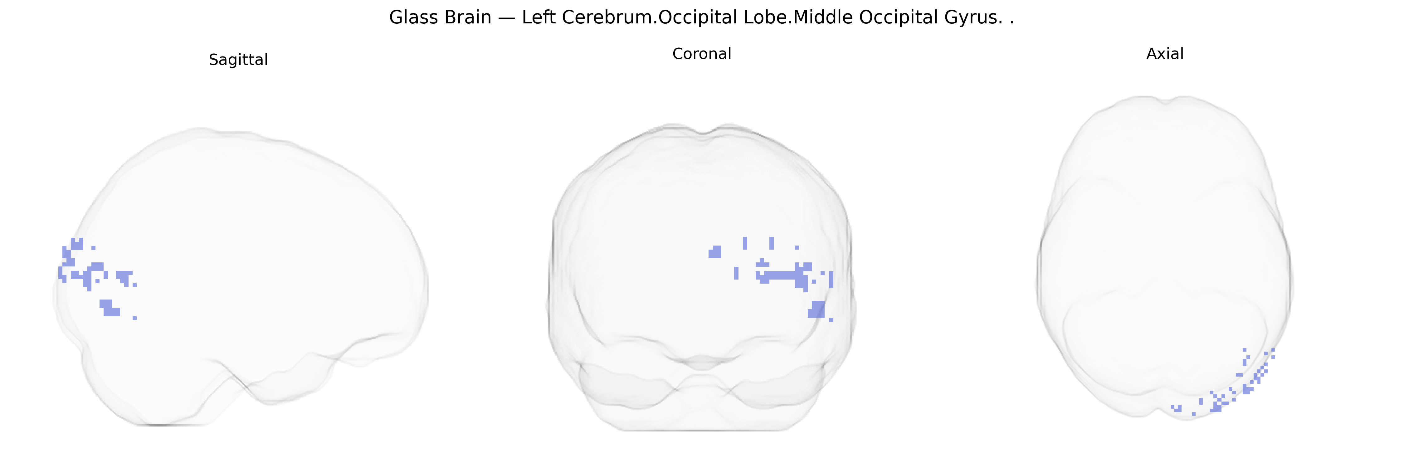
Wali Sidiqyar*, Gaurav Rudravaram*, Elyssa M. McMaster, Trent M. Schwartz, Adam M. Saunders, Kurt G. Schilling, Bennett A. Landman "Introducing SPINS: A Shared Public Visualization Library of Neuroanatomical Structures." Medical Imaging with Deep Learning- short paper
This resource is licensed under CC0 1.0 Universal (Public Domain).