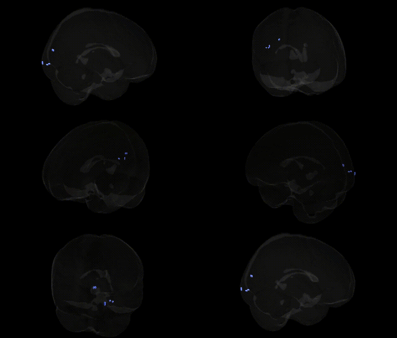
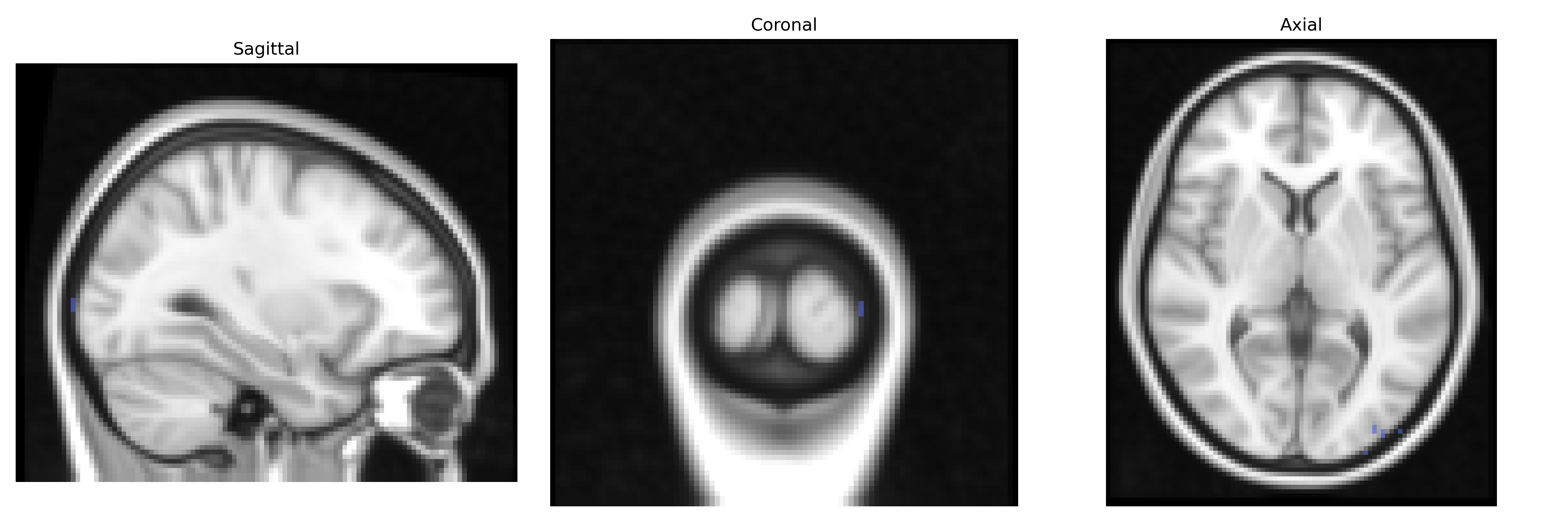
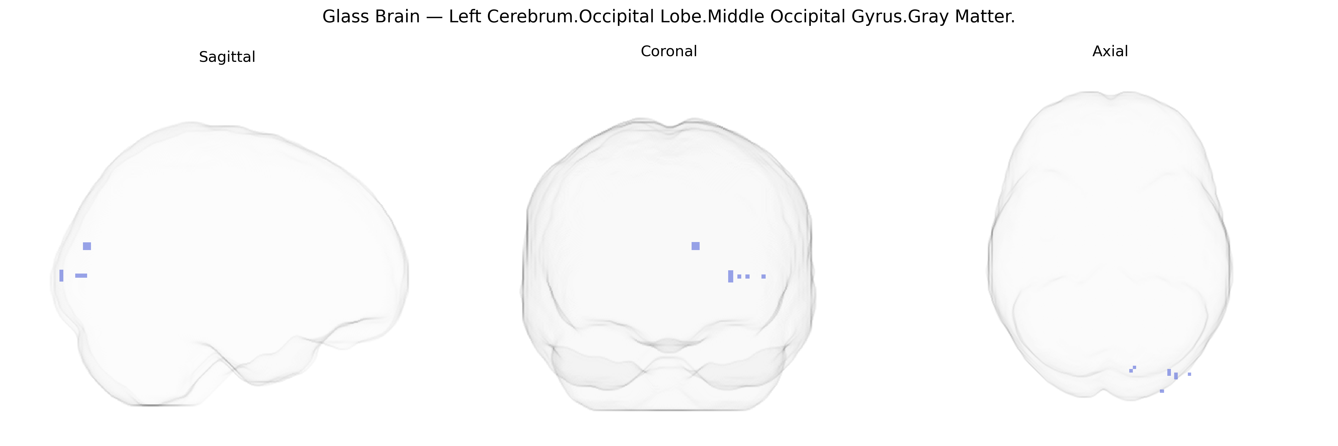
The bilateral Left Cerebrum.Occipital Lobe.Middle Occipital Gyrus.Gray Matter corresponds to cortical tissue in the mid-portion of the occipital lobe, primarily involved in intermediate stages of visual processing. This region participates in the analysis of shape, motion, and spatial relationships, integrating inputs from primary visual cortex (V1) and relaying processed information to higher-order visual and multimodal association areas, including parts of the dorsal and ventral visual streams. Functionally, it contributes to object recognition, visuospatial attention, and the perception of complex visual scenes, and it is interconnected with parietal and temporal cortices as well as subcortical visual structures. There is no direct Wikipedia article for the middle occipital gyrus; a related structure is the Occipital lobe.
The bilateral middle occipital gyrus gray matter (Left Cerebrum.Occipital Lobe.Middle Occipital Gyrus.Gray Matter) has been implicated in genetic studies largely through its role in visual processing and higher-order cognition rather than through region-specific GWAS hits. Imaging genetics work from large consortia such as ENIGMA and UK Biobank has shown that common variants in genes involved in neurodevelopment, synaptic plasticity, and myelination (including loci near MAPT, BDNF, and multiple 3p, 6q, and 10q regions) associate with occipital cortical thickness and surface area, although these effects tend to span broader occipital and visual association cortices rather than the middle occipital gyrus alone. GWAS on total and regional gray-matter volume have identified polygenic influences that overlap with risk variants for schizophrenia, bipolar disorder, major depressive disorder, and autism spectrum disorder, with occipital regions—including middle occipital cortex—showing volumetric or cortical-thickness alterations that co-localize with these psychiatric risk loci. Additional studies link genetic variation affecting white matter and visual pathways (for example, in genes related to axon guidance and myelin such as CNTNAP2, NRXN1, and ERBB4) to functional and structural differences in extrastriate visual areas, including middle occipital gyrus, in conditions like dyslexia, visual hallucinations in psychosis, and migraine with aura, though these associations are typically indirect and embedded within larger networks. Overall, current evidence supports a highly polygenic and distributed genetic architecture influencing gray matter in the middle occipital gyrus, with its alterations emerging as part of broader genetically mediated neurodevelopmental and psychiatric phenotypes rather than as a primary locus of region-specific GWAS signals.
Overview generated by GPT-4o (2026).
Region ID: 520
Hemisphere: bilateral
Atlas: Talairach labels 2mm
Full Quality Version: Download MP4
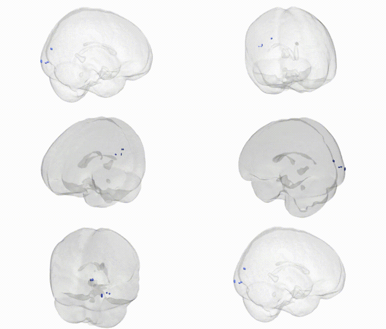
Full Quality Version: Download MP4
Wali Sidiqyar*, Gaurav Rudravaram*, Elyssa M. McMaster, Trent M. Schwartz, Adam M. Saunders, Kurt G. Schilling, Bennett A. Landman "Introducing SPINS: A Shared Public Visualization Library of Neuroanatomical Structures." Medical Imaging with Deep Learning- short paper
This resource is licensed under CC0 1.0 Universal (Public Domain).