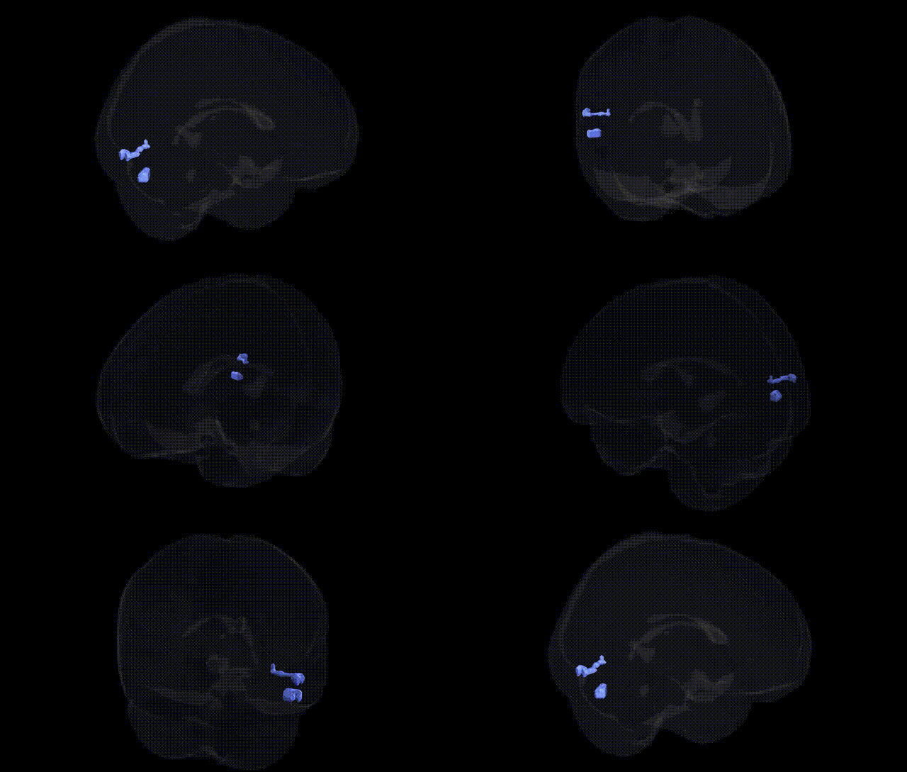
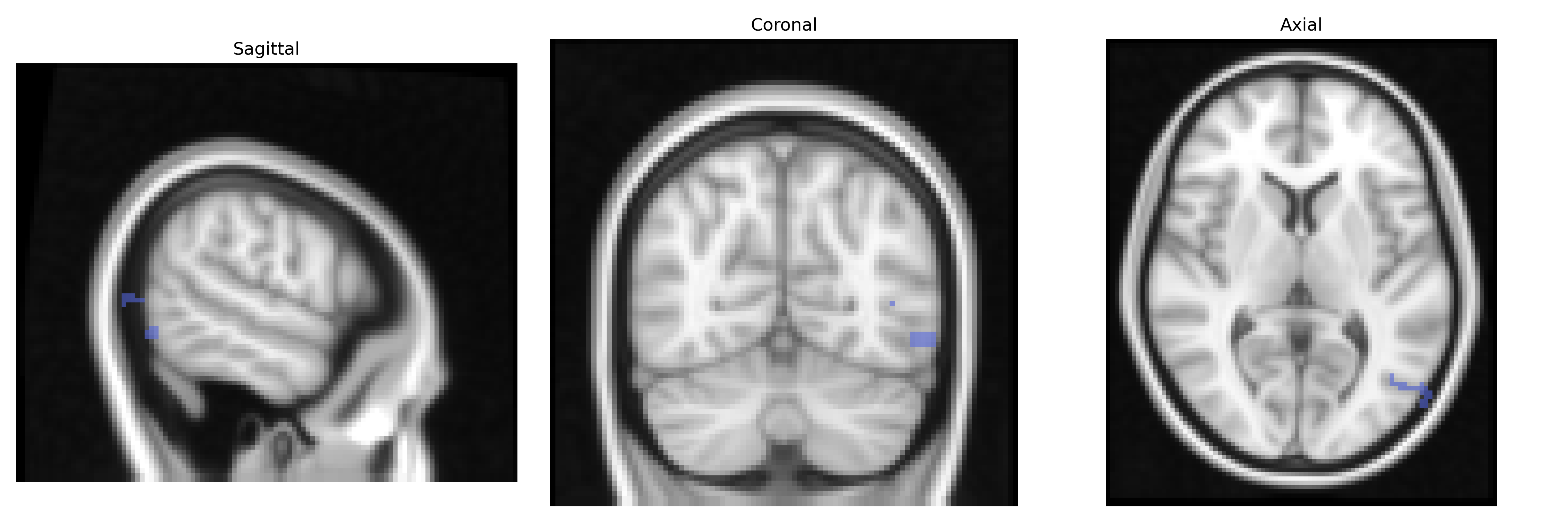
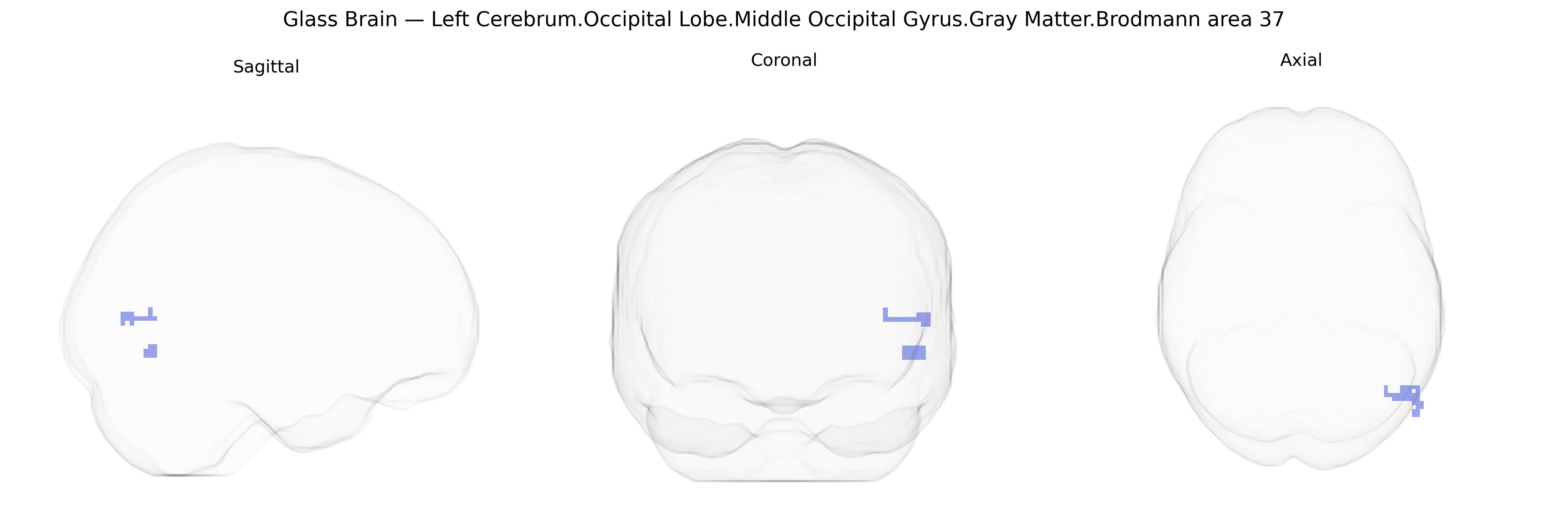
Bilateral Left Cerebrum.Occipital Lobe.Middle Occipital Gyrus.Gray Matter.Brodmann area 37 corresponds primarily to a ventral occipitotemporal associative visual region involved in high-level object recognition, including face and word-form processing, and the integration of visual inputs with semantic memory. Situated at the boundary of the occipital and temporal lobes, this cortex receives processed visual information from earlier occipital areas and participates in transforming complex visual patterns into meaningful representations, supporting functions such as visual category recognition and aspects of language-related visual processing. There is no direct link for “Middle Occipital Gyrus Brodmann area 37”; a closely related structure is the fusiform gyrus within BA37: Fusiform gyrus.
The bilateral left middle occipital gyrus gray matter in Brodmann area 37 (Talairach 2 mm atlas) lies within the ventral visual stream and overlaps key portions of the fusiform/occipitotemporal cortex, a region repeatedly implicated by genetic and GWAS-based imaging studies. Twin and heritability analyses show strong genetic control of occipitotemporal cortical thickness and surface area, with common variants in genes involved in neurodevelopment (e.g., BDNF, NRGN, and synaptic plasticity pathways) associated with structural variation and functional activation in this region during visual, semantic, and face/object processing tasks. Large imaging-genetics consortia (such as ENIGMA and UK Biobank-based GWAS) have reported loci influencing occipital and fusiform cortical morphometry—often near genes regulating neuronal migration, axon guidance, and synaptogenesis—with downstream effects on reading ability, language-related traits, and general cognitive performance. BA37 and adjacent fusiform areas show genetic overlap with neurodevelopmental and neuropsychiatric disorders, including autism spectrum disorder, dyslexia, and schizophrenia, where risk variants (for example in CNTNAP2, DCDC2, and GRIN2A among others) are linked to atypical activation or structural differences in occipitotemporal cortex during face recognition, social perception, or orthographic/lexical processing. Additional GWAS and polygenic score studies connect variation in this region to susceptibility for prosopagnosia-like face-processing deficits, visuospatial skills, and certain forms of temporal lobe epilepsy that propagate through occipitotemporal networks, underscoring BA37 as a genetically sensitive hub for high-level visual and language-related cognition.
Overview generated by GPT-4o (2026).
Region ID: 323
Hemisphere: bilateral
Atlas: Talairach labels 2mm
Full Quality Version: Download MP4
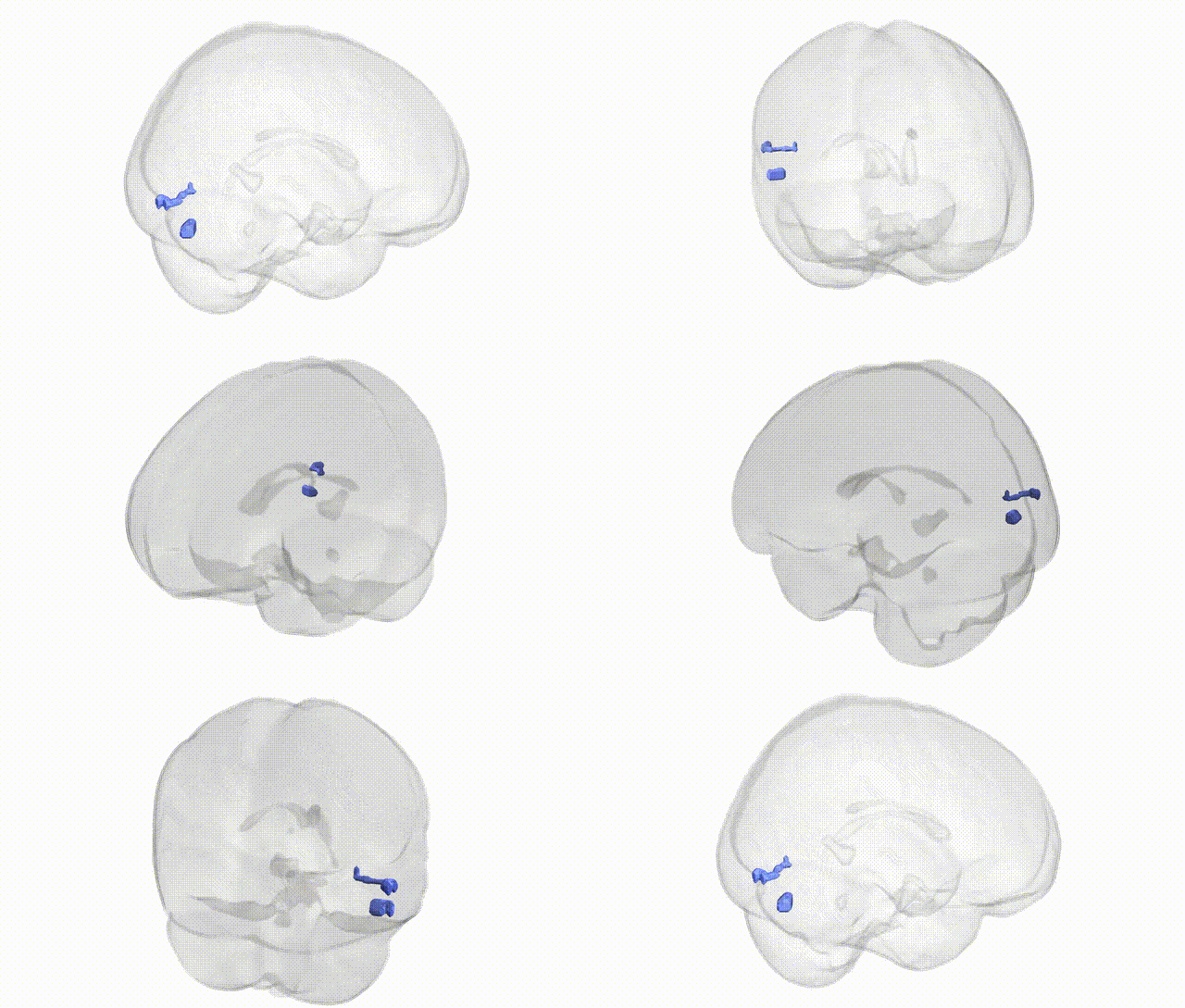
Full Quality Version: Download MP4
Wali Sidiqyar*, Gaurav Rudravaram*, Elyssa M. McMaster, Trent M. Schwartz, Adam M. Saunders, Kurt G. Schilling, Bennett A. Landman "Introducing SPINS: A Shared Public Visualization Library of Neuroanatomical Structures." Medical Imaging with Deep Learning- short paper
This resource is licensed under CC0 1.0 Universal (Public Domain).