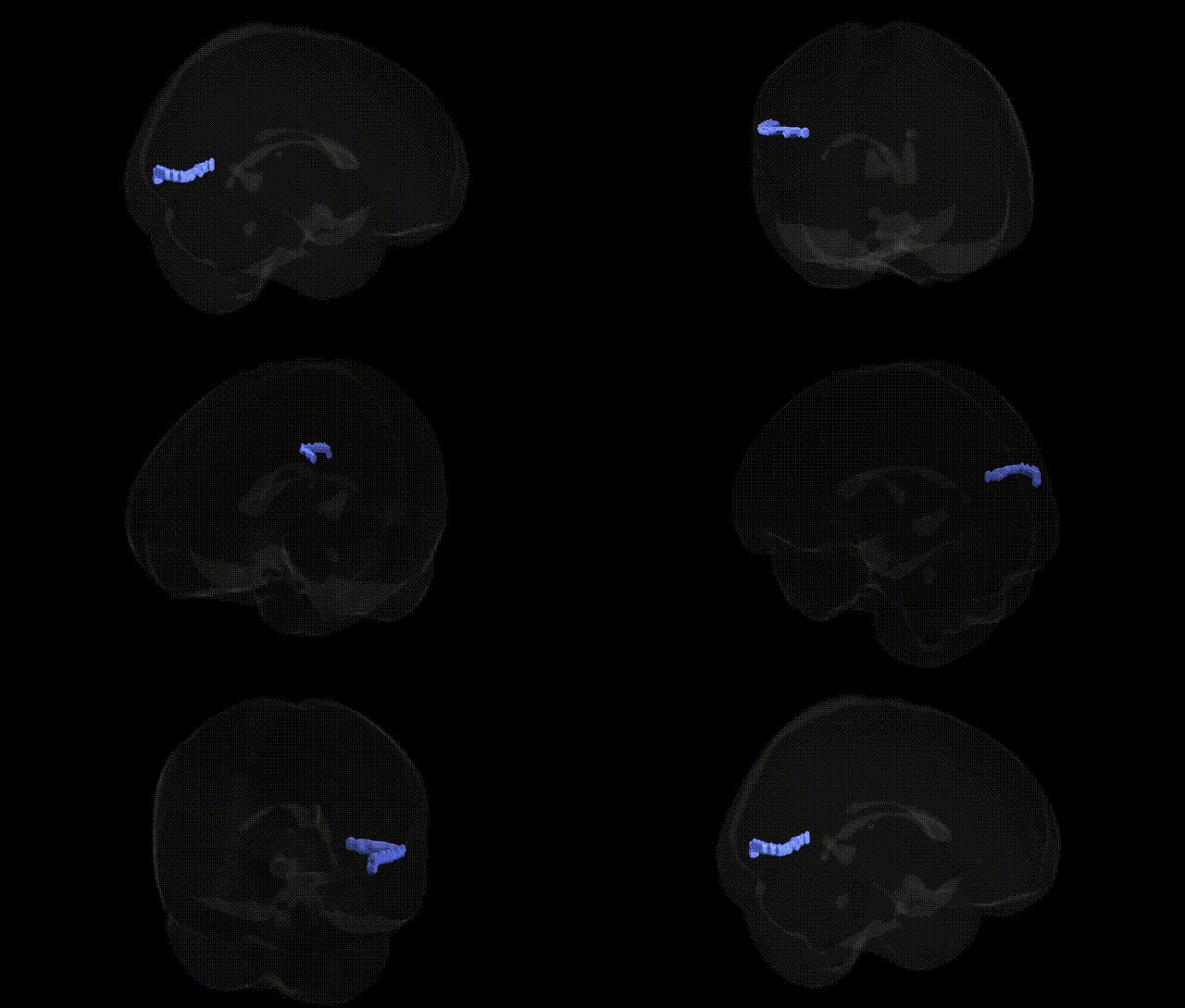
The bilateral Left Cerebrum.Occipital Lobe.Middle Temporal Gyrus.Gray Matter.Brodmann area 19 corresponds to higher-order visual association cortex situated at the occipitotemporal junction within the left hemisphere. Brodmann area 19 receives input from primary (BA17) and secondary (BA18) visual areas and participates in complex processing of visual features, including integration of form, color, motion, and spatial relationships, contributing to object recognition, visual memory, and aspects of reading and language-related visual processing when left-lateralized. Its location in the middle temporal gyrus of the occipital lobe places it within the ventral visual stream (“what” pathway), linking early visual representations to semantic and linguistic networks. There is no direct Wikipedia article for this exact compound region; a closely related structure is Brodmann area 19.
The bilateral left cerebrum occipital lobe middle temporal gyrus gray matter corresponding to Brodmann area 19 (BA19) is a higher-order visual association region implicated in genetic studies of visual processing, reading and language-related traits, and neuropsychiatric disorders. GWAS and imaging-genetics work using voxel-based morphometry and cortical thickness measures have associated structural variation in BA19 and neighboring occipito-temporal regions with common variants in genes affecting neurodevelopment and synaptic function, including BDNF, NRG1, CNTNAP2, and FOXP2-related pathways, particularly in the context of dyslexia, specific language impairment, and reading ability. Occipital-temporal association cortex encompassing BA19 has been implicated in genetic risk for schizophrenia and bipolar disorder through polygenic risk score–brain morphology correlations, with risk alleles in loci such as CACNA1C, ZNF804A, and MIR137 target genes showing relationships to gray matter volume and connectivity in posterior temporal–occipital areas. Autism spectrum disorder risk variants, including those in synaptic genes (e.g., SHANK family, NRXN1), have been linked to altered functional activation and network properties in BA19 during face and object processing tasks, reflecting genetic influence on social-visual integration networks. Neurodegenerative disease studies, especially in Alzheimer’s disease and frontotemporal dementia, have reported APOE ε4- and MAPT-associated differences in atrophy patterns and functional connectivity in posterior temporal and occipital association cortex that overlap BA19, although these effects are typically part of broader posterior cortical networks. GWAS of general cognitive ability and educational attainment have found polygenic influences on occipital and temporal association cortical thickness, including BA19, suggesting that distributed genetic architectures for cognition manifest partly through variation in this visual association region, but to date no single locus shows a uniquely strong or specific association with BA19 independent of wider visual and language-related networks.
Overview generated by GPT-4o (2026).
Region ID: 699
Hemisphere: bilateral
Atlas: Talairach labels 2mm
Full Quality Version: Download MP4
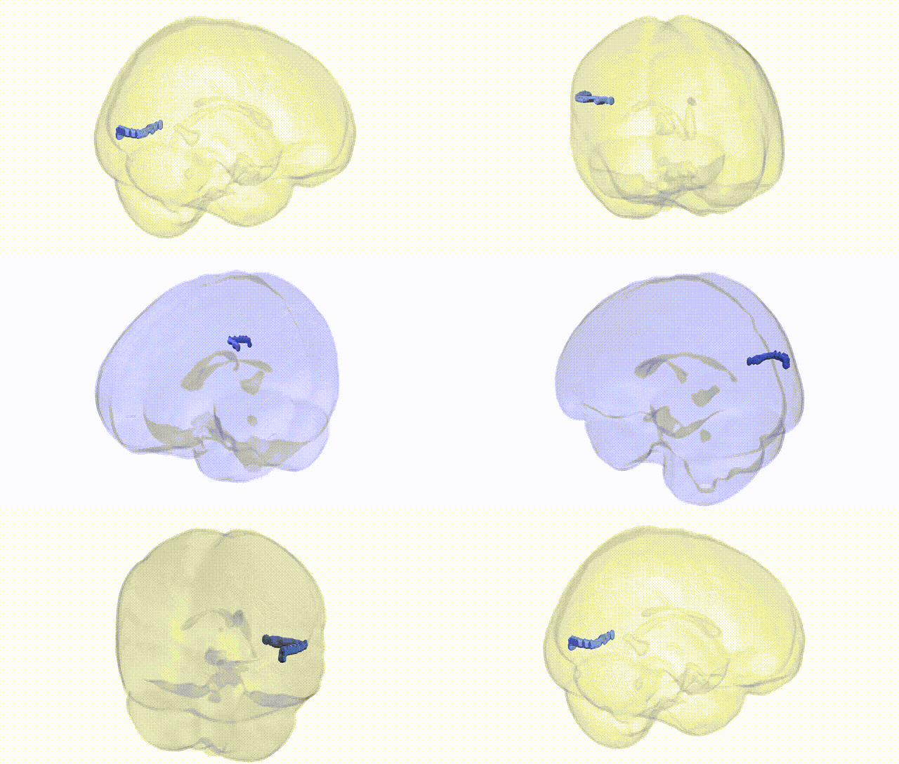
Full Quality Version: Download MP4
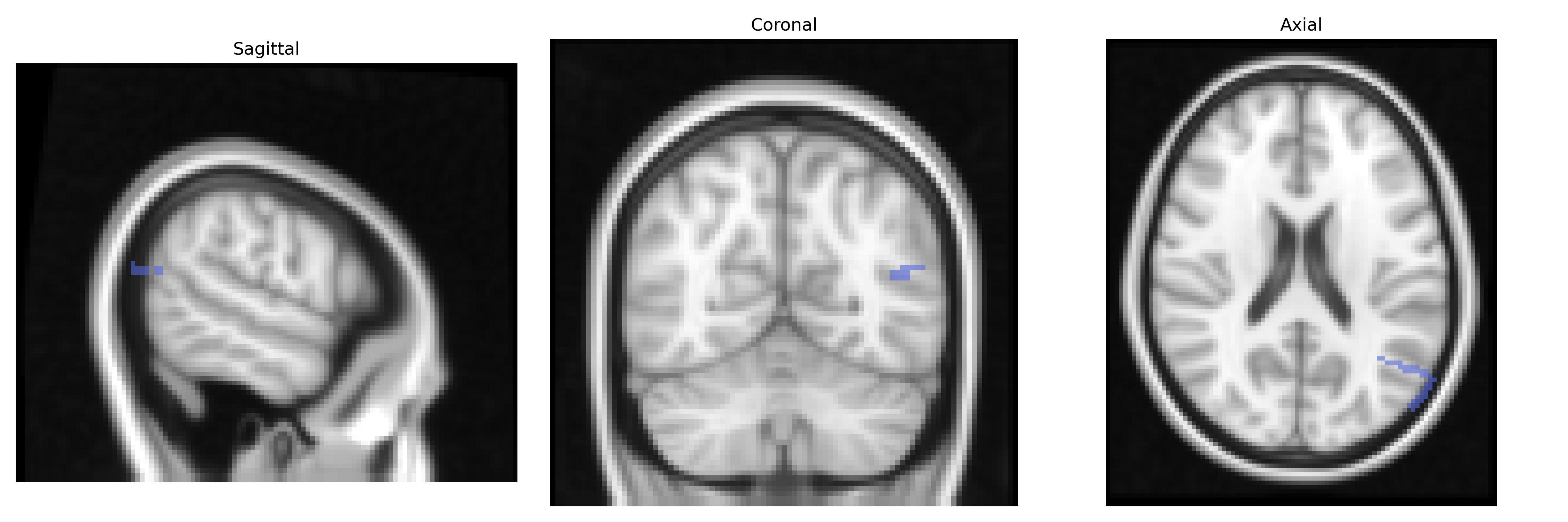
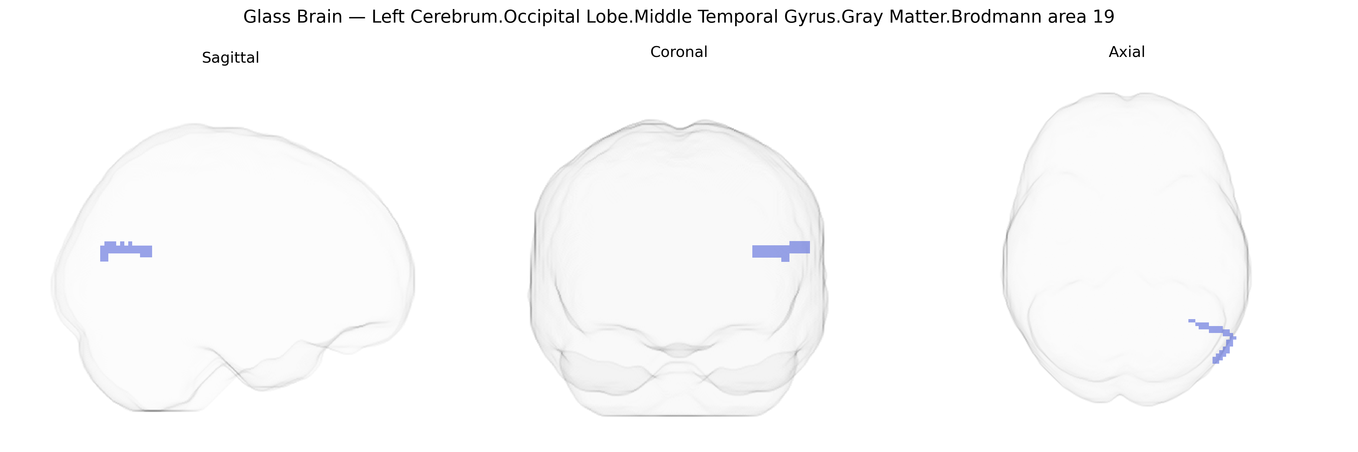
Wali Sidiqyar*, Gaurav Rudravaram*, Elyssa M. McMaster, Trent M. Schwartz, Adam M. Saunders, Kurt G. Schilling, Bennett A. Landman "Introducing SPINS: A Shared Public Visualization Library of Neuroanatomical Structures." Medical Imaging with Deep Learning- short paper
This resource is licensed under CC0 1.0 Universal (Public Domain).