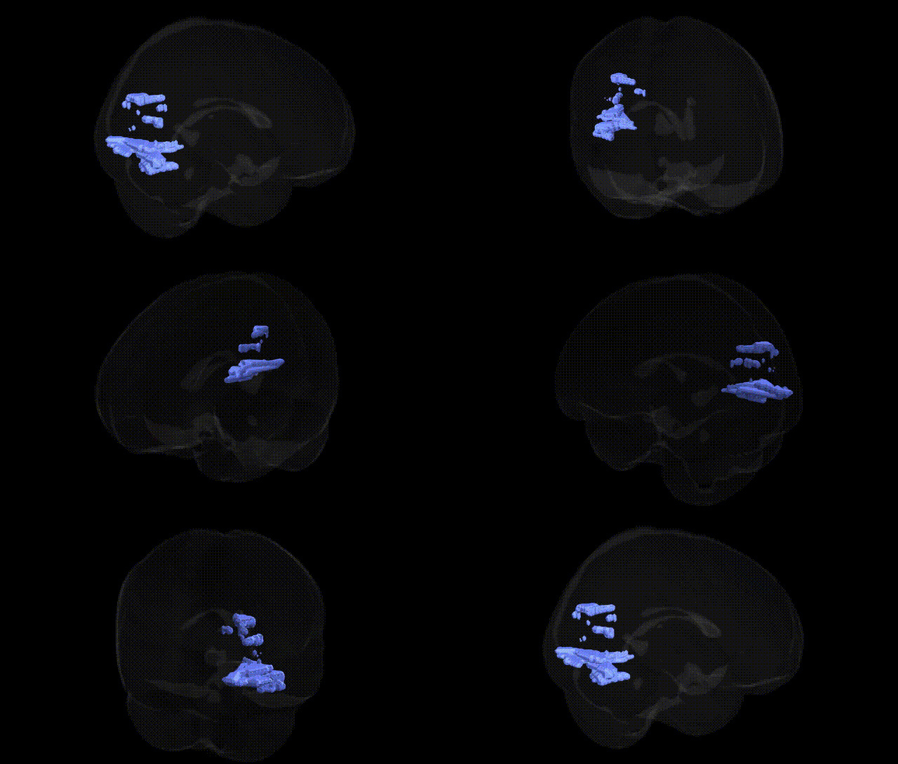
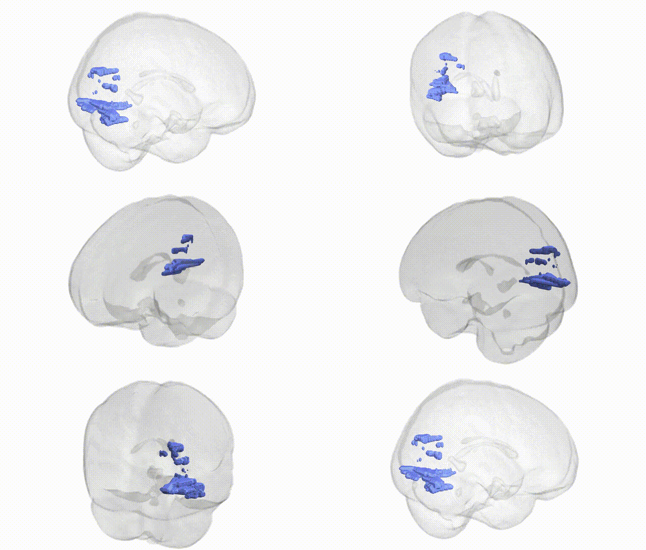
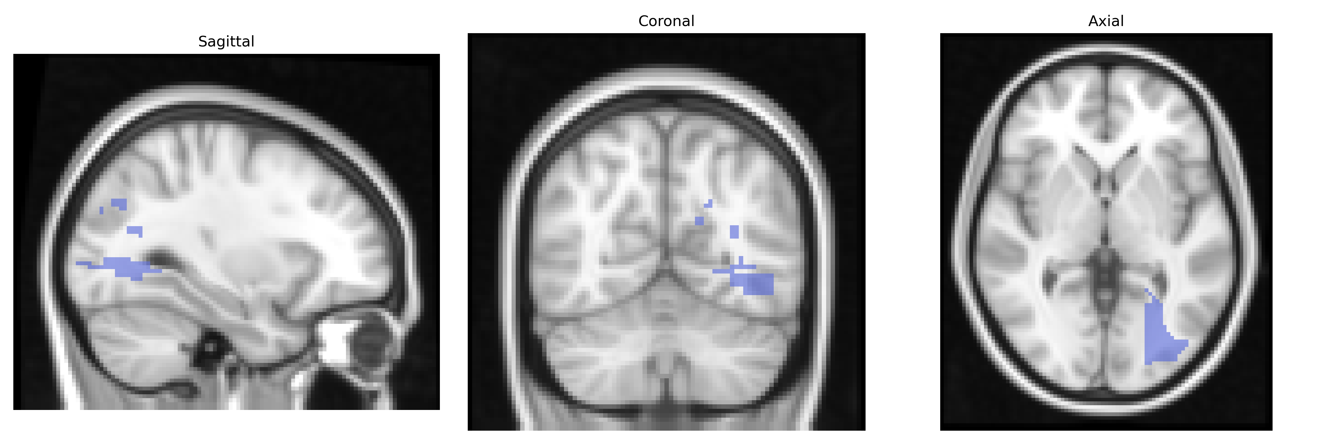
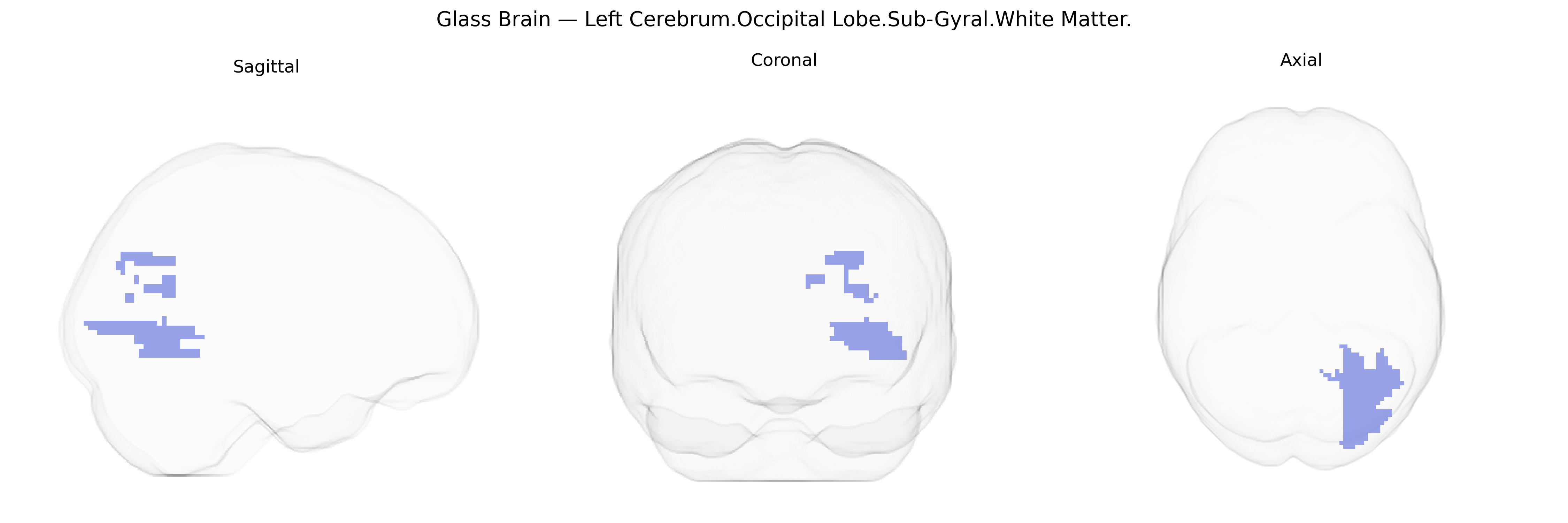
The bilateral Left Cerebrum.Occipital Lobe.Sub-Gyral.White Matter refers to deep white matter tracts located beneath the cortical gyri of the left occipital lobe, within the cerebral hemisphere. This region is composed primarily of myelinated axons that form part of the brain’s visual processing network, interconnecting primary and associative visual cortices with other cortical and subcortical regions, and contributing to the integration and transmission of visual information necessary for perception, spatial orientation, and visually guided behavior. As a sub-gyral white matter zone, it includes portions of major occipital pathways such as segments of the inferior longitudinal fasciculus and optic radiations, supporting communication between occipital cortex, temporal lobe, and thalamic nuclei. There is no direct Wikipedia article for this specific Talairach label; a closely related structure is the Occipital lobe.
Genetic associations specifically targeting the bilateral Left Cerebrum.Occipital Lobe.Sub-Gyral.White Matter, as defined in the Talairach 2 mm atlas, are rarely reported at this fine-grained level, but broader imaging genetics and GWAS studies implicate occipital white matter microstructure and occipital lobe connectivity in several traits and disorders. Variants in genes related to myelination and axonal integrity (e.g., MAG, MBP, PLP1, and broader oligodendrocyte-pathway loci) have been associated with diffusion tensor imaging measures such as fractional anisotropy and mean diffusivity in occipital tracts, including portions of the inferior longitudinal fasciculus and optic radiations that traverse sub-gyral occipital white matter. Large GWAS of brain structural MRI (ENIGMA, UK Biobank) have identified loci near HMGA2 and IGF1 associated with global and regional white matter volumes and cortical surface area, which indirectly encompass occipital regions, and polygenic scores for educational attainment and general cognitive ability correlate with occipital cortical and adjacent white matter measures, reflecting the role of this territory in visual processing and higher-order cognition. In neuropsychiatric disorders such as schizophrenia, bipolar disorder, and major depression, risk variants in synaptic and neurodevelopmental genes (e.g., CACNA1C, ZNF804A, DISC1-related pathways) show imaging endophenotypes that include altered occipital white matter integrity, while in neurodegenerative disease, APOE ε4 and other Alzheimer’s disease risk loci have been linked to widespread white matter changes that extend into occipital sub-gyral regions in tract-based analyses. Additionally, GWAS of migraine and visual aura implicate genes involved in cortical excitability (e.g., CACNA1A, SCN1A) with functional and structural alterations in occipital networks, and rare monogenic conditions affecting visual pathways (such as some leukodystrophies and hereditary optic neuropathies) produce marked pathology in occipital white matter, though these are typically described at the tract or lobe level rather than this specific Talairach-defined subregion.
Overview generated by GPT-4o (2026).
Region ID: 319
Hemisphere: bilateral
Atlas: Talairach labels 2mm
Full Quality Version: Download MP4
Full Quality Version: Download MP4
Wali Sidiqyar*, Gaurav Rudravaram*, Elyssa M. McMaster, Trent M. Schwartz, Adam M. Saunders, Kurt G. Schilling, Bennett A. Landman "Introducing SPINS: A Shared Public Visualization Library of Neuroanatomical Structures." Medical Imaging with Deep Learning- short paper
This resource is licensed under CC0 1.0 Universal (Public Domain).