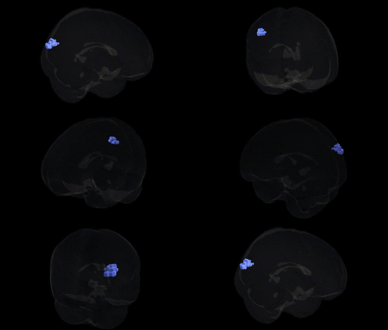
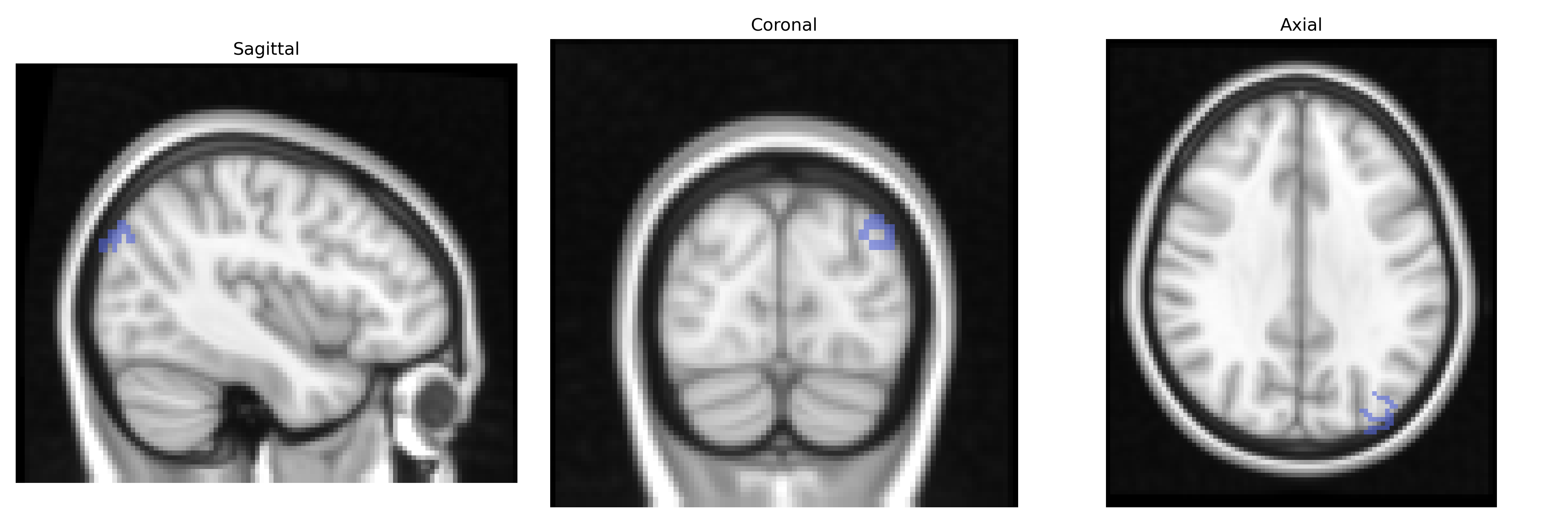
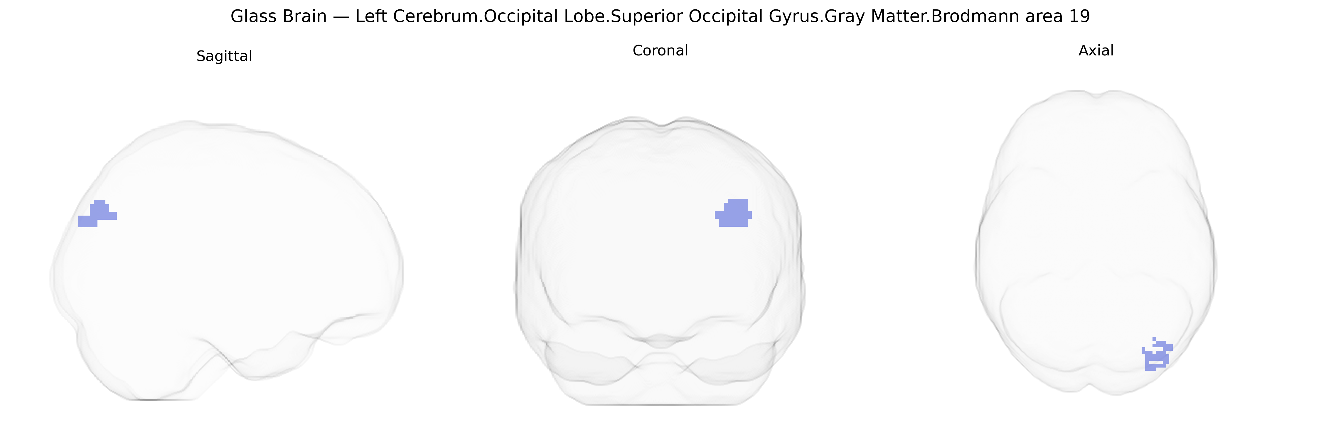
The bilateral Left Cerebrum.Occipital Lobe.Superior Occipital Gyrus.Gray Matter.Brodmann area 19 corresponds to higher-order visual association cortex located dorsally in the occipital lobe, anterior to primary and secondary visual areas. Brodmann area 19 participates in complex visual processing, including integration of shape, color, motion, and spatial relationships, and contributes to object recognition and visuospatial analysis through extensive reciprocal connections with other occipital, parietal, and temporal regions. Neuronal populations here receive convergent input from earlier visual areas (e.g., BA17 and BA18) and project to multimodal association cortices, supporting transformation of basic visual signals into more abstract representations important for perception and visually guided behavior. There is no direct link for “Superior Occipital Gyrus Brodmann area 19”; a related structure is Brodmann area 19.
The bilateral left Brodmann area 19 in the superior occipital gyrus, a key visual association cortex, has been repeatedly implicated in imaging genetics and GWAS of brain structure and function. Large-scale neuroimaging GWAS (e.g., ENIGMA, UK Biobank) have identified common variants in genes involved in synaptic signaling, neuronal development, and axon guidance (such as BDNF, GRIN2B, and genes within the MAPT locus) that are associated with occipital gray matter volume, cortical thickness, or surface area, although findings are often regionally broader than BA19 and not always specific to this Talairach-defined parcel. Functional and task-based imaging genetics studies link polymorphisms in visual and glutamatergic pathway genes (for example, variants in GABAergic and glutamate receptor genes) to altered activation of BA19 during visual processing and attention tasks. Disease-focused GWAS-meta analyses that integrate structural MRI show that genetic risk for neuropsychiatric disorders—particularly schizophrenia, major depressive disorder, and autism spectrum disorder—can be expressed as reduced occipital cortical volume or atypical functional connectivity, with BA19 commonly included in the affected visual association network; however, these effects are typically polygenic and distributed, so BA19 is a node within broader genetic liability patterns rather than a uniquely specified target. In addition, GWAS of visual perception traits and reading-related skills have shown modest associations between risk loci (e.g., in KIAA0319 and other neurodevelopmental genes) and structural or functional variability in occipital association regions, consistent with the role of BA19 in higher-order visual analysis, though precise parcel-level genetic specificity for the bilateral left BA19 region from the Talairach 2 mm atlas remains limited in current literature.
Overview generated by GPT-4o (2026).
Region ID: 848
Hemisphere: bilateral
Atlas: Talairach labels 2mm
Full Quality Version: Download MP4
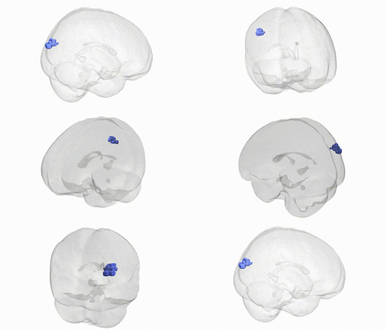
Full Quality Version: Download MP4
Wali Sidiqyar*, Gaurav Rudravaram*, Elyssa M. McMaster, Trent M. Schwartz, Adam M. Saunders, Kurt G. Schilling, Bennett A. Landman "Introducing SPINS: A Shared Public Visualization Library of Neuroanatomical Structures." Medical Imaging with Deep Learning- short paper
This resource is licensed under CC0 1.0 Universal (Public Domain).