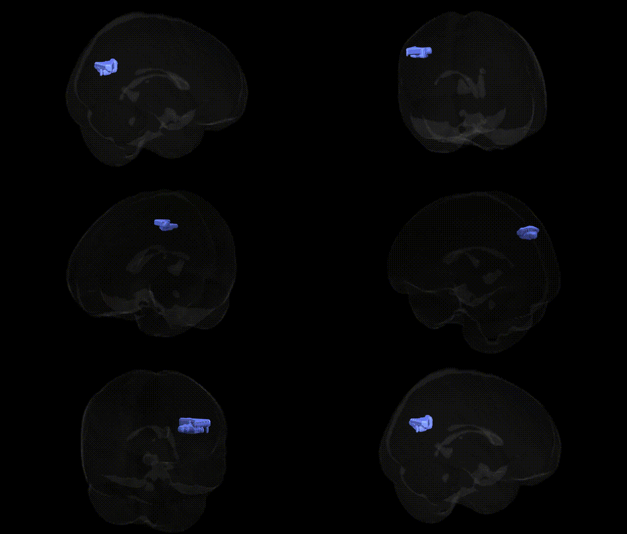
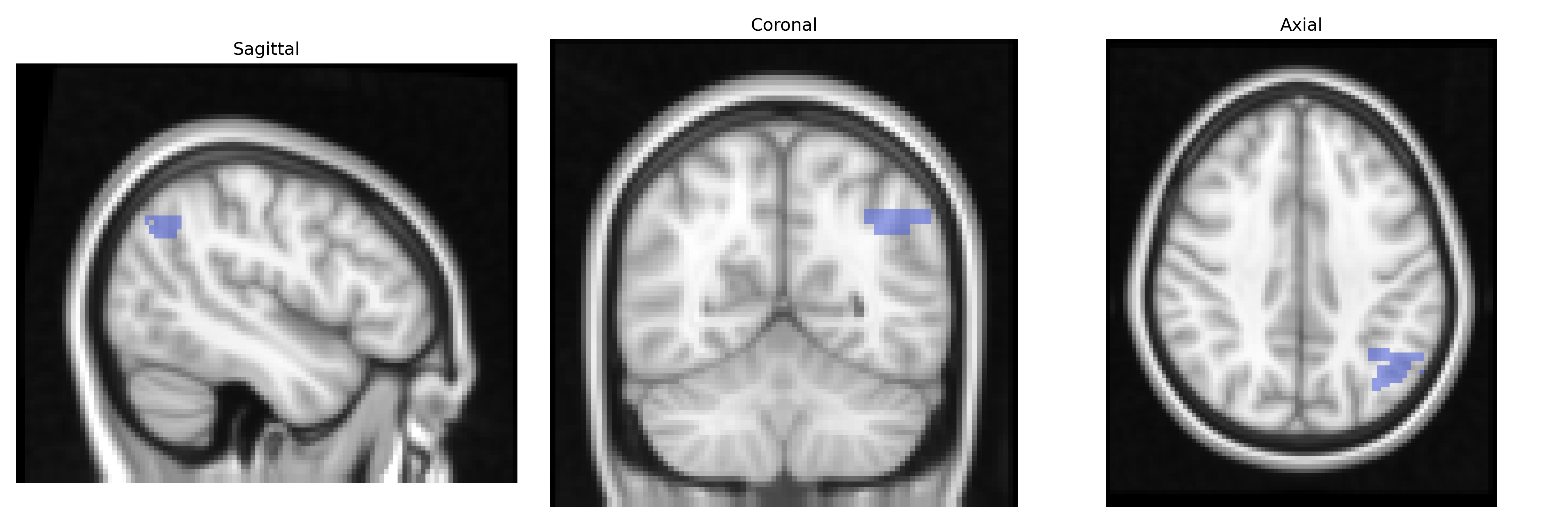
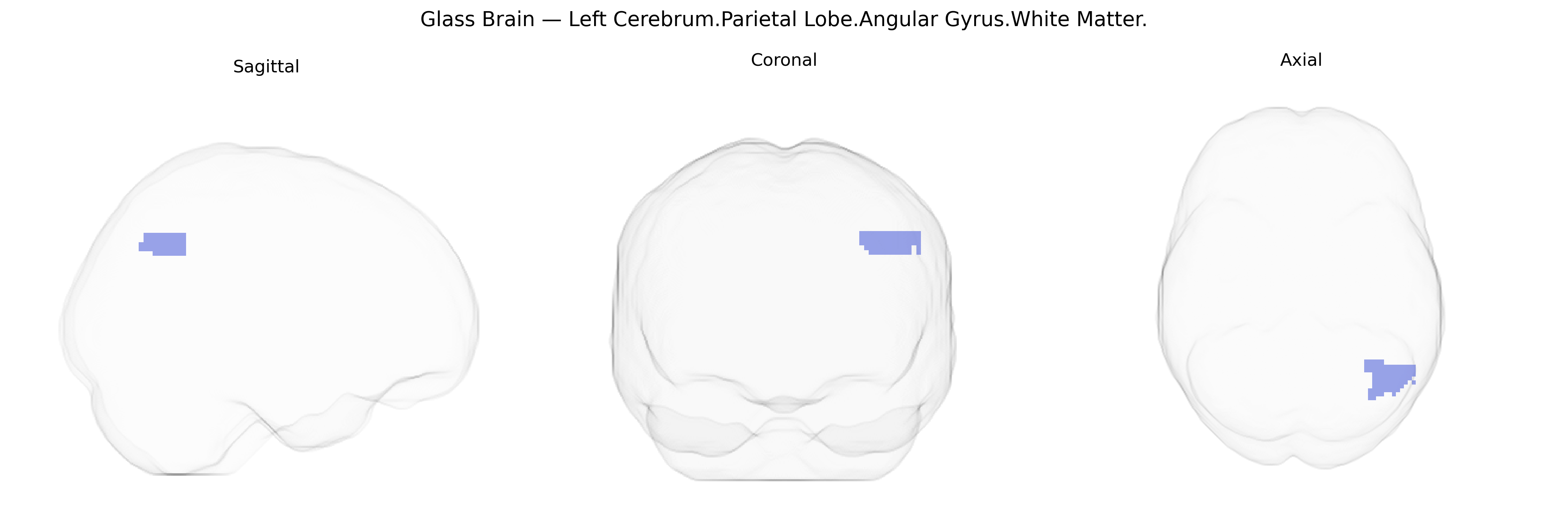
The bilateral Left Cerebrum.Parietal Lobe.Angular Gyrus.White Matter refers to the subcortical fiber pathways underlying the angular gyrus in the inferior parietal lobule of the left cerebral hemisphere. This white matter region contains association fibers that interconnect the angular gyrus cortex with other parietal, temporal, and frontal areas, supporting multimodal integration of visual, auditory, and linguistic information, and contributing to functions such as reading, semantic processing, numerical cognition, and aspects of spatial attention. As part of the broader parietal lobe white matter network, it participates in higher-order cognitive operations by facilitating efficient communication between cortical nodes involved in language and conceptual processing. There is no direct Wikipedia article for the white matter of the angular gyrus; a related cortical structure is the Angular gyrus.
The bilateral Left Cerebrum Parietal Lobe Angular Gyrus white matter, as defined in the Talairach 2 mm atlas, has been implicated in several genetic and neuroimaging genetics findings, though most evidence comes from studies of nearby parietal association and temporo‑parietal regions rather than this specific label. GWAS of brain white matter microstructure (e.g., fractional anisotropy) have identified variants in genes involved in axon guidance, myelination, and oligodendrocyte function (such as CNTN4, NRG1, MAG, and PLP1) that influence diffusion metrics in parietal and temporo‑parietal tracts connecting the angular gyrus to frontal and temporal cortices. Imaging‑genetics work has linked schizophrenia, autism spectrum disorder, and attention‑deficit/hyperactivity disorder risk loci (including variants in CACNA1C, MIR137, and CNTNAP2) to altered structural connectivity and white matter integrity in parietal and temporo‑parietal networks supporting language, number processing, and social cognition, with angular gyrus–adjacent white matter commonly affected. Polygenic risk scores for Alzheimer’s disease and general cognitive ability have also been associated with differences in parietal and angular gyrus–related white matter, consistent with the region’s role in default‑mode and multimodal integration networks. However, most GWAS report findings at the level of tracts, lobes, or cortical networks (e.g., superior longitudinal fasciculus, inferior parietal lobule, default‑mode connectivity), so current genetic associations should be interpreted as involving the broader parietal/angular gyrus white matter system rather than a uniquely specified Talairach 2 mm subregion.
Overview generated by GPT-4o (2026).
Region ID: 965
Hemisphere: bilateral
Atlas: Talairach labels 2mm
Full Quality Version: Download MP4
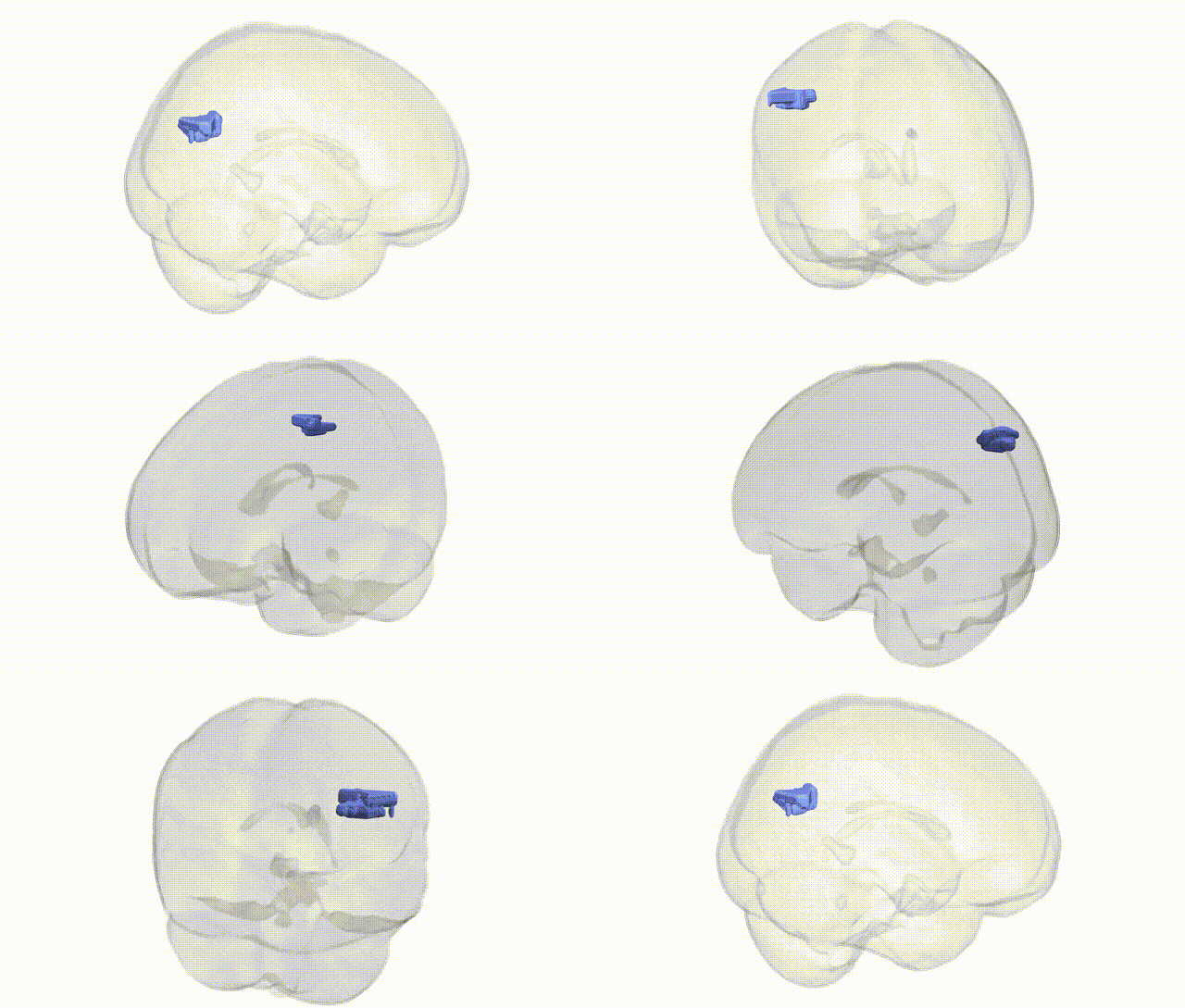
Full Quality Version: Download MP4
Wali Sidiqyar*, Gaurav Rudravaram*, Elyssa M. McMaster, Trent M. Schwartz, Adam M. Saunders, Kurt G. Schilling, Bennett A. Landman "Introducing SPINS: A Shared Public Visualization Library of Neuroanatomical Structures." Medical Imaging with Deep Learning- short paper
This resource is licensed under CC0 1.0 Universal (Public Domain).