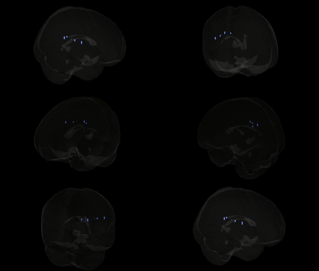
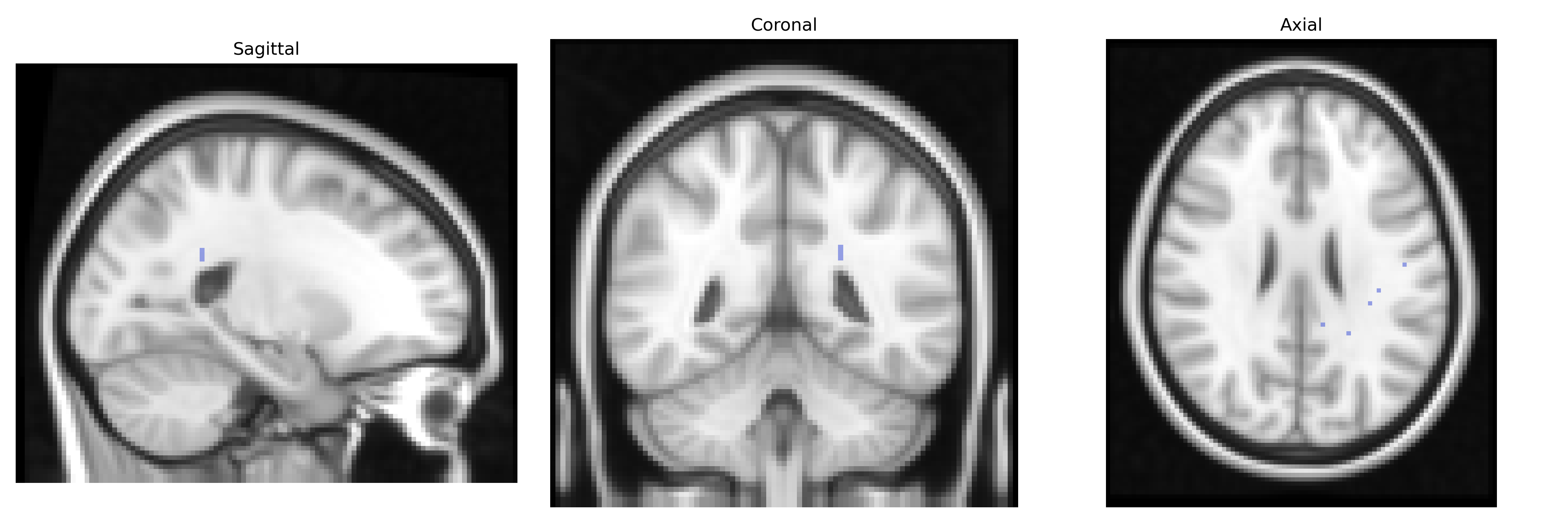
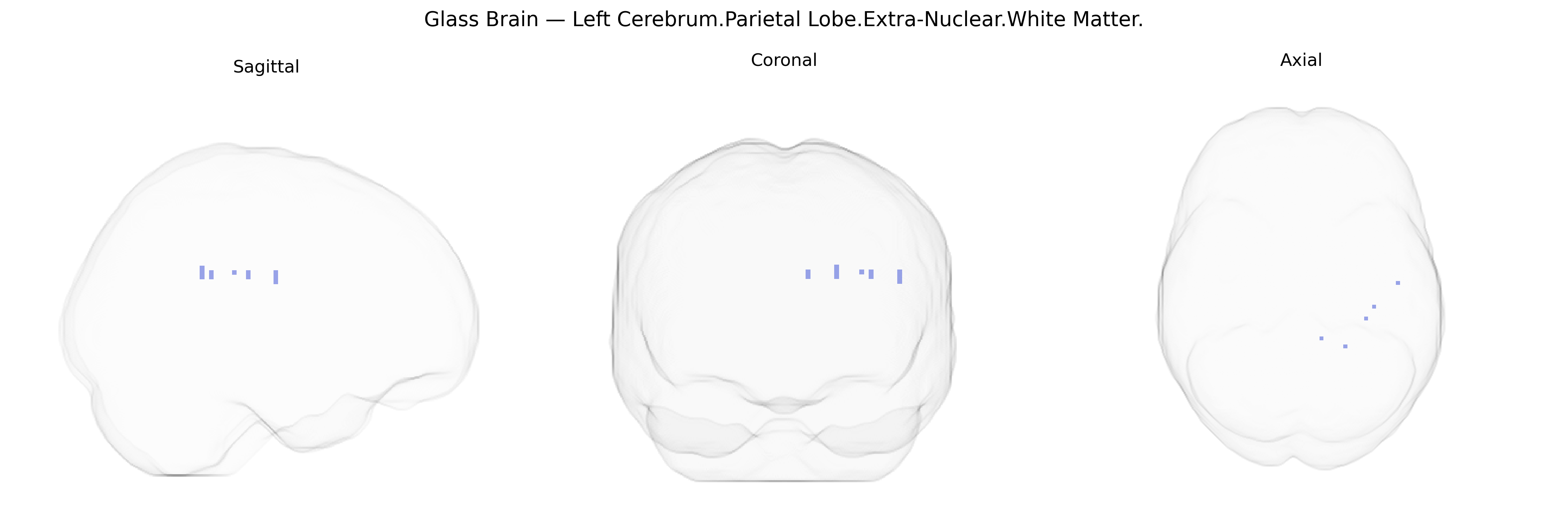
The bilateral Left Cerebrum.Parietal Lobe.Extra-Nuclear.White Matter (Talairach 2 mm Atlas) refers to subcortical association and projection fibers coursing through the white matter deep to the parietal cortex, outside the nuclei of the basal ganglia and thalamus (“extra-nuclear”). These fibers include components of long-range pathways such as the superior longitudinal fasciculus and portions of thalamocortical and corticocortical tracts that interconnect parietal regions with frontal, temporal, and occipital cortices, supporting multimodal integration, spatial processing, attention, and sensorimotor coordination. This area consists primarily of myelinated axons, glial cells, and microvasculature, and does not correspond to a discrete gray matter nucleus. There is no direct Wikipedia article for this exact label; a closely related structure is the Parietal lobe.
The bilateral Left Cerebrum Parietal Lobe extra-nuclear white matter, as defined in the Talairach 2 mm atlas, corresponds largely to association and projection fiber tracts linking parietal cortex with frontal, temporal, and subcortical regions, and genetic associations to this area are typically reported in terms of white matter microstructure (e.g., fractional anisotropy) rather than this atlas label specifically. Large GWAS of brain imaging phenotypes (e.g., ENIGMA, UK Biobank) have identified numerous loci influencing parietal white matter integrity and connectivity, including variants in genes involved in axon guidance and myelination (such as CNTN4, NRG1, and MAG), with pleiotropic effects on cognitive performance, educational attainment, and risk for neuropsychiatric disorders. Parietal white matter tracts—particularly superior longitudinal fasciculus and related fibers—show heritable variation and have been implicated via imaging-genetic studies in attention-deficit/hyperactivity disorder, schizophrenia, autism spectrum disorder, major depressive disorder, and Alzheimer’s disease, often reflecting disrupted frontoparietal networks. Polygenic risk scores for schizophrenia, bipolar disorder, and depression have been associated with altered parietal white matter microstructure, and GWAS of diffusion tensor imaging metrics have revealed shared genetic architectures between parietal white matter organization and traits such as general cognitive ability, processing speed, and risk-taking behavior. Overall, genetic findings indicate that variants influencing neurodevelopmental pathways, synaptic plasticity, and myelin biology contribute to inter-individual differences in the structure and function of parietal association white matter, which in turn modulates vulnerability to a range of cognitive and psychiatric outcomes, although few studies reference the specific Talairach “extra-nuclear” label and instead focus on tract-based or lobar measures.
Overview generated by GPT-4o (2026).
Region ID: 801
Hemisphere: bilateral
Atlas: Talairach labels 2mm
Full Quality Version: Download MP4
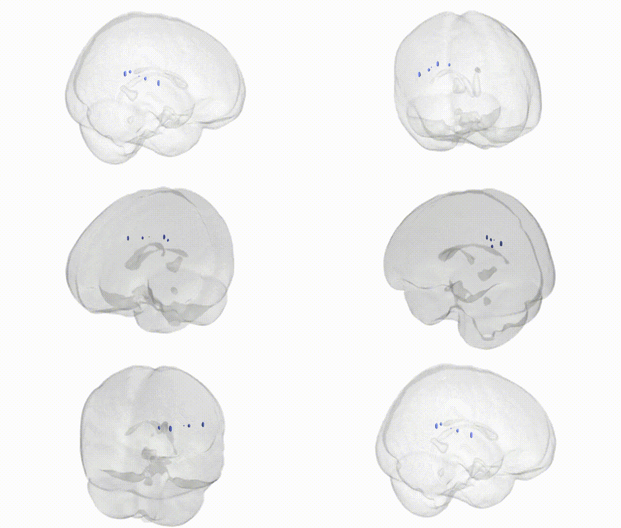
Full Quality Version: Download MP4
Wali Sidiqyar*, Gaurav Rudravaram*, Elyssa M. McMaster, Trent M. Schwartz, Adam M. Saunders, Kurt G. Schilling, Bennett A. Landman "Introducing SPINS: A Shared Public Visualization Library of Neuroanatomical Structures." Medical Imaging with Deep Learning- short paper
This resource is licensed under CC0 1.0 Universal (Public Domain).