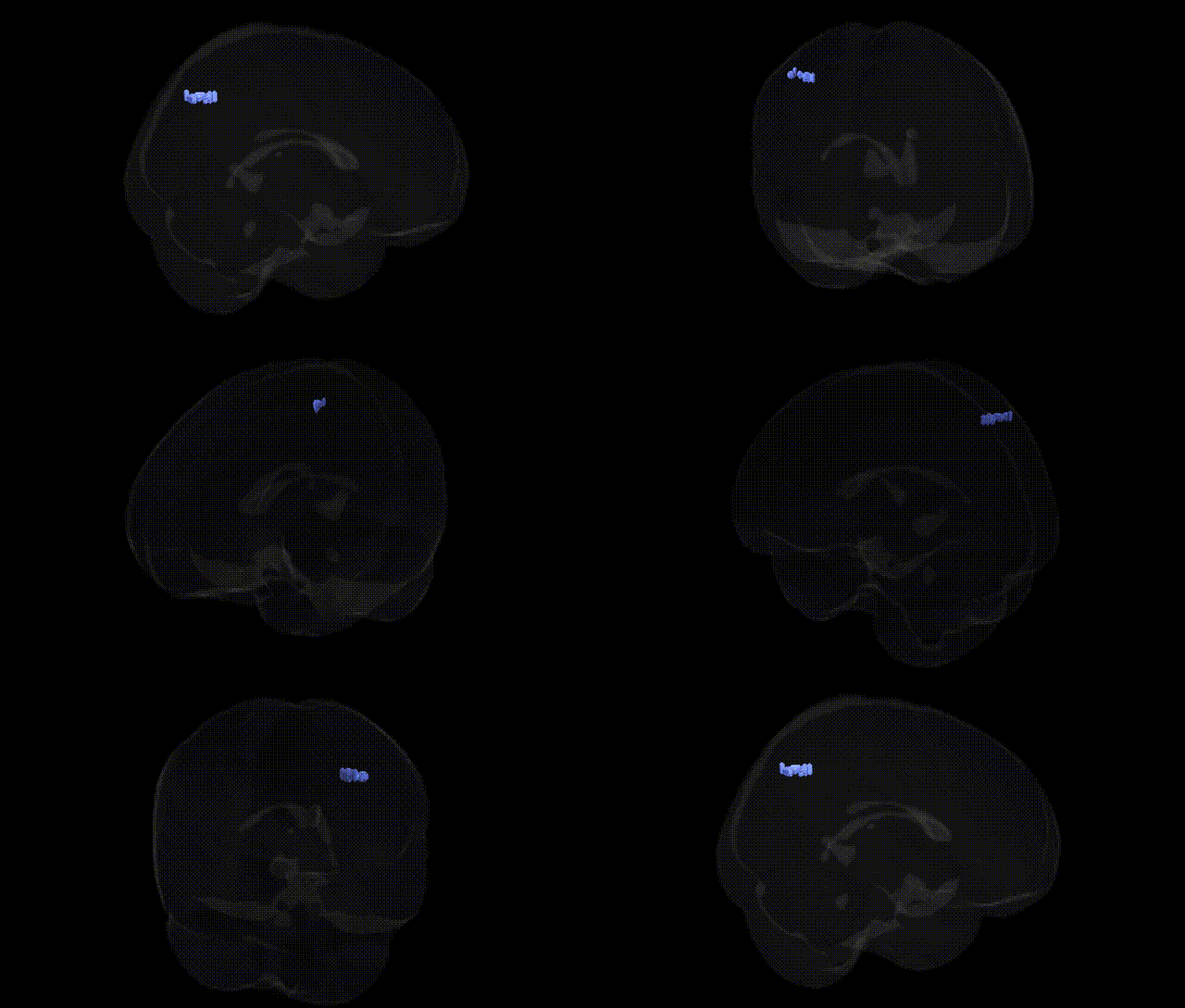
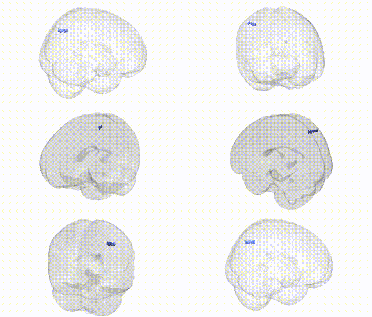
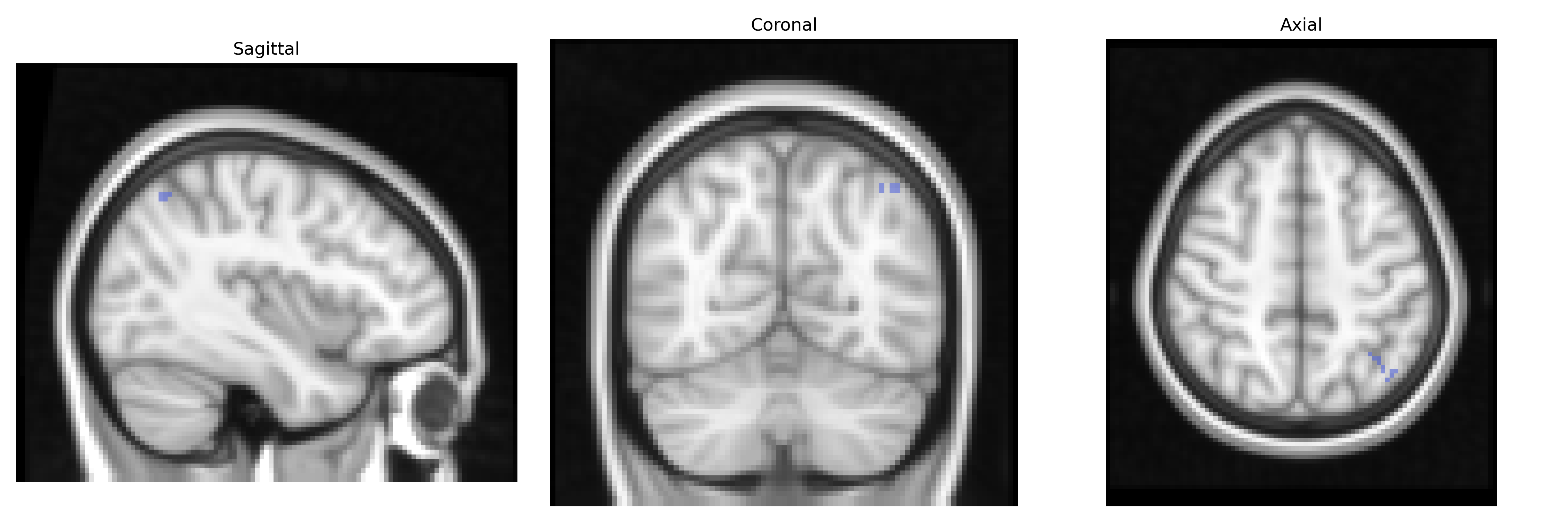
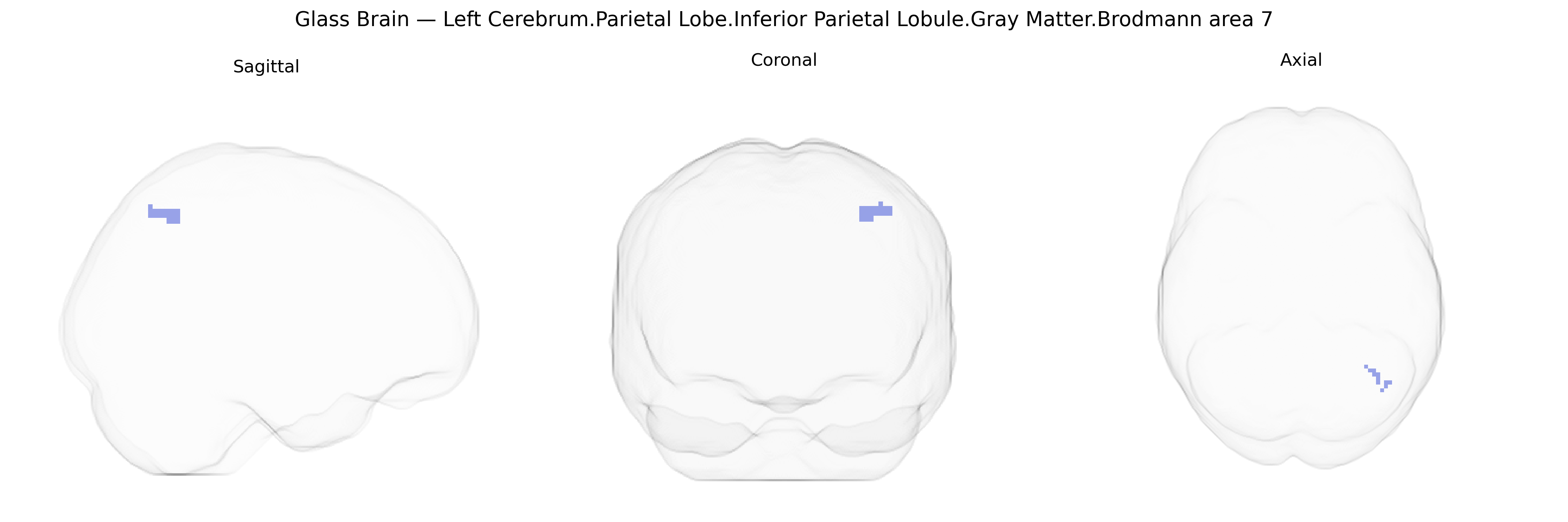
Bilateral Left Cerebrum.Parietal Lobe.Inferior Parietal Lobule.Gray Matter.Brodmann area 7 corresponds to a portion of the superior posterior parietal cortex involved in higher-order visuospatial processing, visuomotor integration, and the transformation of sensory input into coordinated actions. This association cortex receives multimodal input from visual, somatosensory, and premotor regions, contributing to spatial attention, eye–hand coordination, and the representation of extrapersonal space. Neuronal populations within this area participate in constructing internal models of spatial relationships and body position, supporting functions such as reaching, object manipulation, and navigation. Functionally, Brodmann area 7 forms part of the dorsal visual stream and parietal networks that link perception to action. There is no direct link for this exact Talairach label; a closely related structure is Brodmann area 7.
The bilateral Left Cerebrum Parietal Lobe Inferior Parietal Lobule (IPL) gray matter Brodmann area 7, as defined in the Talairach 2 mm atlas, has been repeatedly implicated in genetic studies of higher-order cognition, attention, visuospatial processing, and social cognition. GWAS of cortical thickness and surface area have identified common variants near genes such as HMGA2, KIAA0586, and IGF1 associated with parietal and specifically inferior parietal or superior parietal morphometry, with several large ENIGMA consortium studies showing heritable effects on IPL structure and BA7-linked cortical measures. Variants in genes involved in synaptic function and neurodevelopment (e.g., CNTNAP2, DISC1, NRGN, and GRIN2B) have been associated, via imaging genetics, with altered gray-matter volume, activation, or connectivity in IPL/BA7 during tasks requiring working memory, language, or social cognition. Neuropsychiatric GWAS for schizophrenia, major depression, ADHD, and autism spectrum disorder have shown that polygenic risk scores for these conditions correlate with structural and functional alterations in BA7/IPL, suggesting that distributed genetic risk influences this parietal hub rather than single locus–region specificity. In Alzheimer’s disease and other dementias, risk variants in APOE, CLU, PICALM, and BIN1 have been related to accelerated thinning and hypometabolism in parietal association cortices including BA7, consistent with the region’s vulnerability in posterior cortical atrophy. GWAS of traits such as general intelligence, mathematical ability, reading skill, and attentional performance also demonstrate that polygenic scores for cognitive ability and educational attainment predict individual differences in IPL/BA7 volume and activation, supporting a role for genetically influenced variation in this parietal association area in complex cognitive and neurodevelopmental phenotypes.
Overview generated by GPT-4o (2026).
Region ID: 1043
Hemisphere: bilateral
Atlas: Talairach labels 2mm
Full Quality Version: Download MP4
Full Quality Version: Download MP4
Wali Sidiqyar*, Gaurav Rudravaram*, Elyssa M. McMaster, Trent M. Schwartz, Adam M. Saunders, Kurt G. Schilling, Bennett A. Landman "Introducing SPINS: A Shared Public Visualization Library of Neuroanatomical Structures." Medical Imaging with Deep Learning- short paper
This resource is licensed under CC0 1.0 Universal (Public Domain).