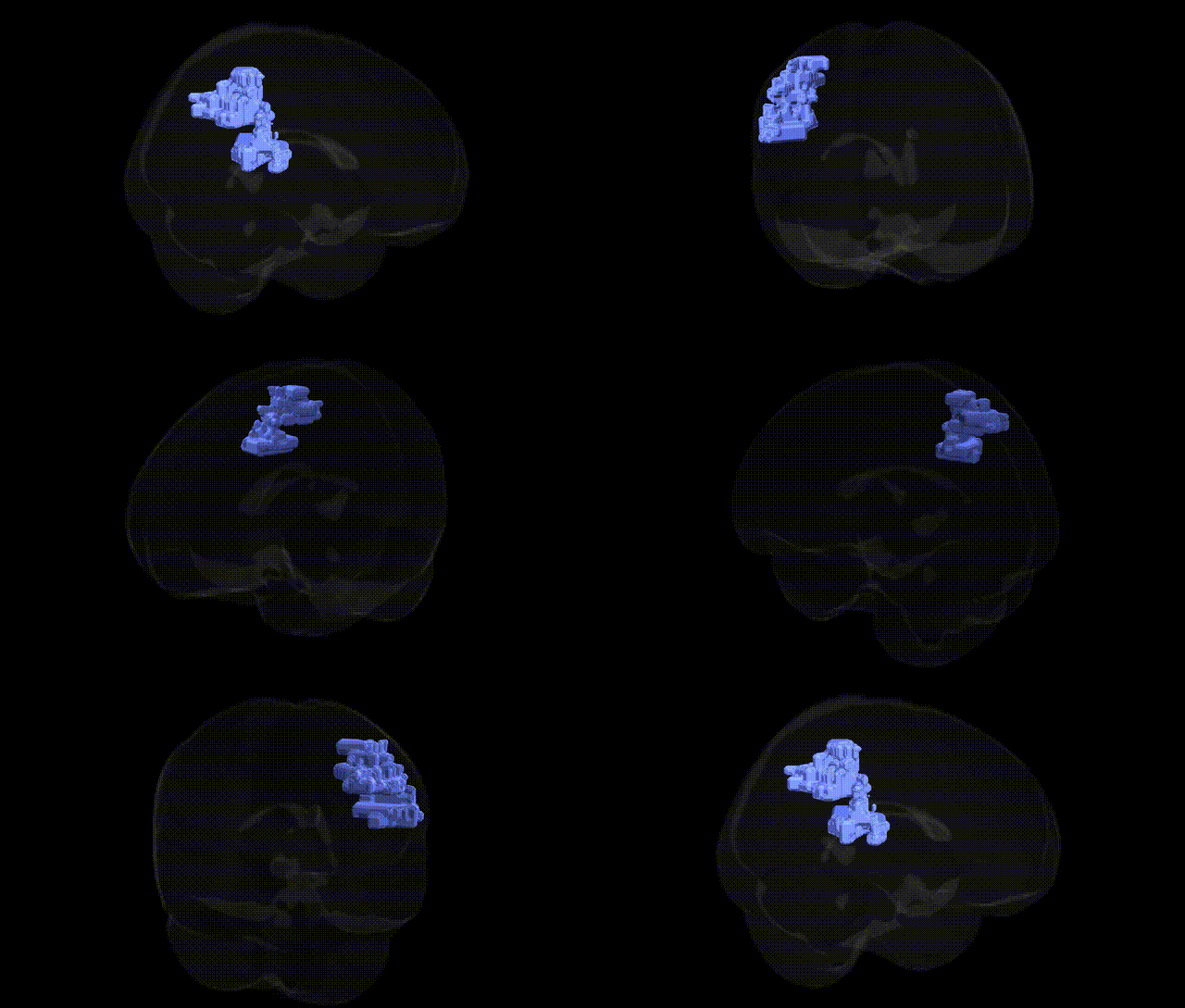
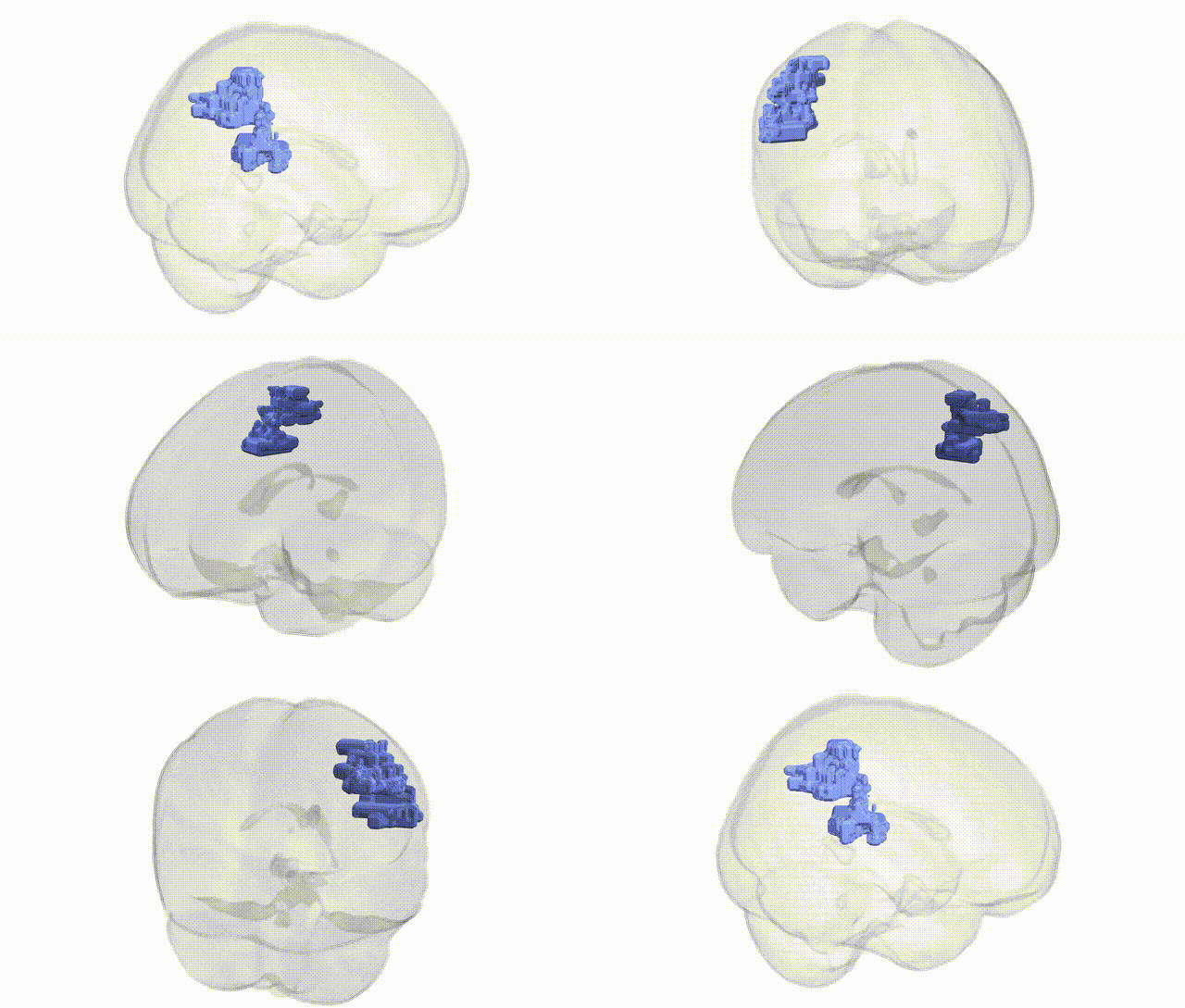
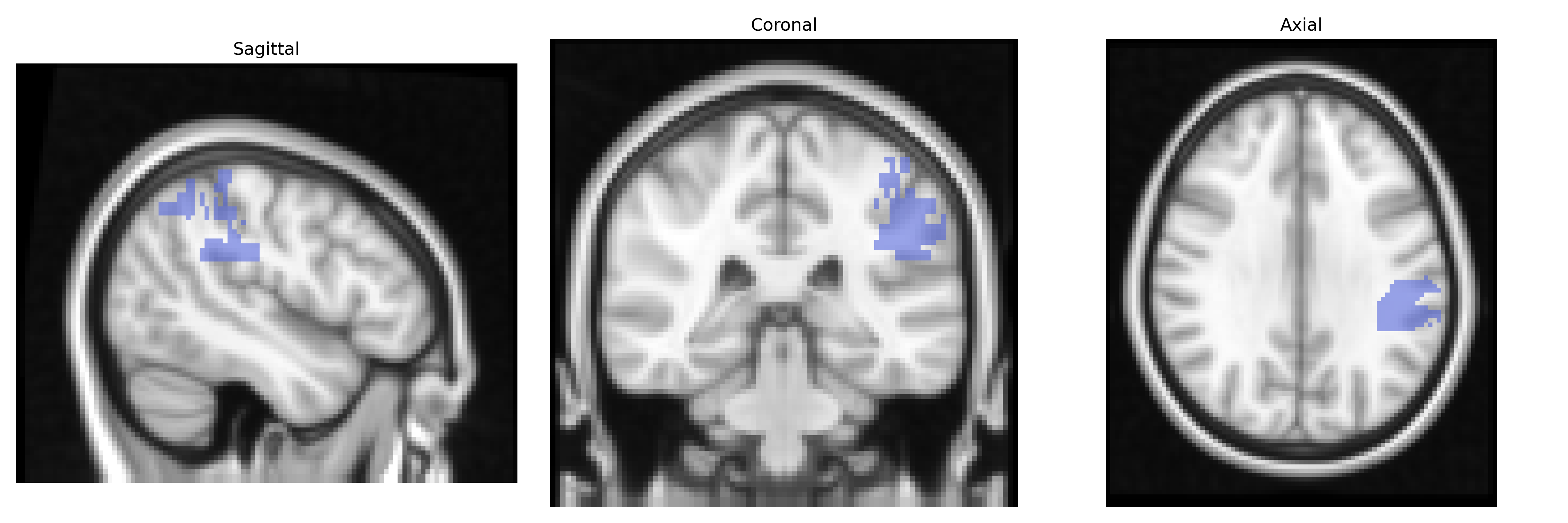
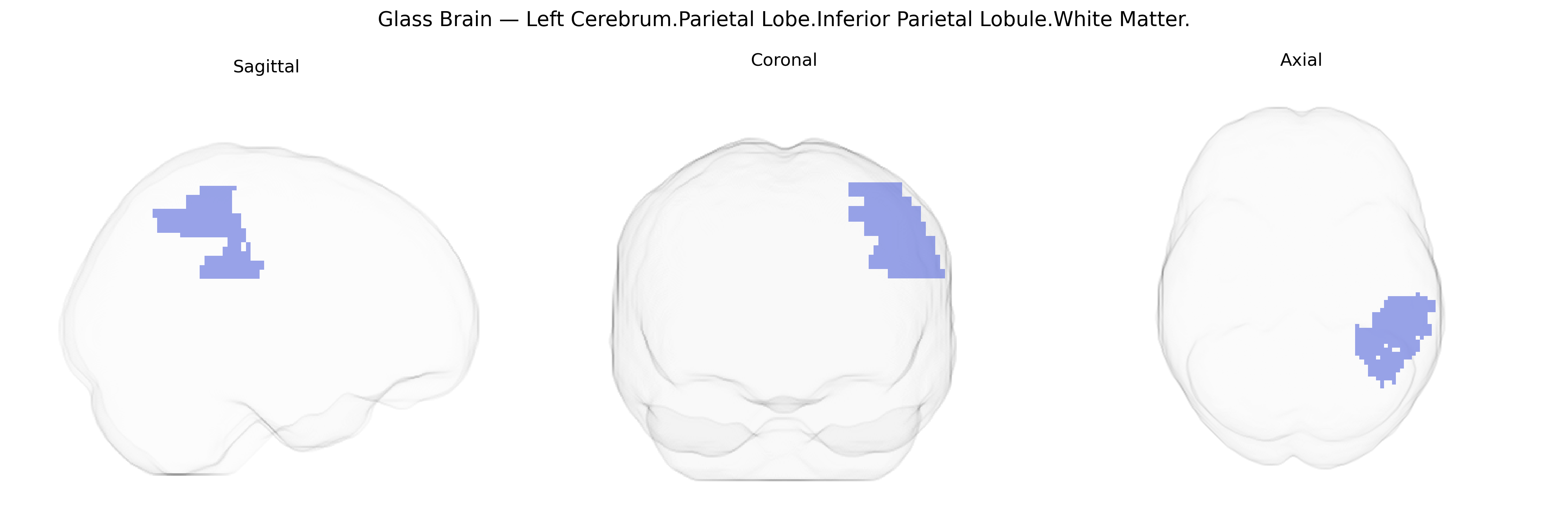
The bilateral Left Cerebrum.Parietal Lobe.Inferior Parietal Lobule.White Matter corresponds to subcortical fiber tracts underlying the inferior parietal lobule, a heteromodal association region of the parietal lobe involved in multimodal sensory integration, spatial attention, language, and aspects of body representation. This white matter compartment contains short association fibers connecting inferior parietal cortex with adjacent parietal and temporal areas, as well as portions of long-range association pathways (such as components of the superior longitudinal fasciculus) that link parietal regions with frontal and other cortical territories. Functionally, these fibers support the coordination and transfer of information necessary for higher-order cognitive operations including praxis, numerical processing, and semantic aspects of language. There is no direct link for the specific white matter compartment; a closely related cortical structure is the Inferior parietal lobule.
The bilateral left Inferior Parietal Lobule white matter, as defined in the Talairach 2 mm Atlas, has been indirectly implicated in genetic studies through its role in large-scale parietal association networks rather than via region-specific GWAS hits. Variants in genes involved in axonal guidance and myelination (for example, CNTNAP2, NRG1, and various oligodendrocyte-related loci) have been associated with microstructural properties of parietal lobe white matter on diffusion MRI, influencing fractional anisotropy and connectivity measures that encompass the inferior parietal regions. GWAS of brain imaging phenotypes from large cohorts such as ENIGMA and UK Biobank have identified multiple loci (including variants near genes such as HMGA2, FOXO3, and MAPT) associated with global and regional cortical volumes, surface area, and white matter integrity, with parietal association areas frequently among the regions showing significant effects, though not typically resolved to the “Left Cerebrum.Parietal Lobe.Inferior Parietal Lobule.White Matter” label. Genetically influenced alterations of this parietal white matter territory have been reported in neurodevelopmental and psychiatric conditions such as schizophrenia, autism spectrum disorder, and attention-deficit/hyperactivity disorder, where risk variants in synaptic and neurodevelopmental genes (e.g., CACNA1C, DISC1, and several common polygenic risk loci) are associated with disrupted fronto-parietal and temporo-parietal connectivity that traverse the inferior parietal lobule. Additionally, polygenic risk scores for Alzheimer’s disease and other dementias show associations with degeneration of parietal association cortex and underlying white matter tracts, including pathways passing through inferior parietal regions, linking APOE and other AD-related loci to structural and functional changes in this area. Overall, genetic associations tend to target network-level traits—parietal connectivity, association-fiber microstructure, and cognition (language, numerical processing, attention)—rather than the specific Talairach-labeled inferior parietal lobule white matter region, but converging evidence indicates that its structure and function are modulated by a broad polygenic architecture underlying neurodevelopment, cognition, and risk for major neuropsychiatric and neurodegenerative disorders.
Overview generated by GPT-4o (2026).
Region ID: 879
Hemisphere: bilateral
Atlas: Talairach labels 2mm
Full Quality Version: Download MP4
Full Quality Version: Download MP4
Wali Sidiqyar*, Gaurav Rudravaram*, Elyssa M. McMaster, Trent M. Schwartz, Adam M. Saunders, Kurt G. Schilling, Bennett A. Landman "Introducing SPINS: A Shared Public Visualization Library of Neuroanatomical Structures." Medical Imaging with Deep Learning- short paper
This resource is licensed under CC0 1.0 Universal (Public Domain).