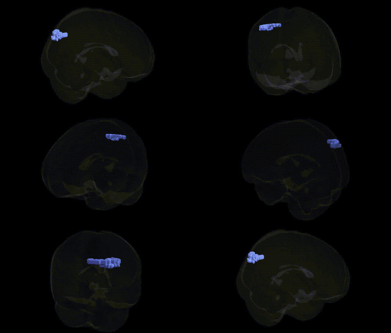
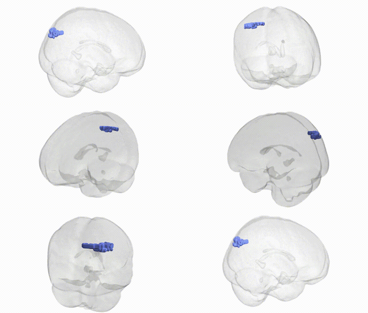
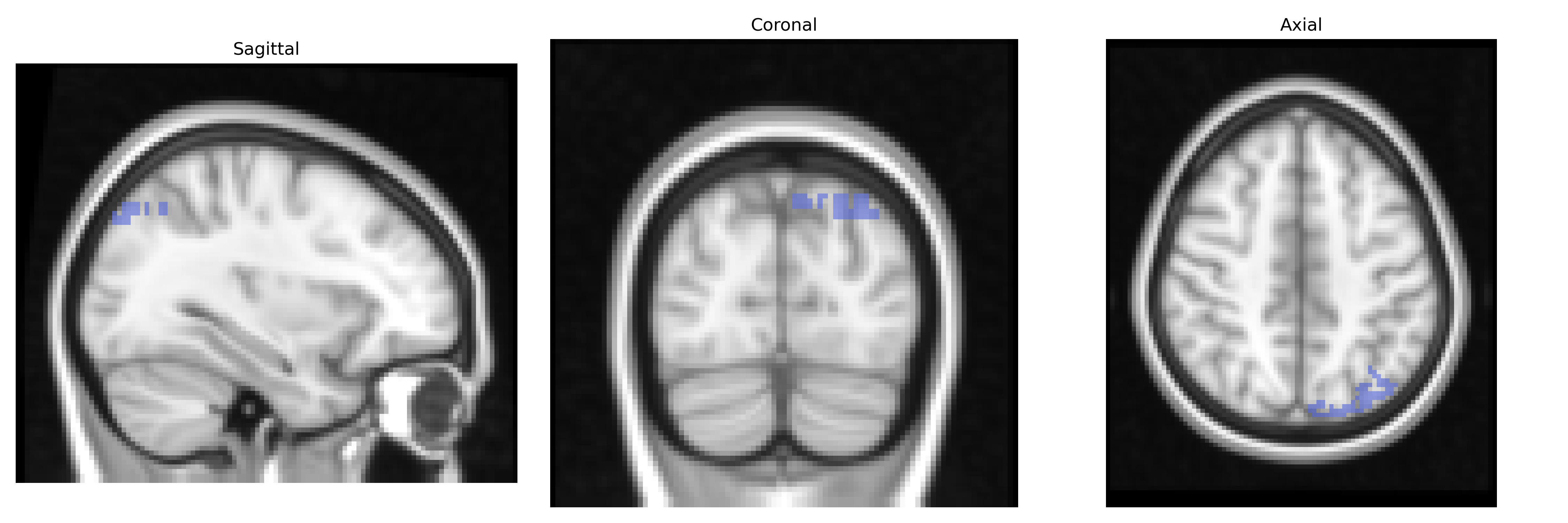
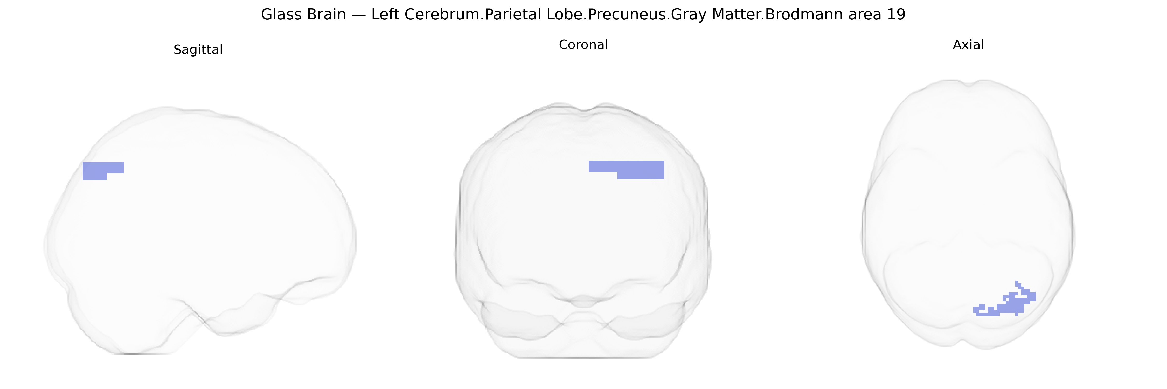
The bilateral Left Cerebrum.Parietal Lobe.Precuneus.Gray Matter.Brodmann area 19 corresponds to a dorsal extension of the associative visual cortex (BA19) within the medial parietal lobe, specifically the precuneus. This region forms part of the higher-order visual processing network, integrating complex visual information with spatial representation, visuospatial imagery, and aspects of episodic memory and self-referential processing. Functionally, it participates in multimodal integration of visual input with somatosensory and proprioceptive signals, contributing to visuomotor coordination and navigation. As BA19, it is linked to processing of motion, form, and visual scenes beyond primary and secondary visual cortices, and, within the precuneus, it is also involved in default mode network activity and internal mentation. There is no direct Wikipedia article for this exact Talairach label; a closely related structure is the Precuneus.
The bilateral precuneus gray matter within Brodmann area 19 (posterior medial parietal/occipital association cortex) has been implicated in several genetic and GWAS-based findings, largely through imaging genetics and neuropsychiatric studies rather than region-specific candidate gene work. Variants in APOE, CLU, and BIN1, among other Alzheimer’s disease–risk genes, have been associated with structural and functional alterations in the precuneus–posterior cingulate network, reflecting its role in early amyloid deposition and default mode network disruption. Large-scale GWAS of cortical thickness and surface area (e.g., ENIGMA and UK Biobank studies) have identified polygenic influences on parietal and occipital association cortices, including loci near genes involved in neurodevelopmental processes (such as MAPT, HMGA2, and WNT pathway components), though these findings are usually reported at the lobe or network level rather than specifically at Talairach BA19. Imaging genetics research links common variants in dopaminergic, glutamatergic, and synaptic plasticity genes (e.g., COMT, BDNF Val66Met) to task-related activation and connectivity in precuneus and surrounding association cortex during episodic memory, visuospatial processing, and self-referential cognition. GWAS and polygenic risk studies for schizophrenia, major depression, autism spectrum disorder, and ADHD repeatedly implicate alterations in the precuneus/default mode network, with risk alleles in synaptic, immune, and calcium-channel genes (such as CACNA1C, GRIN2A, and complement pathway genes) associated with changes in gray matter volume and functional connectivity that encompass BA19. Additionally, neurodevelopmental and cognitive GWAS (for intelligence, educational attainment, and reading/visual processing traits) report associations with genetic variants that modulate occipital-parietal association cortex morphology and activation, indirectly linking polygenic cognitive profiles to the structure and function of the bilateral precuneus in Brodmann area 19.
Overview generated by GPT-4o (2026).
Region ID: 986
Hemisphere: bilateral
Atlas: Talairach labels 2mm
Full Quality Version: Download MP4
Full Quality Version: Download MP4
Wali Sidiqyar*, Gaurav Rudravaram*, Elyssa M. McMaster, Trent M. Schwartz, Adam M. Saunders, Kurt G. Schilling, Bennett A. Landman "Introducing SPINS: A Shared Public Visualization Library of Neuroanatomical Structures." Medical Imaging with Deep Learning- short paper
This resource is licensed under CC0 1.0 Universal (Public Domain).