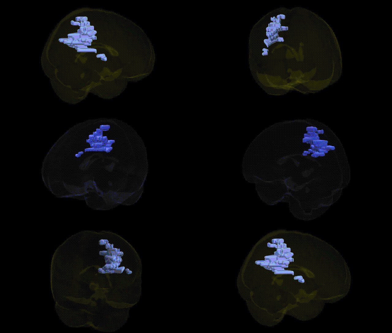
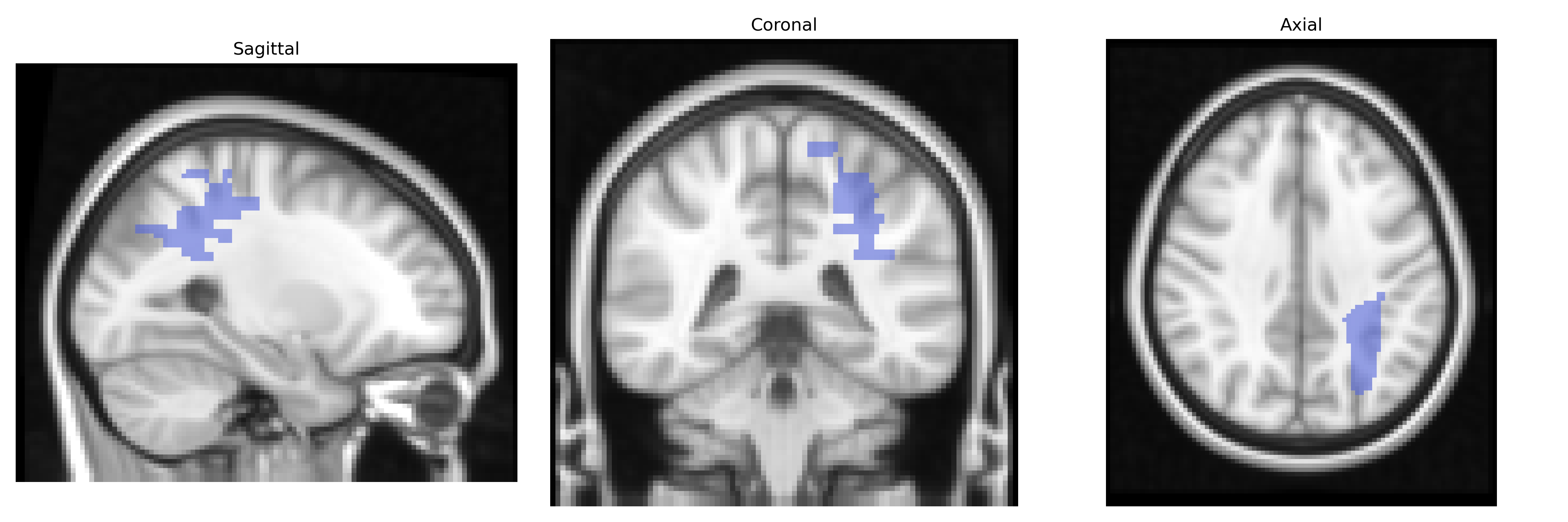
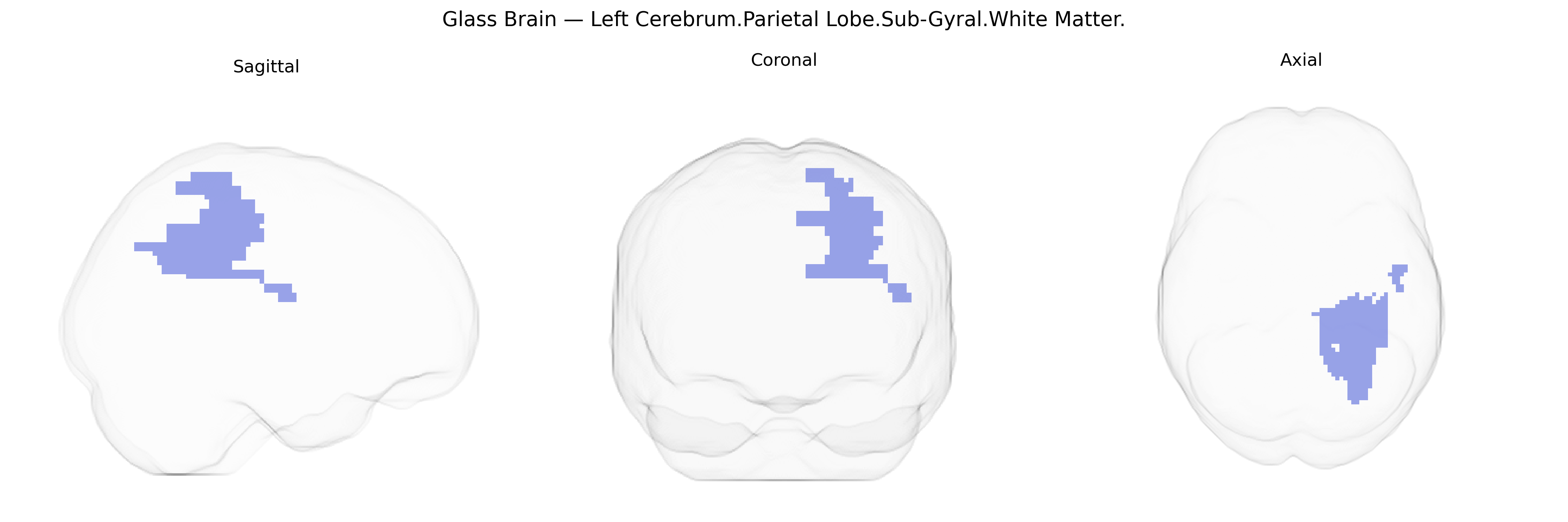
The bilateral Left Cerebrum.Parietal Lobe.Sub-Gyral.White Matter corresponds to deep parietal lobe white matter lying beneath the cortical gyri of the left cerebral hemisphere, as defined in the Talairach 2 mm Atlas. This region contains association and projection fibers that interconnect parietal cortical areas (such as primary somatosensory and posterior parietal regions) with frontal, temporal, and occipital cortices, as well as thalamic nuclei, supporting multimodal sensory integration, spatial processing, attention, and sensorimotor coordination. Its sub-gyral position reflects mainly intralobar and interlobar pathways rather than specific nuclei, and it forms part of larger white matter tracts such as components of the superior longitudinal fasciculus and other parietal association fibers. There is no direct Wikipedia article for this specific atlas label; see instead the related structure Parietal lobe.
Genetic associations specifically targeting the bilateral Left Cerebrum.Parietal Lobe.Sub-Gyral.White Matter (as defined in the Talairach 2 mm atlas) are not commonly reported at that exact anatomical resolution, but GWAS and imaging‑genetics studies implicating parietal lobe white matter more broadly provide relevant context. Variants in genes affecting myelination and axonal integrity (e.g., MAG, MBP, PLP1, and broader oligodendrocyte‑related pathways) have been associated with regional white matter microstructure measures such as fractional anisotropy and mean diffusivity in parietal tracts, and large neuroimaging GWAS (e.g., ENIGMA, UK Biobank) have linked loci near DNM1L, PCDH gene clusters, and other neuronal adhesion and cytoskeletal genes to parietal white matter volume and connectivity. Parietal white matter abnormalities influenced by genetic risk scores have been observed in schizophrenia, bipolar disorder, and major depressive disorder, where polygenic risk for these conditions correlates with reduced integrity in frontoparietal and parietal association fibers. Similarly, ADHD, autism spectrum disorder, and dyslexia show heritable alterations in parietal and temporoparietal white matter pathways, with candidate and GWAS-identified variants in genes such as CNTNAP2, FOXP2, and synaptic scaffolding or neurotransmission genes contributing to disrupted connectivity underlying attention, language, and visuospatial processing. Neurodegenerative and cerebrovascular GWAS (e.g., for Alzheimer’s disease risk loci like APOE, CLU, PICALM, and small vessel disease loci affecting COL4A1/2 and NOTCH3) have been linked to white matter hyperintensities and microstructural changes that prominently involve parietal regions, suggesting a genetically mediated vulnerability of parietal sub-gyral white matter to demyelination, ischemia, and neurodegeneration, even though most findings are reported at lobar or tract-level rather than in the exact Talairach-defined subregion.
Overview generated by GPT-4o (2026).
Region ID: 796
Hemisphere: bilateral
Atlas: Talairach labels 2mm
Full Quality Version: Download MP4
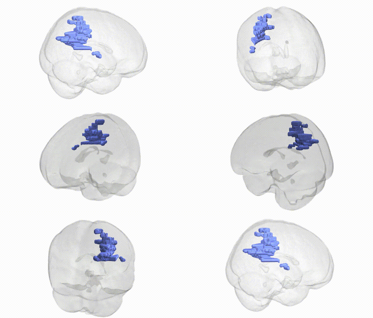
Full Quality Version: Download MP4
Wali Sidiqyar*, Gaurav Rudravaram*, Elyssa M. McMaster, Trent M. Schwartz, Adam M. Saunders, Kurt G. Schilling, Bennett A. Landman "Introducing SPINS: A Shared Public Visualization Library of Neuroanatomical Structures." Medical Imaging with Deep Learning- short paper
This resource is licensed under CC0 1.0 Universal (Public Domain).