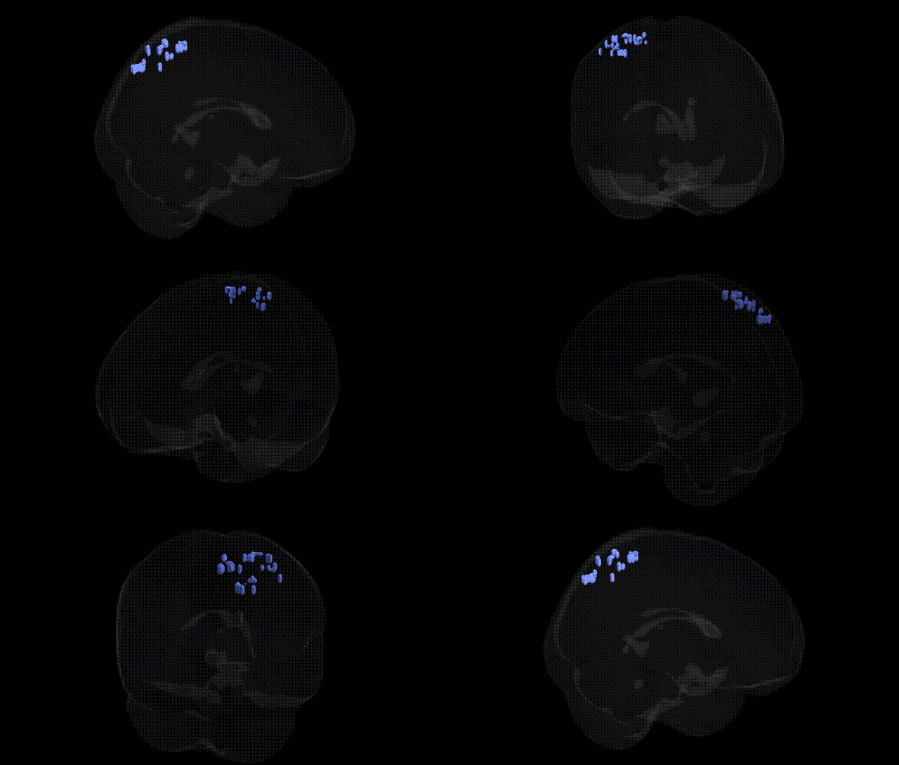
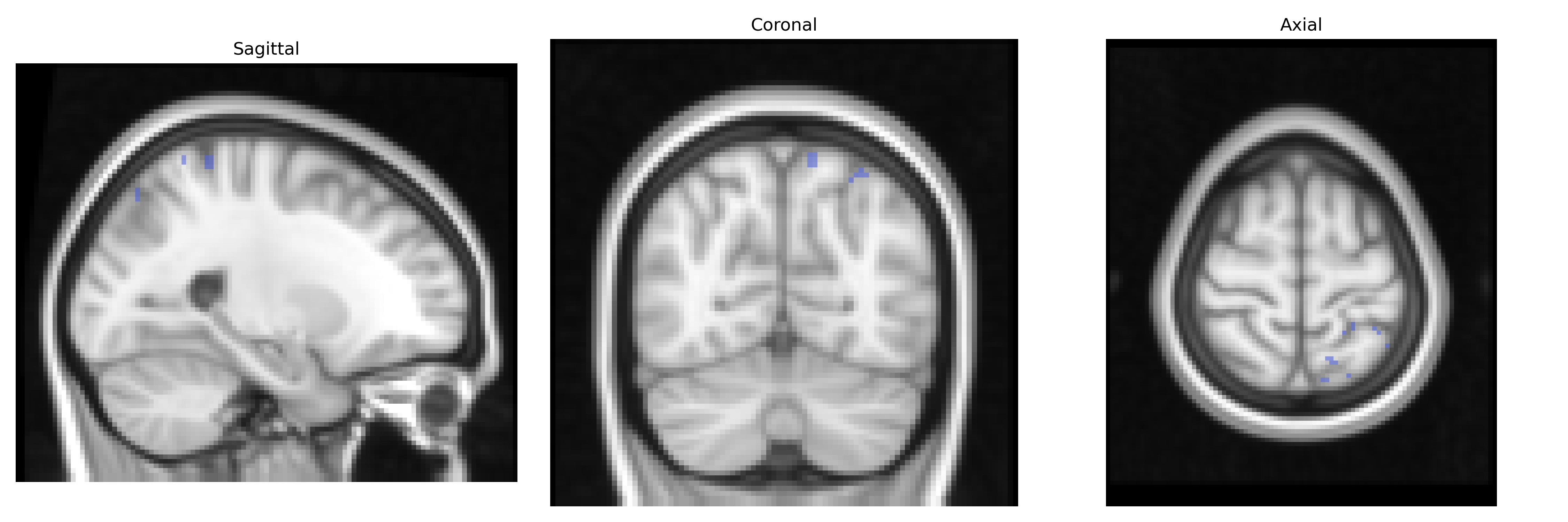
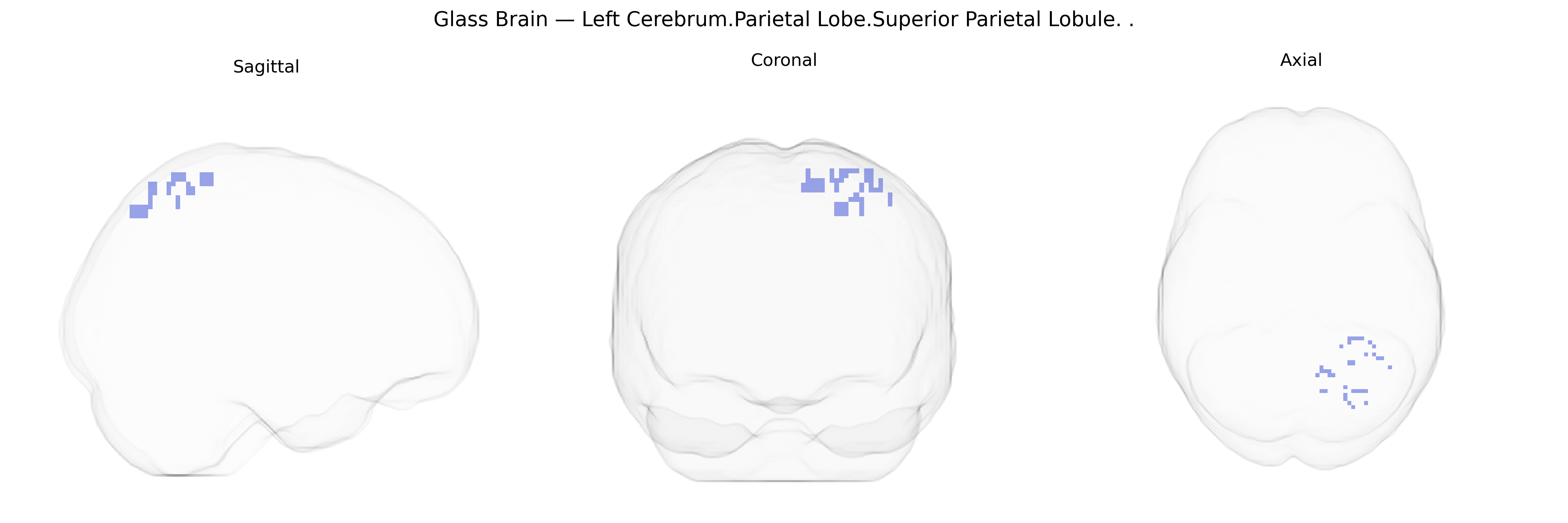
The bilateral Left Cerebrum.Parietal Lobe.Superior Parietal Lobule corresponds to cortical tissue in the dorsal parietal lobe, situated posterior to the primary somatosensory cortex and superior to the intraparietal sulcus. This region is involved in higher-order somatosensory processing, spatial orientation, and sensorimotor integration, contributing to the perception of body position in space, visuospatial attention, and the coordination of visually guided movements. Neuronal populations here integrate tactile, proprioceptive, and visual information, supporting functions such as object manipulation, mental rotation, and aspects of planned motor actions. Lesions in the superior parietal lobule can result in deficits in spatial awareness, impaired tactile discrimination, and disorders of body schema.
Superior parietal lobule
The bilateral superior parietal lobule (SPL) of the left cerebrum has been implicated in a range of genetic associations primarily through GWAS of cognitive, neuropsychiatric, and neurodevelopmental traits that show structural or functional variation in this region. Polygenic influences on general cognitive ability and intelligence, including loci near genes such as KIBRA (WWC1), BDNF, and genes involved in synaptic plasticity and neuronal development, have been associated with individual differences in SPL cortical thickness, surface area, or activation during attention and working memory tasks. Large imaging-genetics consortia (e.g., ENIGMA) have identified heritable variation in parietal lobe morphology linked to common variants in pathways related to axon guidance, calcium signaling, and neurogenesis. In neuropsychiatric GWAS, risk variants for schizophrenia (e.g., near CACNA1C and other calcium channel genes), ADHD (dopaminergic and synaptic genes), and autism spectrum disorder (synaptic and chromatin remodeling genes) have been connected to altered SPL volume or connectivity, consistent with this region’s role in visuospatial integration, attention, and social cognition. GWAS of Alzheimer’s disease, frontotemporal dementia, and other neurodegenerative disorders (including APOE and multiple immune-related loci) have also shown downstream effects on parietal atrophy patterns that prominently involve the superior parietal lobule. Additionally, genetic associations with traits such as mathematical ability, spatial navigation, and motor coordination often map onto networks that include the SPL, highlighting that many polygenic influences on complex cognition and behavior manifest partly through structural and functional variation in this parietal subregion.
Overview generated by GPT-4o (2026).
Region ID: 1037
Hemisphere: bilateral
Atlas: Talairach labels 2mm
Full Quality Version: Download MP4
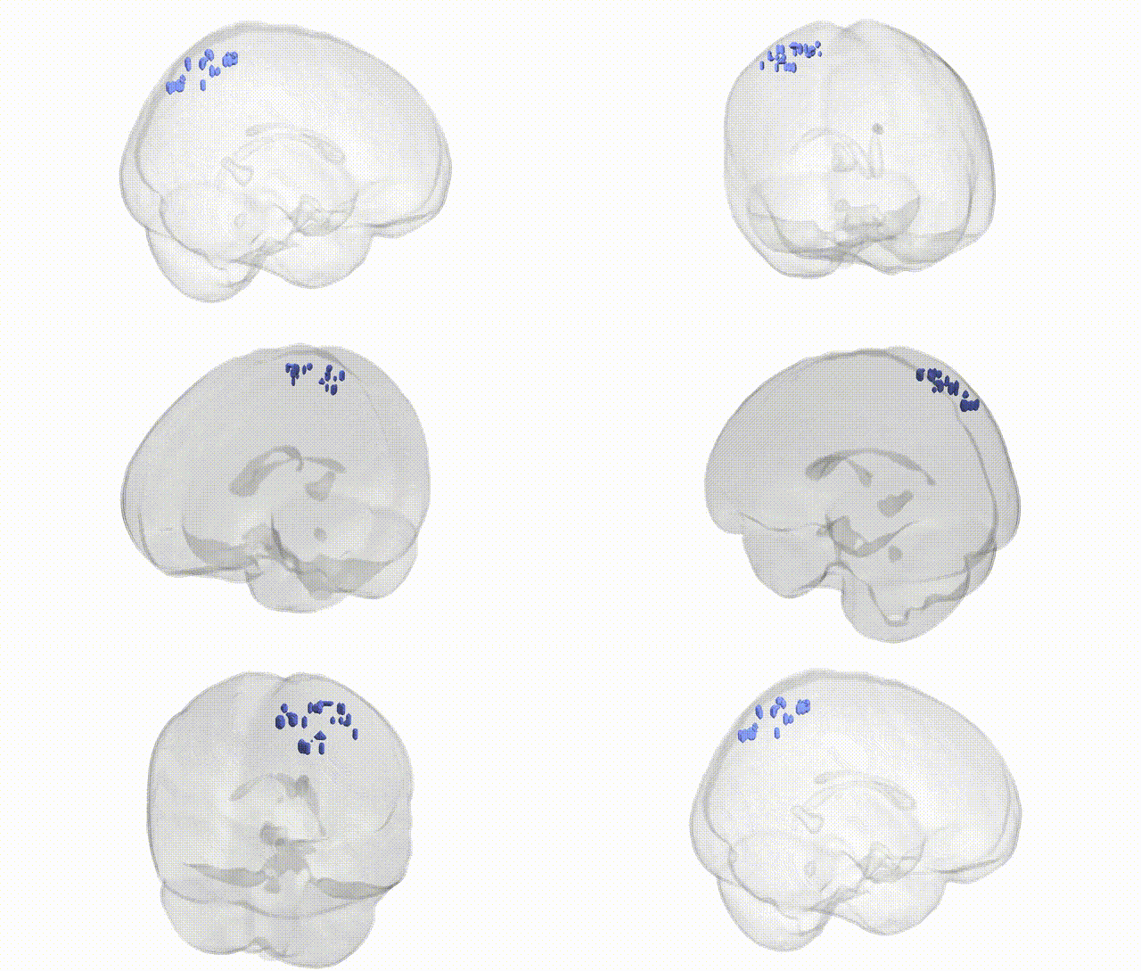
Full Quality Version: Download MP4
Wali Sidiqyar*, Gaurav Rudravaram*, Elyssa M. McMaster, Trent M. Schwartz, Adam M. Saunders, Kurt G. Schilling, Bennett A. Landman "Introducing SPINS: A Shared Public Visualization Library of Neuroanatomical Structures." Medical Imaging with Deep Learning- short paper
This resource is licensed under CC0 1.0 Universal (Public Domain).