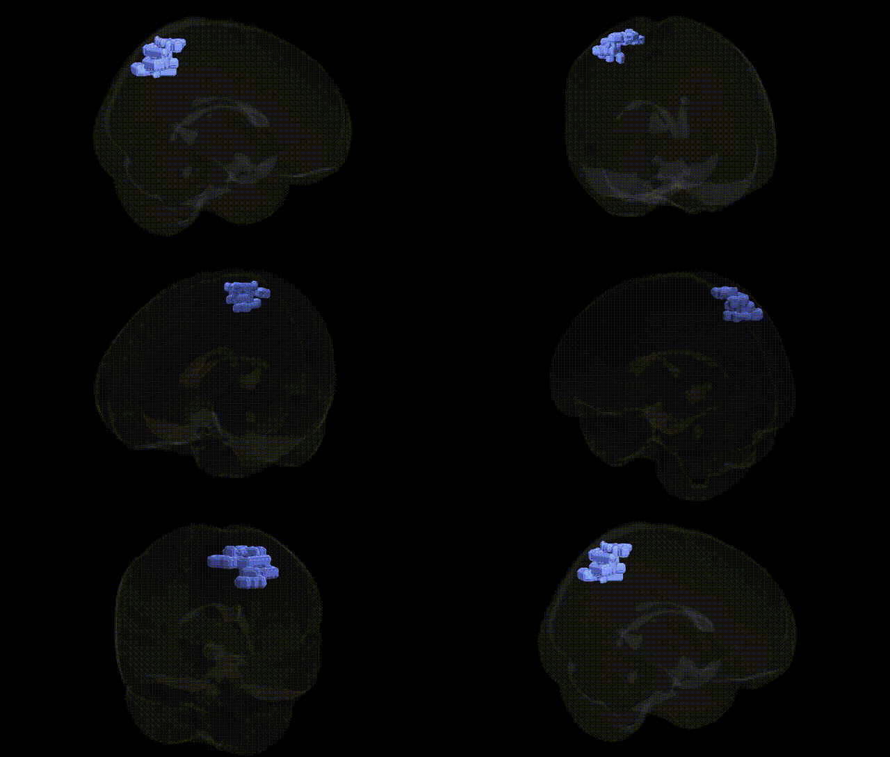
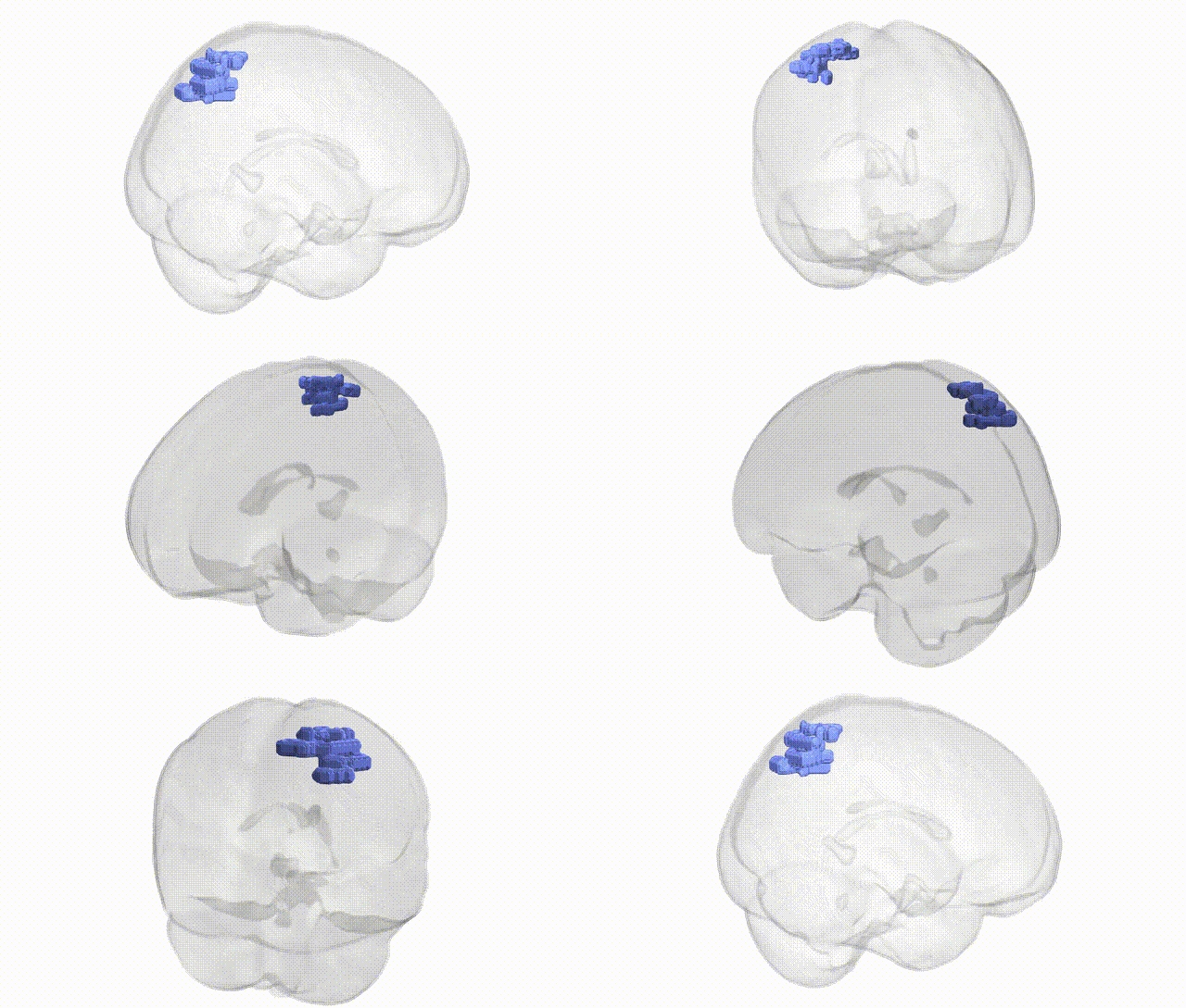
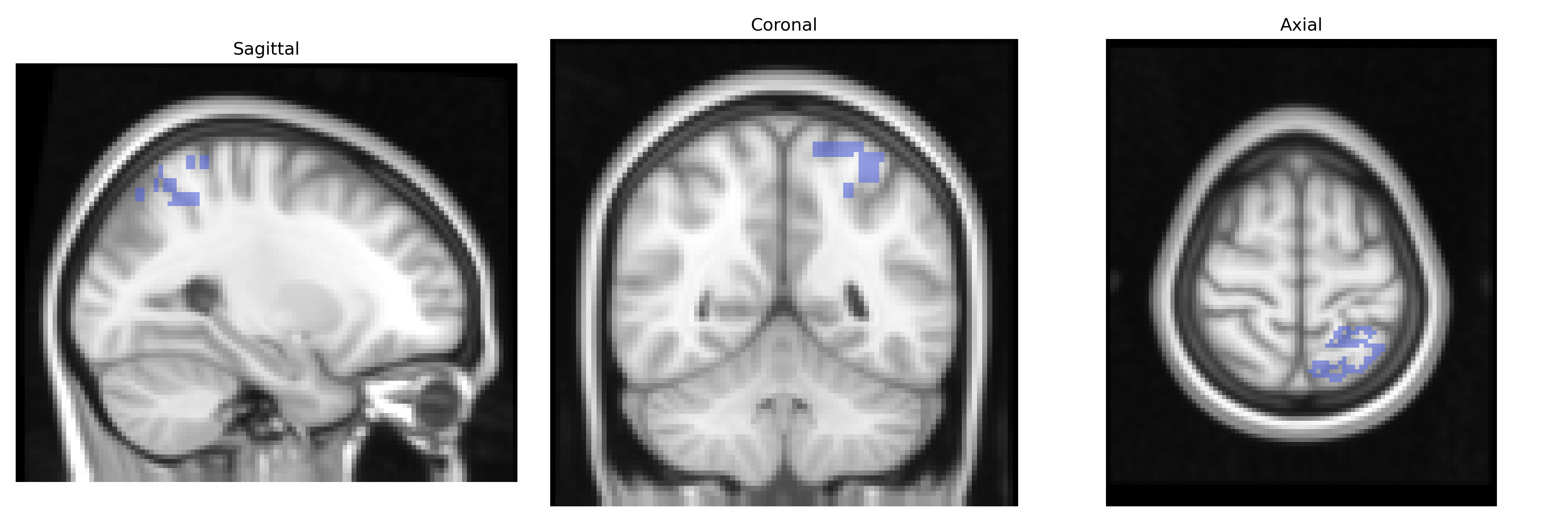
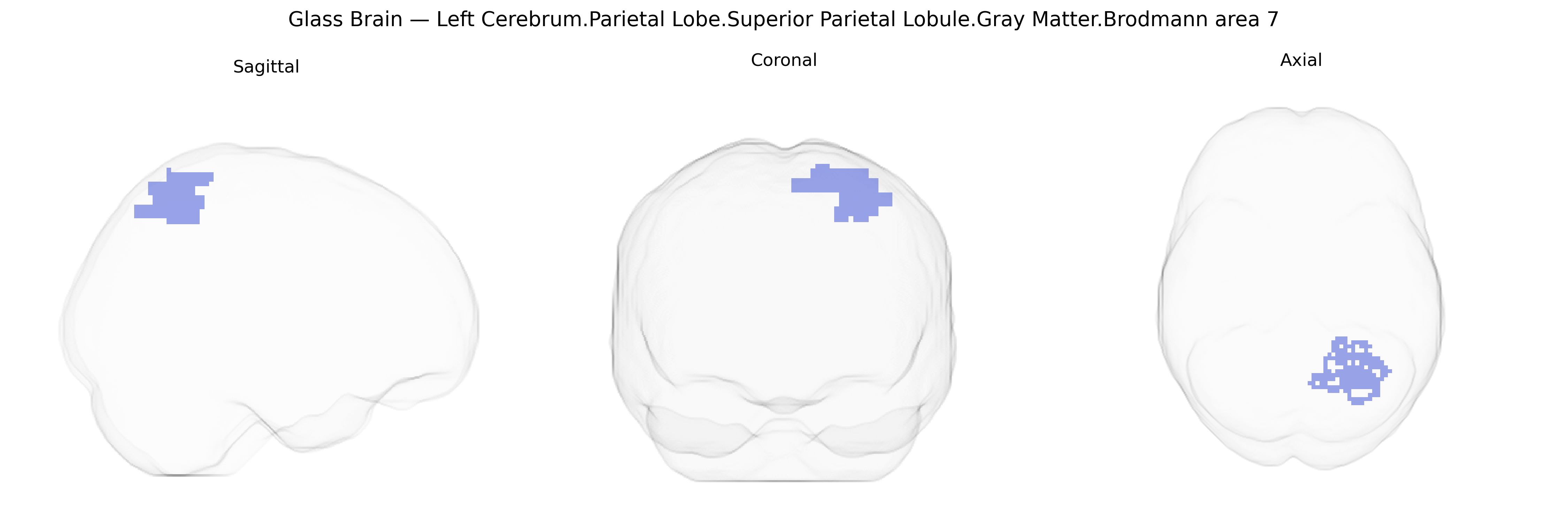
The bilateral Left Cerebrum.Parietal Lobe.Superior Parietal Lobule.Gray Matter.Brodmann area 7 corresponds to association cortex located in the dorsal parietal lobe, bordering the precuneus medially and extending laterally within the superior parietal lobule. This agranular to dysgranular heteromodal region integrates visual, somatosensory, and proprioceptive inputs to support spatial orientation, visuomotor coordination, and higher-order aspects of attention and body schema. Neuronal populations in Brodmann area 7 contribute to transforming sensory information into action-related representations, including reaching and grasping in peripersonal space, and participate in the neural substrates of visuospatial working memory and navigation. Functionally, this area forms part of the dorsal visual stream and is heavily interconnected with premotor, frontal eye field, and occipital visual areas, enabling coordinated sensorimotor behavior. Brodmann area 7
The bilateral left superior parietal lobule (Brodmann area 7) has been implicated in multiple genetic and GWAS-based associations through its roles in visuospatial processing, attention, sensorimotor integration, and higher-order cognition. Imaging genetics studies repeatedly link variation in genes affecting synaptic plasticity and neurodevelopment—such as COMT, BDNF, DISC1, and NRG1—to structural and functional differences in parietal cortex, including area 7, often in the context of working memory, attentional control, and cognitive flexibility. Large neuroimaging GWAS (e.g., ENIGMA and UK Biobank) have identified numerous common variants associated with cortical thickness and surface area in parietal regions, many mapping to genes involved in neuronal migration, axon guidance, and calcium signaling, with polygenic influences shared with general cognitive ability, educational attainment, and intelligence. Parietal lobe morphology, including superior parietal regions, shows genetic correlations with attention-deficit/hyperactivity disorder, schizophrenia, and major depression, reflecting overlapping polygenic architectures for brain structure and psychiatric risk. GWAS of neurodevelopmental and motor coordination traits (e.g., developmental coordination disorder and fine motor skills) have highlighted variants in genes governing cerebellar–parietal networks that include area 7. In addition, risk alleles for Alzheimer’s disease and other dementias (such as APOE ε4 and loci near CLU, PICALM, and BIN1) are associated with accelerated atrophy and hypometabolism in parietal association cortex, including superior parietal regions, consistent with their role in episodic memory networks and visuospatial dysfunction. Overall, the genetic architecture of this Brodmann area 7 region is highly polygenic, with shared variants contributing to individual differences in cognition, susceptibility to psychiatric and neurodegenerative disorders, and variation in gray matter volume and connectivity.
Overview generated by GPT-4o (2026).
Region ID: 1039
Hemisphere: bilateral
Atlas: Talairach labels 2mm
Full Quality Version: Download MP4
Full Quality Version: Download MP4
Wali Sidiqyar*, Gaurav Rudravaram*, Elyssa M. McMaster, Trent M. Schwartz, Adam M. Saunders, Kurt G. Schilling, Bennett A. Landman "Introducing SPINS: A Shared Public Visualization Library of Neuroanatomical Structures." Medical Imaging with Deep Learning- short paper
This resource is licensed under CC0 1.0 Universal (Public Domain).