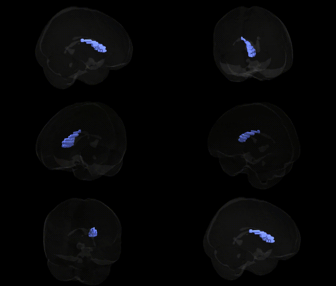
The bilateral Left Cerebrum.Sub-lobar.Caudate.Gray Matter.Caudate Body corresponds to the main portion of the caudate nucleus, a C-shaped gray matter structure within the dorsal striatum of the basal ganglia, situated sub-lobar to the cerebral cortex. The caudate body receives extensive glutamatergic input from frontal and association cortices and dopaminergic projections from the midbrain, integrating motor, cognitive, and limbic information. Through its GABAergic outputs to the internal and external segments of the globus pallidus and substantia nigra, it contributes critically to action selection, procedural and habit learning, executive functions, and aspects of reward-based behavior. Functional and structural abnormalities in the caudate body are implicated in disorders such as Parkinson’s disease, Huntington’s disease, obsessive-compulsive disorder, and attention-deficit/hyperactivity disorder. Caudate nucleus
The bilateral Left Cerebrum Sub-lobar Caudate Gray Matter (Caudate Body) region has been repeatedly implicated in genetic studies of brain structure and neuropsychiatric traits, primarily through GWAS of subcortical volumes and imaging genetics. Large-scale consortia such as ENIGMA and UK Biobank have identified multiple loci associated with caudate volume, including variants near genes involved in neurodevelopment, synaptic function, and cell adhesion (e.g., CRTC1, FAT3, SLC39A8, and several intergenic regions), with polygenic influences shared across other subcortical structures. The caudate is a key node in frontostriatal and cortico–basal ganglia loops, and genetic liability to disorders such as schizophrenia, bipolar disorder, major depression, ADHD, and obsessive–compulsive disorder has been linked to altered caudate morphology and function, often via polygenic risk scores that correlate with caudate volume or activation patterns. Huntington’s disease, caused by CAG-repeat expansion in HTT, produces prominent neurodegeneration in the caudate, and rare variant studies in movement disorders and dystonia also highlight genes expressed in striatal medium spiny neurons. In addition, GWAS of cognitive performance, educational attainment, and general intelligence show overlap between genetic variants influencing caudate volume and those associated with cognitive and executive functions, suggesting that heritable differences in this caudate subregion contribute to both neuropsychiatric risk and normal variation in cognition and personality.
Overview generated by GPT-4o (2026).
Region ID: 692
Hemisphere: bilateral
Atlas: Talairach labels 2mm
Full Quality Version: Download MP4
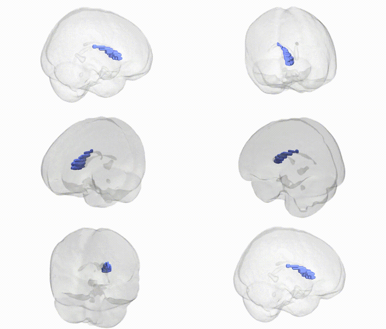
Full Quality Version: Download MP4
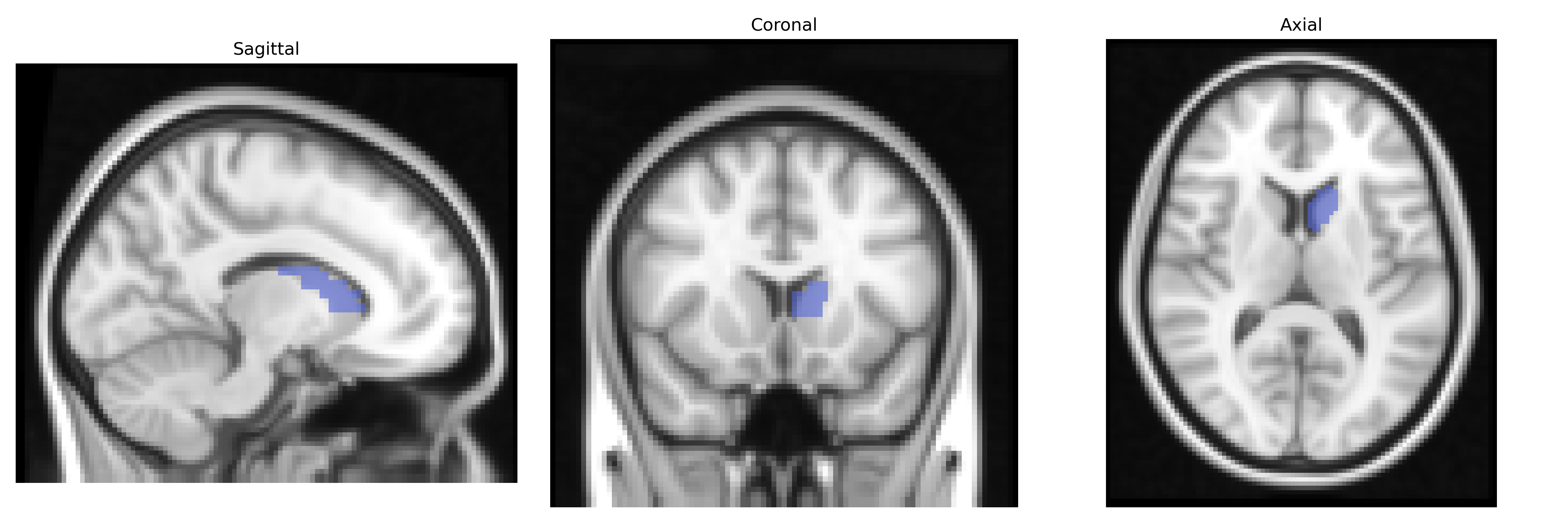
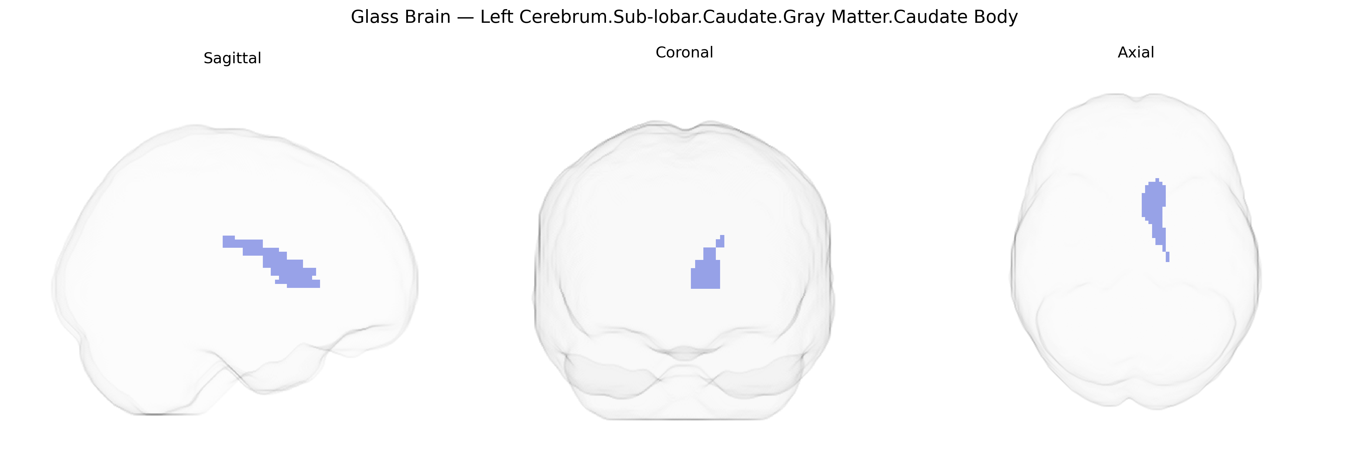
Wali Sidiqyar*, Gaurav Rudravaram*, Elyssa M. McMaster, Trent M. Schwartz, Adam M. Saunders, Kurt G. Schilling, Bennett A. Landman "Introducing SPINS: A Shared Public Visualization Library of Neuroanatomical Structures." Medical Imaging with Deep Learning- short paper
This resource is licensed under CC0 1.0 Universal (Public Domain).