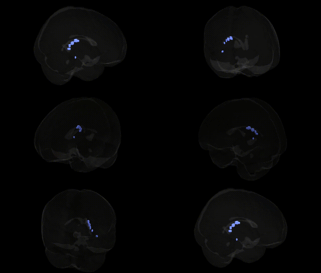
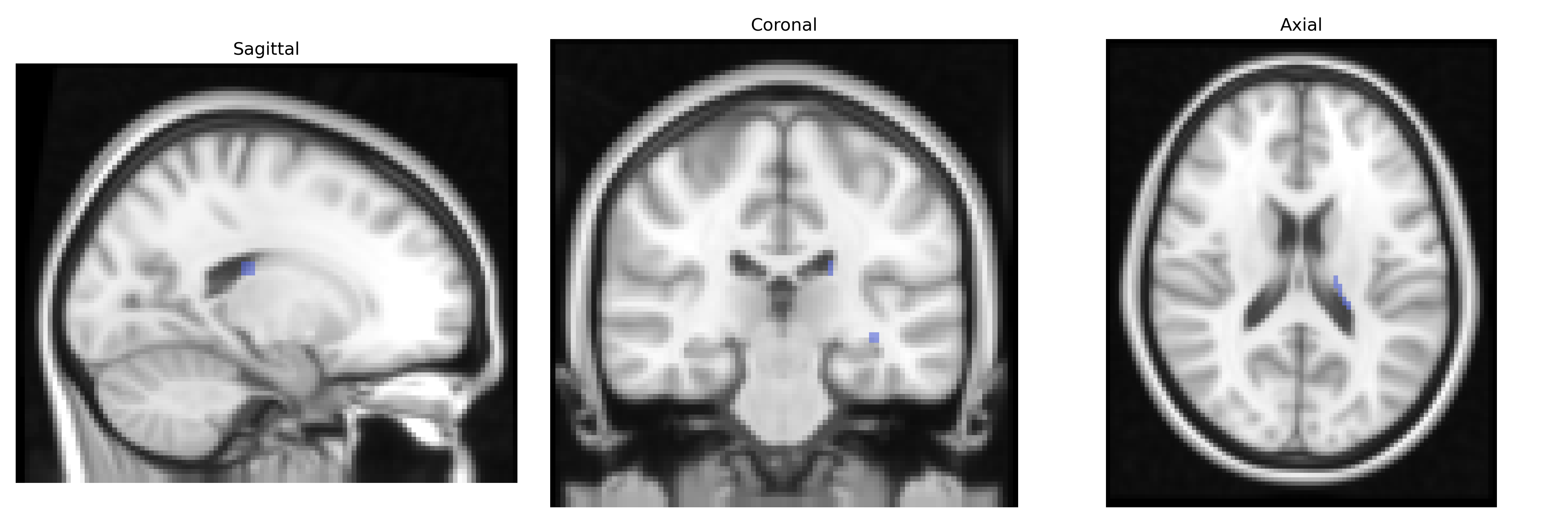
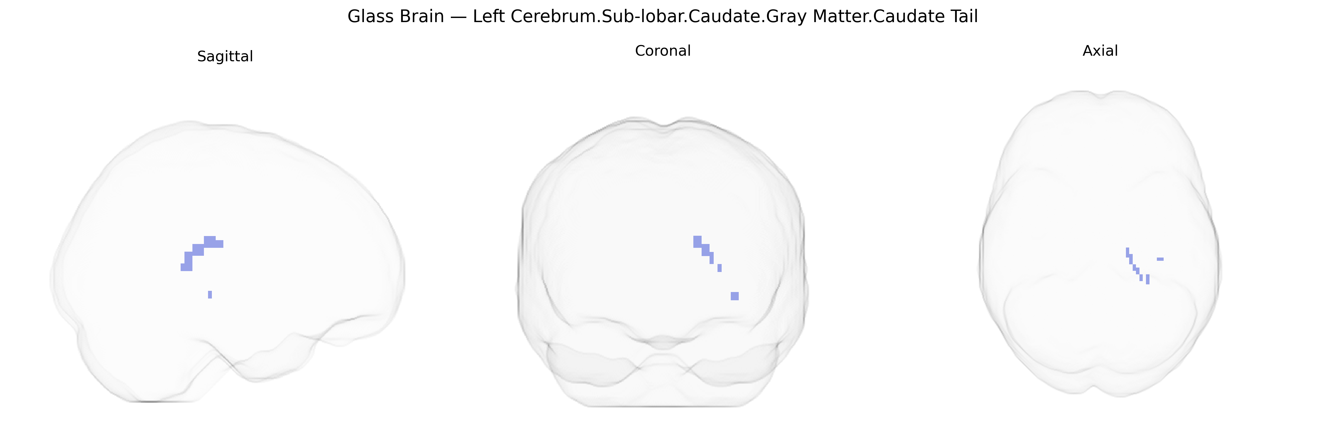
The bilateral Left Cerebrum.Sub-lobar.Caudate.Gray Matter.Caudate Tail corresponds to the posterior portion of the caudate nucleus, a gray matter structure within the sub-lobar (deep) region of the cerebral hemispheres and a major component of the dorsal striatum in the basal ganglia. The caudate tail curves posteriorly and ventrally from the body of the caudate, following the lateral ventricle into temporal regions, and is involved in integrating cortical information for motor control, habit formation, and certain aspects of associative and limbic processing, including learning, reward-related behaviors, and some dimensions of cognition and emotion. As part of the basal ganglia circuitry, it receives extensive glutamatergic input from cortex and dopaminergic input from the midbrain, and sends GABAergic projections to globus pallidus and substantia nigra, contributing to action selection and the regulation of movement and behavioral routines. There is no direct Wikipedia article for the caudate tail; see instead the related structure Caudate nucleus.
The caudate tail, a posterior extension of the caudate nucleus within the sub-lobar gray matter of the left cerebrum, participates in cortico-striatal circuits implicated in motor control, habit learning, reward processing, and aspects of cognition, and genetic influences on its structure and function have been partially characterized in large-scale imaging genetics and GWAS studies. Common variants in several genes involved in dopaminergic signaling and neurodevelopment—such as DRD2, DAT1 (SLC6A3), and COMT—have been repeatedly associated with caudate volume or activity, and polygenic scores for traits like schizophrenia, attention-deficit/hyperactivity disorder (ADHD), and bipolar disorder show correlations with caudate morphology in UK Biobank and ENIGMA consortium analyses. GWAS of subcortical brain volumes have identified loci near genes such as DCC, TOX3, and KTN1 as associated with caudate size, suggesting roles for axon guidance and cytoskeletal dynamics in caudate development. In psychiatric genetics, risk variants for schizophrenia (e.g., in CACNA1C, ZNF804A, and other synaptic genes) and obsessive-compulsive disorder have been linked to altered caudate activation and connectivity, particularly in fronto-striatal loops, while ADHD-related alleles show associations with reduced caudate volume and altered reward-related activation. Neurodegenerative disorders also implicate this region: Huntington’s disease, caused by CAG expansion in HTT, features early atrophy of caudate structures including the tail, and Parkinson’s disease risk loci affecting dopaminergic pathways are associated with functional changes in caudate-based motor and cognitive circuits. Beyond clinical diagnoses, GWAS of cognitive performance, educational attainment, and personality traits (e.g., neuroticism) demonstrate polygenic associations with caudate volume and connectivity, indicating that many small-effect variants across neurodevelopmental, synaptic, and neurotransmitter-related genes collectively shape inter-individual differences in the structure and function of the caudate tail region.
Overview generated by GPT-4o (2026).
Region ID: 435
Hemisphere: bilateral
Atlas: Talairach labels 2mm
Full Quality Version: Download MP4
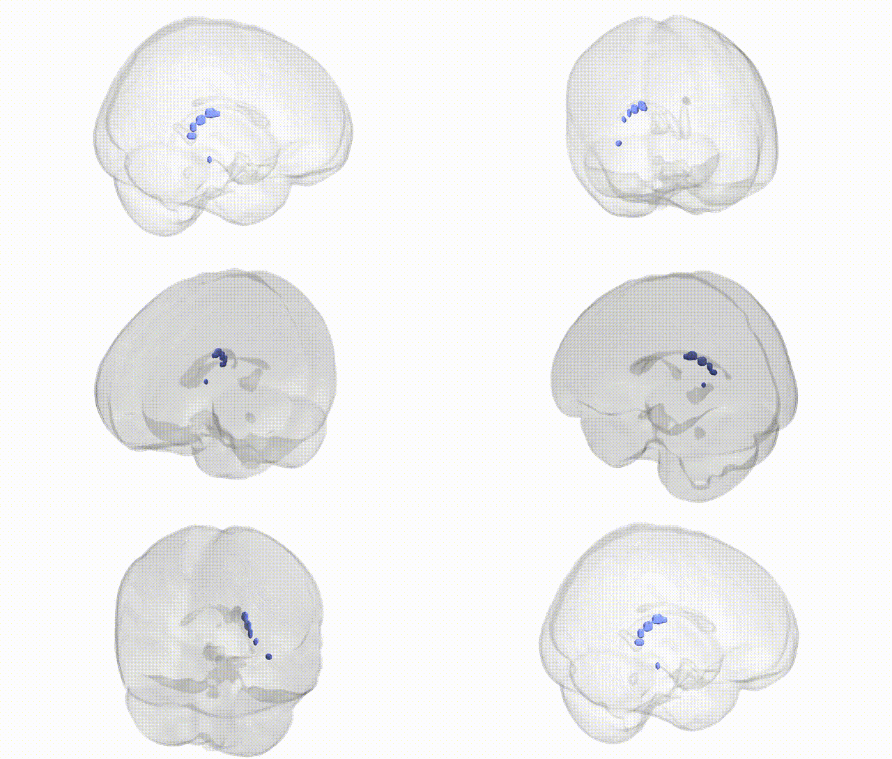
Full Quality Version: Download MP4
Wali Sidiqyar*, Gaurav Rudravaram*, Elyssa M. McMaster, Trent M. Schwartz, Adam M. Saunders, Kurt G. Schilling, Bennett A. Landman "Introducing SPINS: A Shared Public Visualization Library of Neuroanatomical Structures." Medical Imaging with Deep Learning- short paper
This resource is licensed under CC0 1.0 Universal (Public Domain).