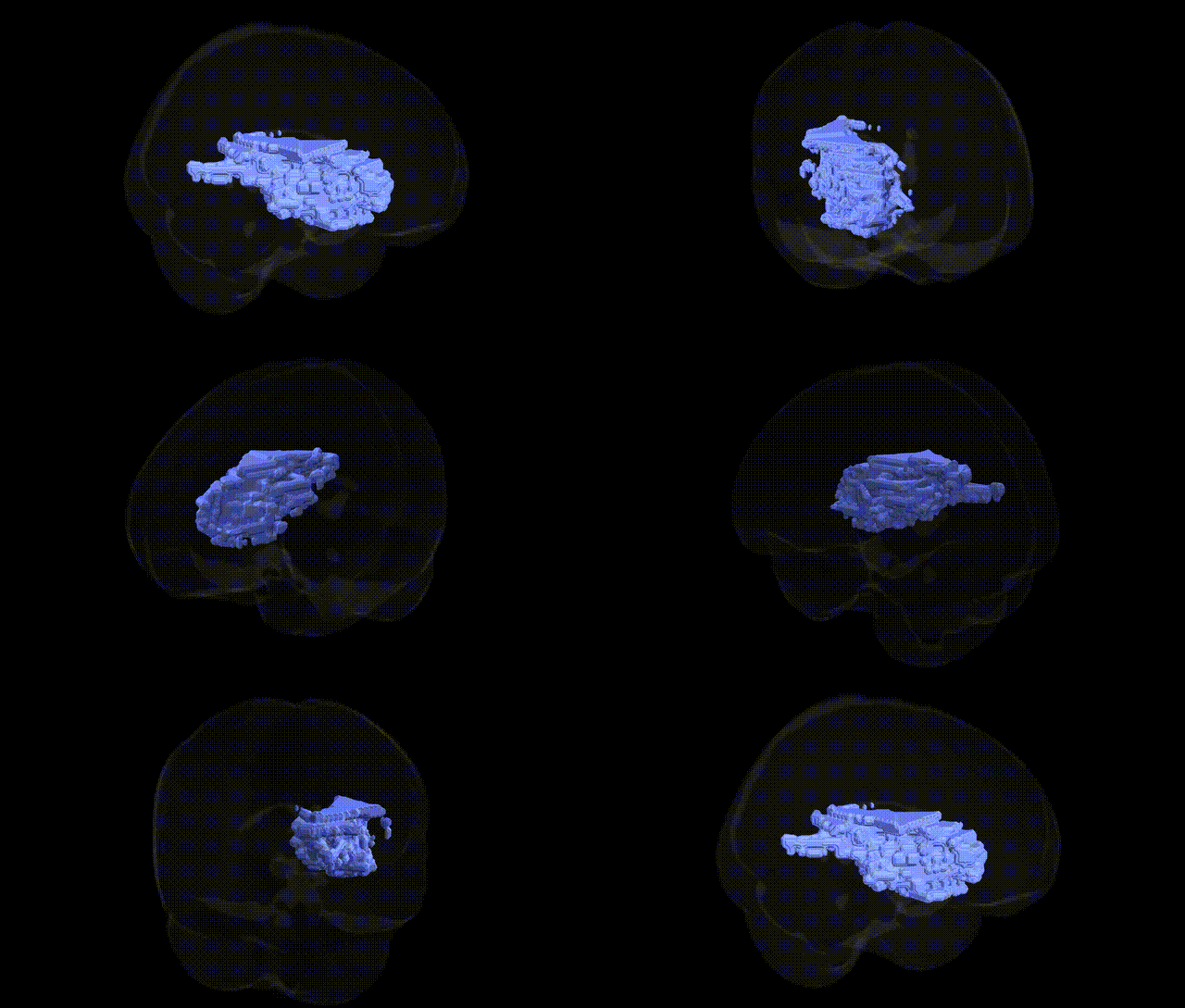
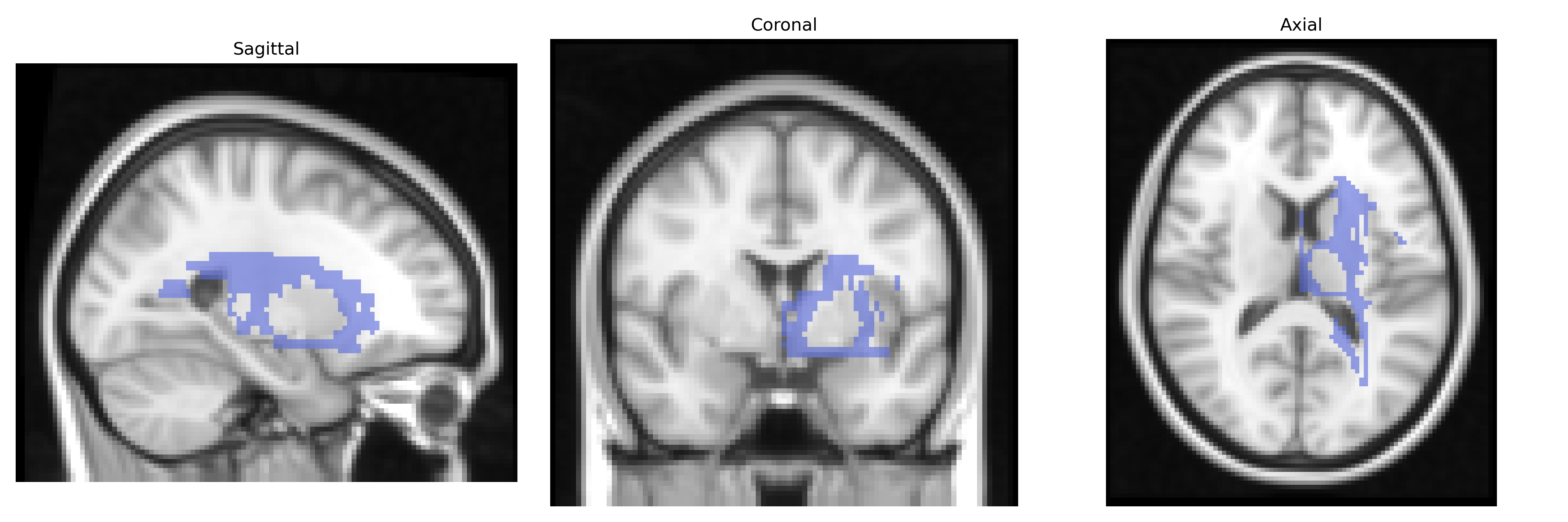
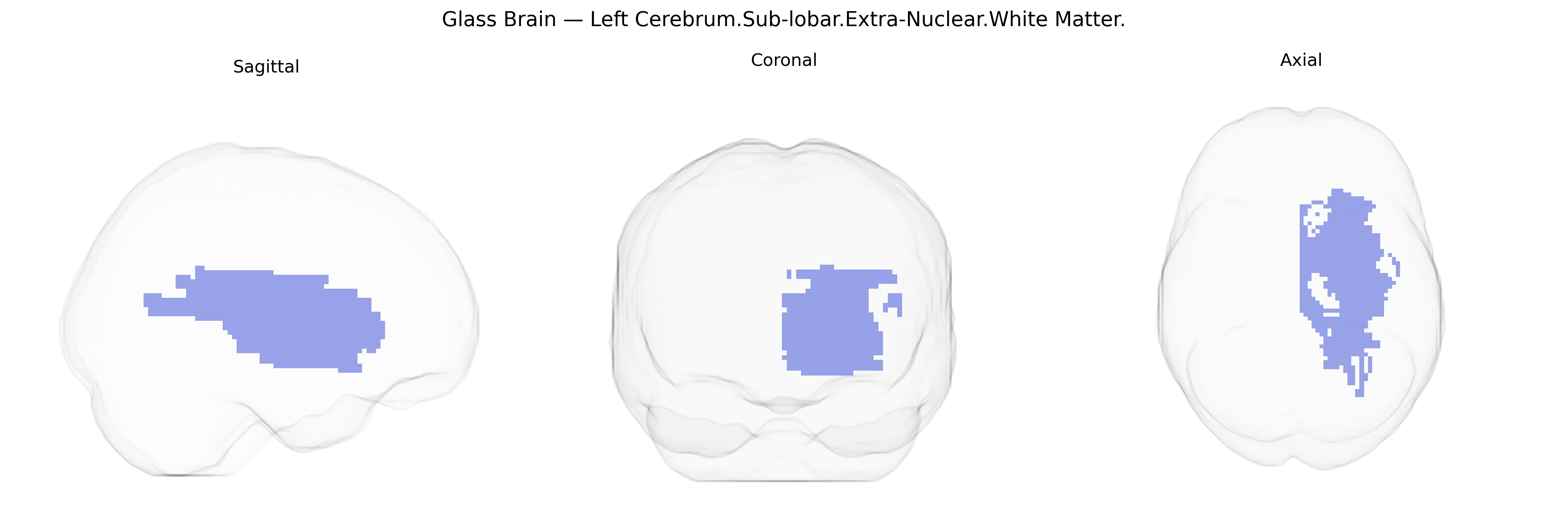
The bilateral Left Cerebrum.Sub-lobar.Extra-Nuclear.White Matter refers to deep, subcortical white matter tracts located within the left cerebral hemisphere but outside the major nuclear gray-matter structures such as the basal ganglia and thalamus. These regions consist primarily of myelinated axons that form association, commissural, and projection fibers, supporting the integration and transmission of information between cortical areas and between the cortex and subcortical structures. Functionally, this extra-nuclear white matter contributes to large-scale brain networks subserving cognition, sensorimotor processing, and language, and is particularly relevant to the integrity of connectivity assessed by diffusion MRI. Focal or diffuse pathology in this region, such as demyelination, ischemia, or traumatic injury, can disrupt network communication and lead to a broad spectrum of neurological and neuropsychological deficits. There is no direct link for “Extra-Nuclear White Matter”; a closely related structure is the Cerebral white matter.
The bilateral left cerebrum sub-lobar extra-nuclear white matter, as defined in the Talairach 2 mm atlas, corresponds largely to deep frontal and subcortical white matter tracts whose genetic architecture has been examined in large-scale neuroimaging GWAS using diffusion MRI and structural connectivity measures rather than this specific atlas label. Variants in genes involved in myelination, axonal guidance, and oligodendrocyte function—such as MAG, MBP, CNTN4, DNM1L, and loci near NRXN1 and NTRK3—have been associated with white matter microstructure (e.g., fractional anisotropy, mean diffusivity) in fronto-subcortical pathways that traverse extra-nuclear territories. Polygenic influences on white matter in this region overlap strongly with risk alleles for schizophrenia, bipolar disorder, major depressive disorder, ADHD, and autism spectrum disorder, consistent with repeatedly observed disorder-related alterations in deep frontal and periventricular white matter. GWAS of general cognitive ability, processing speed, educational attainment, and brain age acceleration have also implicated common variants that modulate broad white matter integrity, including tracts in the left frontal and sub-lobar zones, suggesting that genetic determinants of neurodevelopment and neuroinflammation contribute to both clinical and normative variability in these pathways. While no GWAS targets the Talairach “extra-nuclear white matter” label specifically, convergent evidence from tract-based and voxelwise imaging-genetics studies supports a shared polygenic basis for white matter characteristics in this region, involving overlapping risk architecture for psychosis, mood and neurodevelopmental disorders, and cognitive traits.
Overview generated by GPT-4o (2026).
Region ID: 347
Hemisphere: bilateral
Atlas: Talairach labels 2mm
Full Quality Version: Download MP4
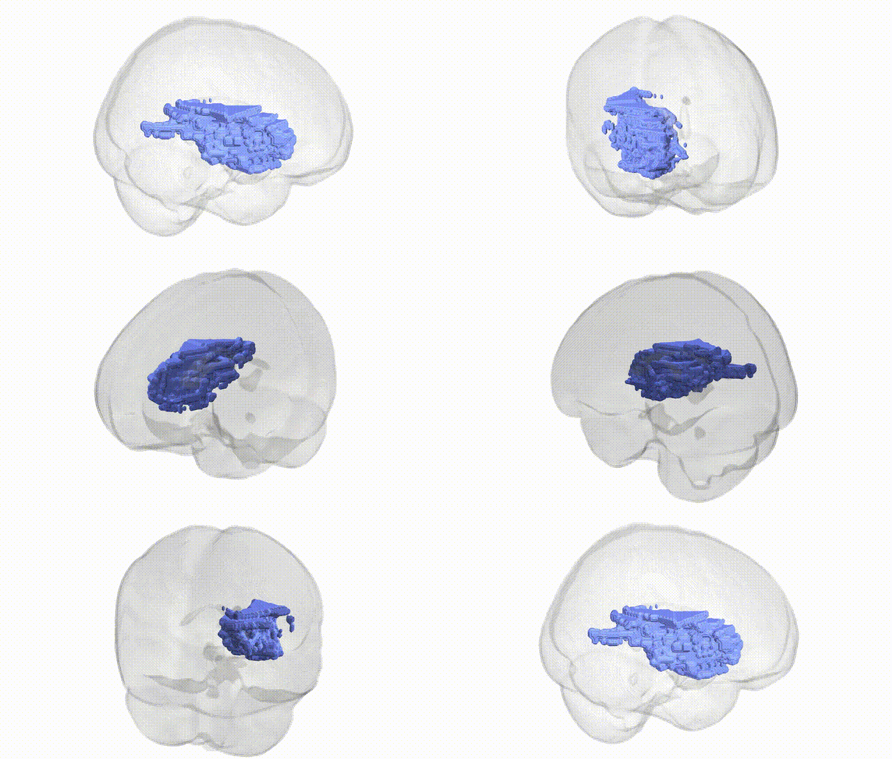
Full Quality Version: Download MP4
Wali Sidiqyar*, Gaurav Rudravaram*, Elyssa M. McMaster, Trent M. Schwartz, Adam M. Saunders, Kurt G. Schilling, Bennett A. Landman "Introducing SPINS: A Shared Public Visualization Library of Neuroanatomical Structures." Medical Imaging with Deep Learning- short paper
This resource is licensed under CC0 1.0 Universal (Public Domain).