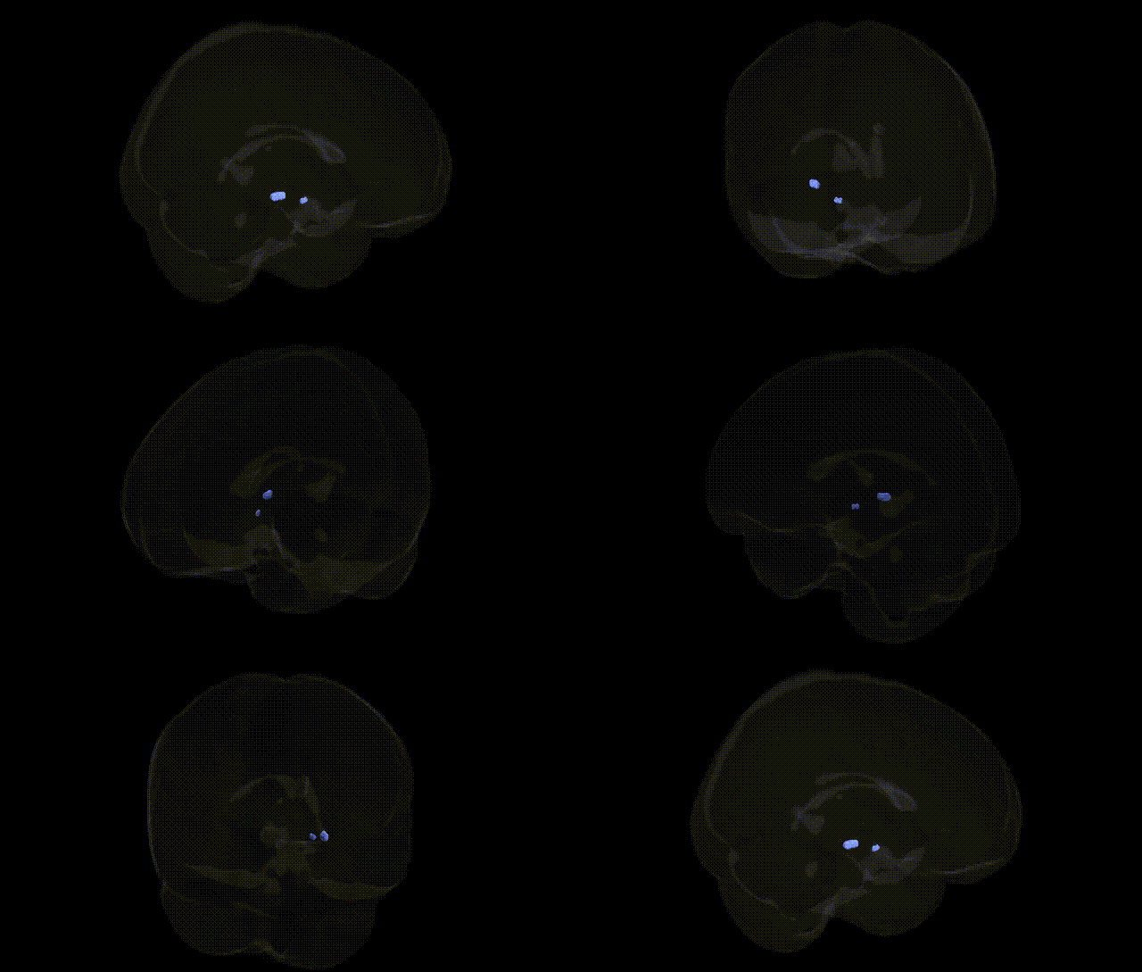
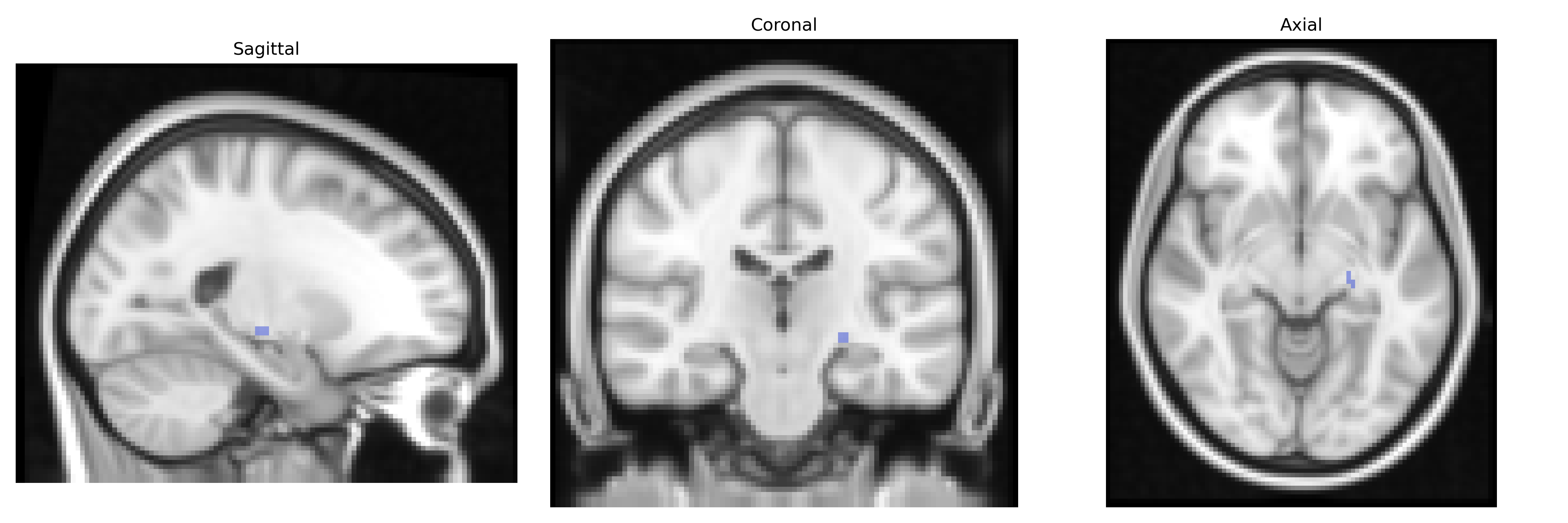
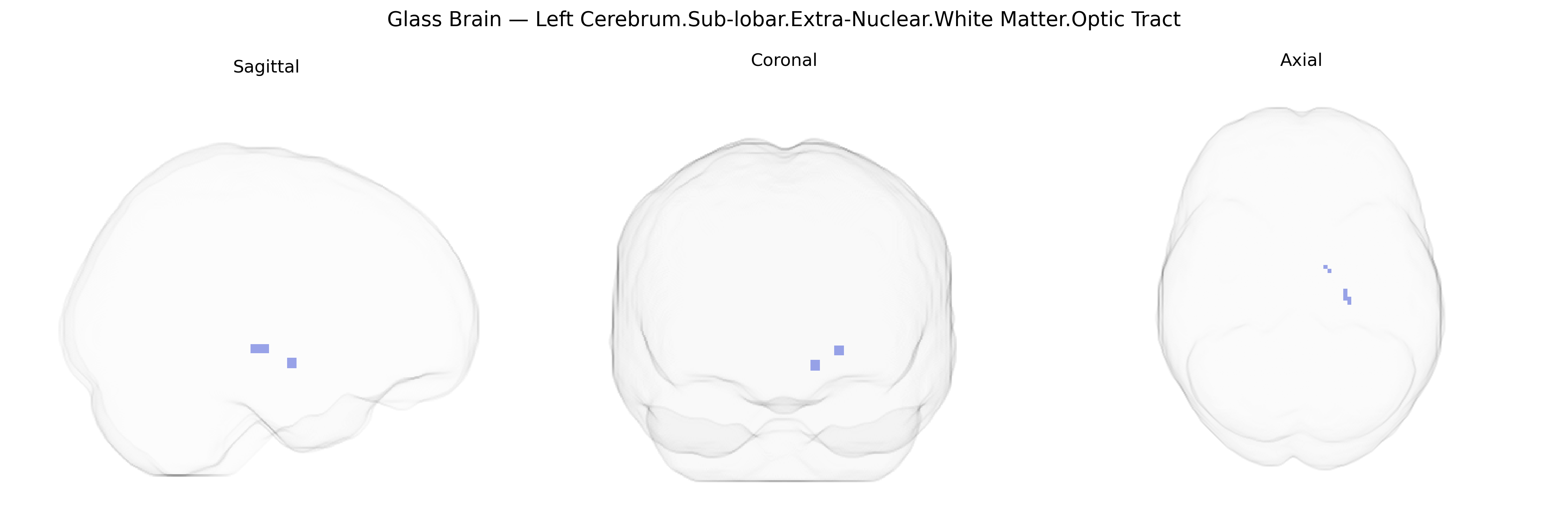
The bilateral Left Cerebrum.Sub-lobar.Extra-Nuclear.White Matter.Optic Tract region in the Talairach 2 mm atlas corresponds to the left optic tract, a white matter pathway carrying visual information from the optic chiasm to the lateral geniculate nucleus, and further contributing to projections toward visual cortical areas. Composed of heavily myelinated axons originating mainly from retinal ganglion cells, the optic tract is essential for relaying signals underlying conscious vision, reflexive eye movements, and pupillary light responses. Although categorized as “sub-lobar extra-nuclear white matter” in the atlas, functionally it is a key component of the central visual pathway, integrating binocular input and preserving retinotopic organization as it courses around the cerebral peduncle toward the thalamus. There is no direct Wikipedia article for this specific Talairach composite label; a closely related structure is the Optic tract.
The optic tract, located in the sub-lobar extra-nuclear white matter of the left cerebrum as defined in the Talairach 2mm atlas, has been primarily implicated in genetic studies through its role in visual pathway integrity rather than region-specific GWAS hits, with most findings emerging from broader neuroimaging-genetics work on visual system structures and white matter microstructure. Variants in genes associated with myelination and axonal guidance—such as MYRF, NRG1, CNTN4, and EPHA family members—have been linked to global and tract-specific white matter properties (e.g., fractional anisotropy) in diffusion MRI studies, which often show effects in optic radiations and adjacent visual pathways that may encompass or parallel the optic tract. GWAS of retinal and optic nerve traits (e.g., optic disc area, intraocular pressure, retinal nerve fiber layer thickness) have identified risk loci in genes like SIX6, ATOH7, CDKN2B-AS1, and TMCO1, many of which are also implicated in glaucoma and optic neuropathies; these disorders involve degeneration of retinal ganglion cell axons that course through the optic nerve and tract, implying genetic influence on the integrity of this pathway. Additionally, variants associated with demyelinating and neuroinflammatory disorders such as multiple sclerosis (e.g., HLA-DRB1, IL2RA) are strongly linked to optic neuritis and optic pathway lesions, including the optic tract, and imaging-genetics work in MS demonstrates that risk alleles correlate with lesion burden and microstructural damage in visual white matter tracts. More exploratory GWAS of brain connectivity and tractography-based measures have suggested polygenic influences on the structural connectivity of occipital and subcortical visual pathways, though these effects are typically reported at the level of broad tracts or networks rather than the Talairach-defined optic tract region itself. Overall, genetic associations for this specific atlas-defined region are indirect, arising from studies of visual pathway structure, white matter integrity, and optic nerve-related disorders, rather than from GWAS focused explicitly on the left extra-nuclear optic tract label.
Overview generated by GPT-4o (2026).
Region ID: 359
Hemisphere: bilateral
Atlas: Talairach labels 2mm
Full Quality Version: Download MP4
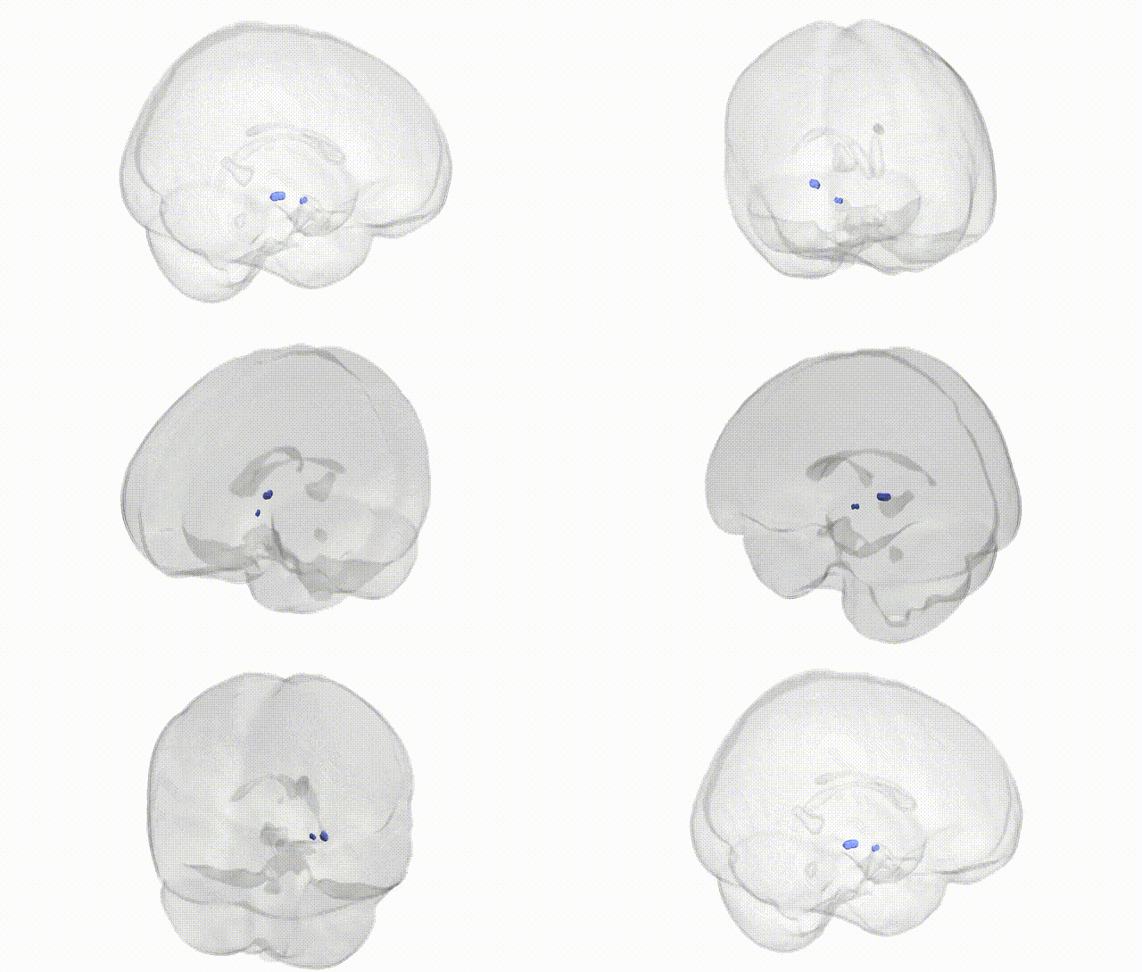
Full Quality Version: Download MP4
Wali Sidiqyar*, Gaurav Rudravaram*, Elyssa M. McMaster, Trent M. Schwartz, Adam M. Saunders, Kurt G. Schilling, Bennett A. Landman "Introducing SPINS: A Shared Public Visualization Library of Neuroanatomical Structures." Medical Imaging with Deep Learning- short paper
This resource is licensed under CC0 1.0 Universal (Public Domain).