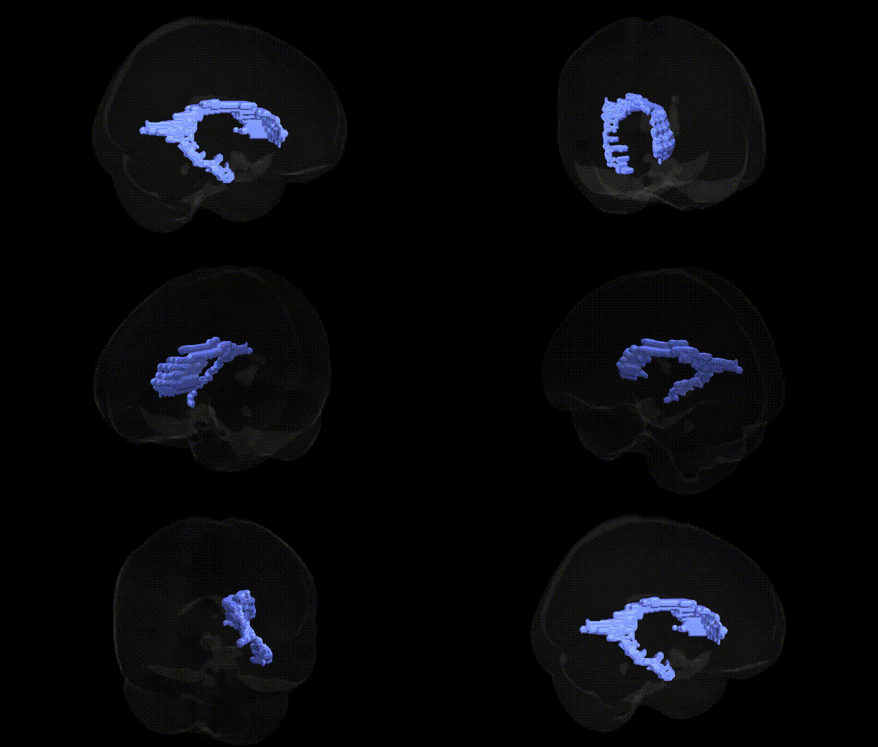
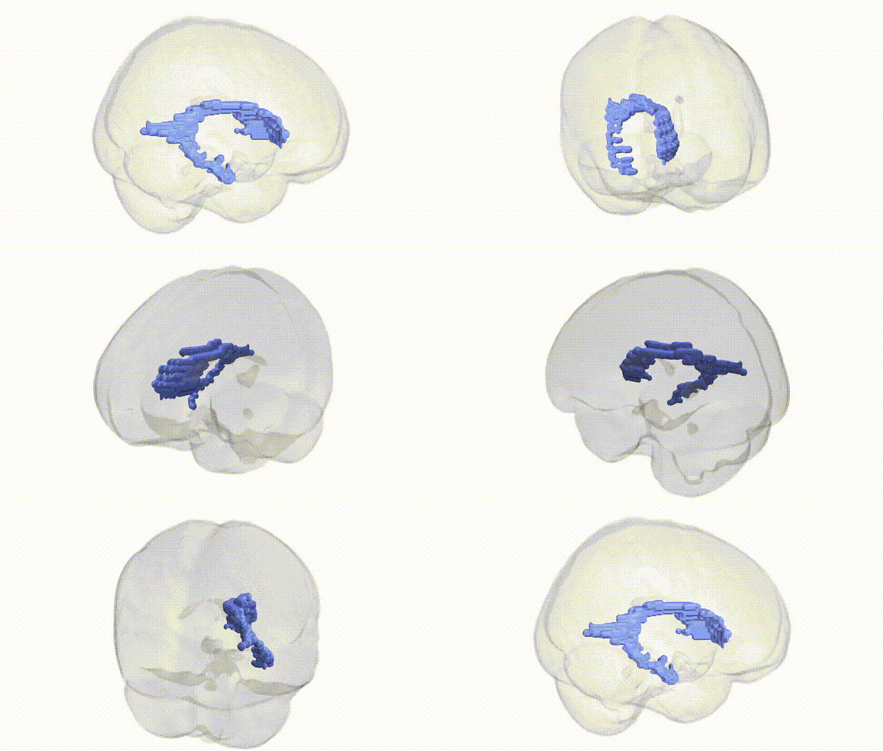
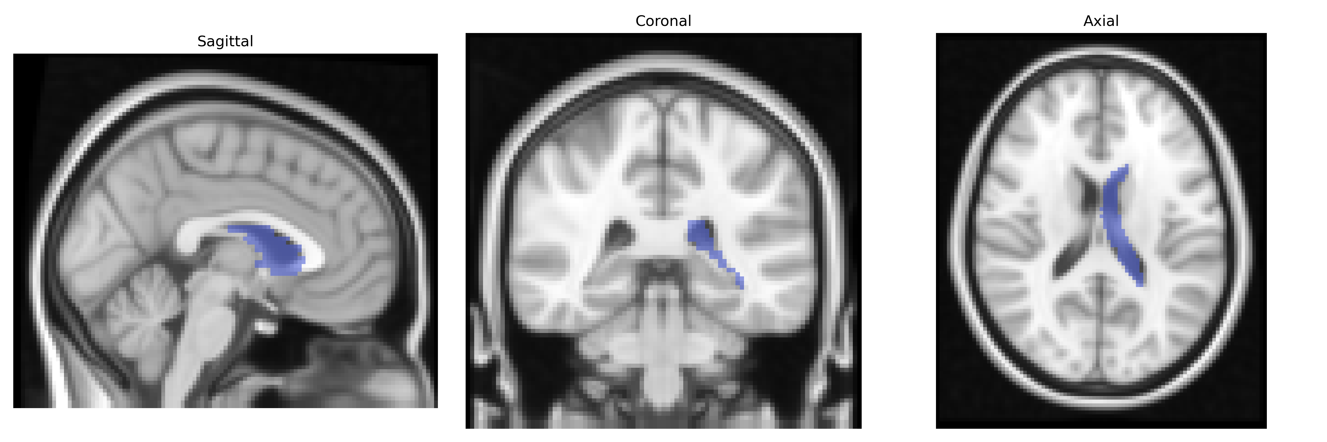
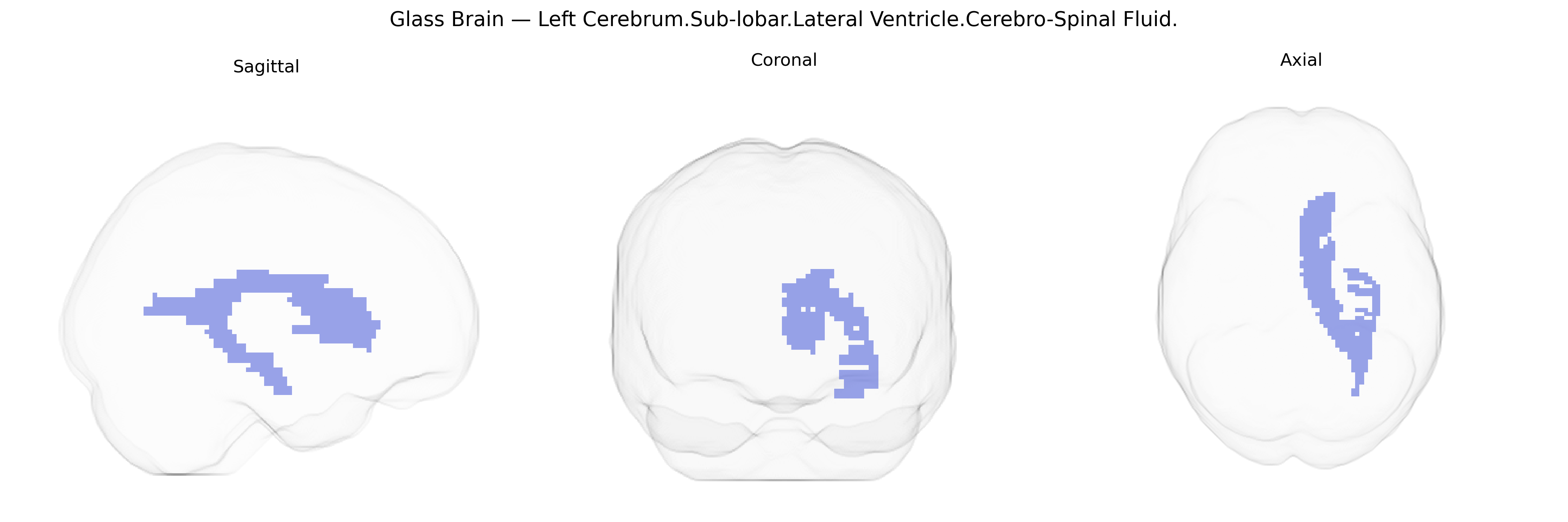
The bilateral Left Cerebrum.Sub-lobar.Lateral Ventricle.Cerebro-Spinal Fluid region refers to the cerebrospinal fluid (CSF) occupying the lateral ventricles within the left cerebral hemisphere, as defined in the Talairach 2 mm Atlas. The lateral ventricles are paired C-shaped cavities located deep within the cerebrum, extending through frontal, parietal, temporal, and occipital lobes. They are lined by ependymal cells and continuous with the third ventricle via the interventricular foramina, forming part of the ventricular system through which CSF circulates to cushion the brain, maintain intracranial pressure, and support metabolic waste clearance. This atlas label thus denotes ventricular CSF rather than neural parenchyma, highlighting a fluid-filled anatomical compartment critical for central nervous system homeostasis. No direct link: related structure: Lateral ventricle.
The bilateral brain region corresponding to the Talairach label “Left Cerebrum.Sub-lobar.Lateral Ventricle.Cerebro-Spinal Fluid” represents cerebrospinal fluid (CSF) volume within the lateral ventricles, and genetic associations identified for this area largely reflect influences on ventricular size and surrounding brain structures rather than the CSF itself. Genome-wide association studies of ventricular volume and related CSF spaces consistently implicate variants affecting neurodevelopment, neurodegeneration, and brain atrophy, including loci in or near genes such as APOE (particularly ε4) in Alzheimer’s disease, which is strongly associated with enlarged ventricles due to medial temporal and global cortical atrophy, and genes like MAPT and GRN in frontotemporal dementia, where ventricular expansion reflects regional tissue loss. Polygenic influences from schizophrenia- and bipolar disorder–associated loci (e.g., in CACNA1C, MIR137-related regions, and other synaptic/neurodevelopmental genes) have also been linked to increased lateral ventricular volume, interpreted as a marker of neurodevelopmental disturbance or gray matter reduction. Large-scale imaging genetics consortia (e.g., ENIGMA, UK Biobank–based studies) report that lateral ventricular volume is highly heritable and associated with multiple common variants spread across the genome, overlapping with genetic architectures for cortical thickness, subcortical volumes (hippocampus, caudate, thalamus), cognitive performance, and general brain aging indices, positioning this CSF region as a genetically influenced macrostructural phenotype that integrates risk for neurodegenerative and major psychiatric disorders as well as normal variation in brain development and aging.
Overview generated by GPT-4o (2026).
Region ID: 173
Hemisphere: bilateral
Atlas: Talairach labels 2mm
Full Quality Version: Download MP4
Full Quality Version: Download MP4
Wali Sidiqyar*, Gaurav Rudravaram*, Elyssa M. McMaster, Trent M. Schwartz, Adam M. Saunders, Kurt G. Schilling, Bennett A. Landman "Introducing SPINS: A Shared Public Visualization Library of Neuroanatomical Structures." Medical Imaging with Deep Learning- short paper
This resource is licensed under CC0 1.0 Universal (Public Domain).