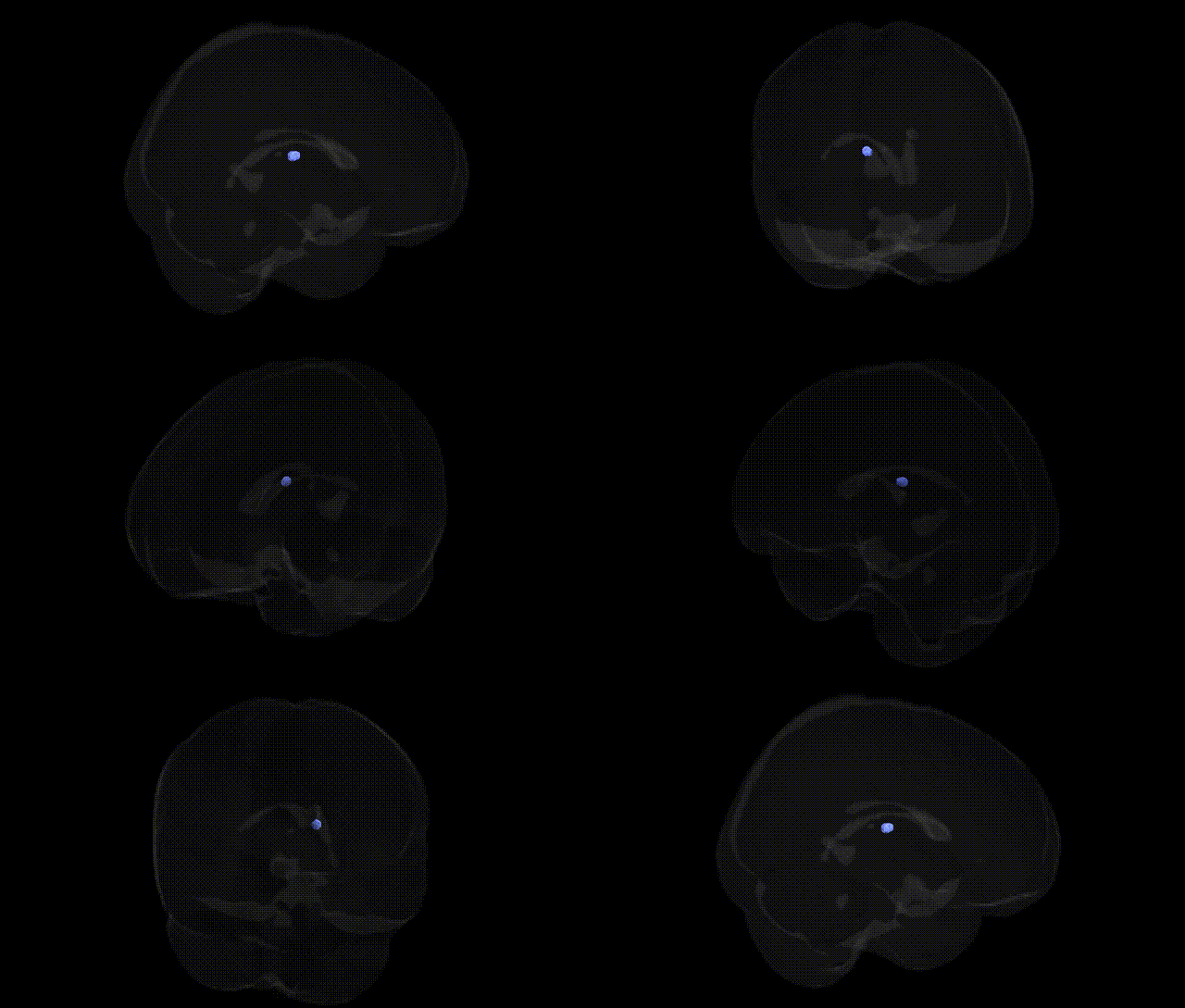
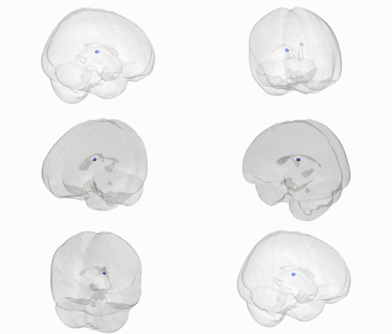
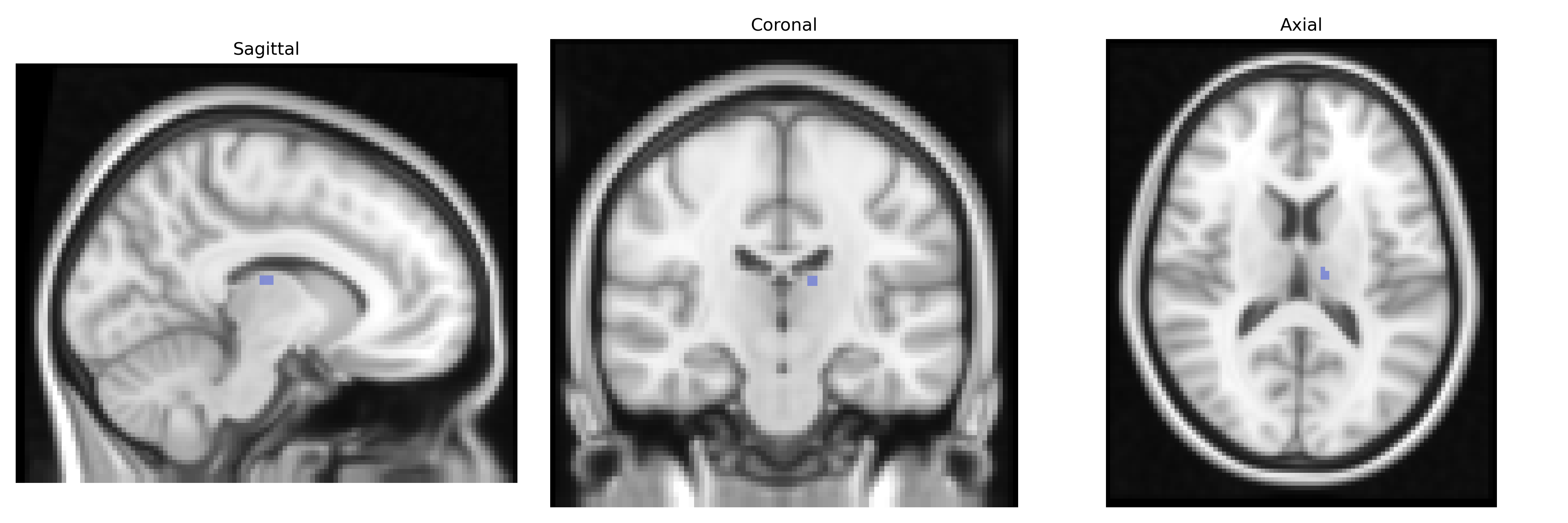
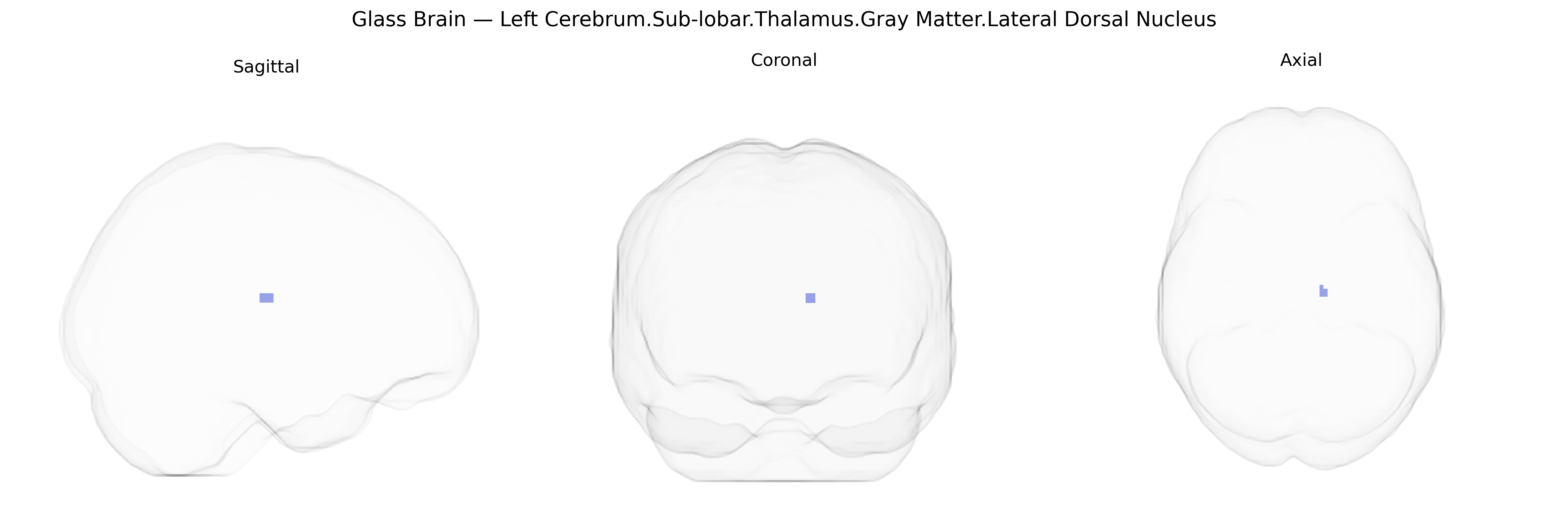
The bilateral Left Cerebrum.Sub-lobar.Thalamus.Gray Matter.Lateral Dorsal Nucleus corresponds to the lateral dorsal (LD) nucleus of the thalamus, a gray matter structure situated within the dorsal thalamus and typically considered part of the lateral nuclear group. The LD nucleus is strongly interconnected with limbic and associative cortical regions, including the cingulate gyrus and parietal association cortex, and is implicated in functions related to spatial cognition, memory, and emotional processing. It contributes to thalamo-cortical circuits that integrate multimodal sensory information with higher-order cognitive and affective states, supporting navigation, contextual memory, and aspects of attention. In primates and humans, although less extensively characterized than major relay nuclei, the LD nucleus is thought to participate in broader limbic networks and may influence behavior through its connections with the posterior cingulate and retrosplenial cortices. There is no direct Wikipedia article for the lateral dorsal thalamic nucleus; a related structure is the Thalamus.
The lateral dorsal nucleus of the thalamus, a sub-lobar gray matter structure within the left cerebrum as defined in the Talairach 2 mm atlas, participates in higher-order cognitive, limbic, and visuospatial networks, and its genetic associations generally emerge from broader imaging genetics and GWAS frameworks targeting thalamic volume, connectivity, or thalamic-cortical networks rather than this nucleus in isolation. Large-scale neuroimaging GWAS, such as those from the ENIGMA consortium and UK Biobank, have identified common variants in genes related to neurodevelopment, synaptic function, and axon guidance (e.g., variants near genes like NPTX2, PTPRD, and others involved in thalamocortical circuitry) that influence thalamic morphology and microstructure; these thalamic-related loci often show pleiotropic associations with psychiatric and neurodevelopmental disorders including schizophrenia, bipolar disorder, major depressive disorder, ADHD, and autism spectrum disorder, as well as cognitive traits such as general intelligence and educational attainment. Studies integrating polygenic risk scores with MRI have linked higher genetic liability for schizophrenia, depression, and neuroticism to altered thalamic volumes or network properties, findings that likely involve the lateral dorsal nucleus given its participation in frontoparietal and limbic circuits. Structural and functional imaging genetics research in Alzheimer’s disease, Parkinson’s disease, and epilepsy has also implicated thalamic nuclei broadly through risk variants in genes such as APOE, MAPT, and ion channel or synaptic genes, with disease-associated alleles correlating with thalamic atrophy or connectivity changes that may include lateral dorsal subdivisions. However, current GWAS catalogs and nucleus-specific segmentation studies provide limited resolution for the lateral dorsal nucleus itself, so genetic associations are best understood as applying to thalamic or thalamocortical systems broadly, with the lateral dorsal nucleus contributing to these genetically influenced network phenotypes rather than serving as a uniquely established GWAS locus on its own.
Overview generated by GPT-4o (2026).
Region ID: 787
Hemisphere: bilateral
Atlas: Talairach labels 2mm
Full Quality Version: Download MP4
Full Quality Version: Download MP4
Wali Sidiqyar*, Gaurav Rudravaram*, Elyssa M. McMaster, Trent M. Schwartz, Adam M. Saunders, Kurt G. Schilling, Bennett A. Landman "Introducing SPINS: A Shared Public Visualization Library of Neuroanatomical Structures." Medical Imaging with Deep Learning- short paper
This resource is licensed under CC0 1.0 Universal (Public Domain).