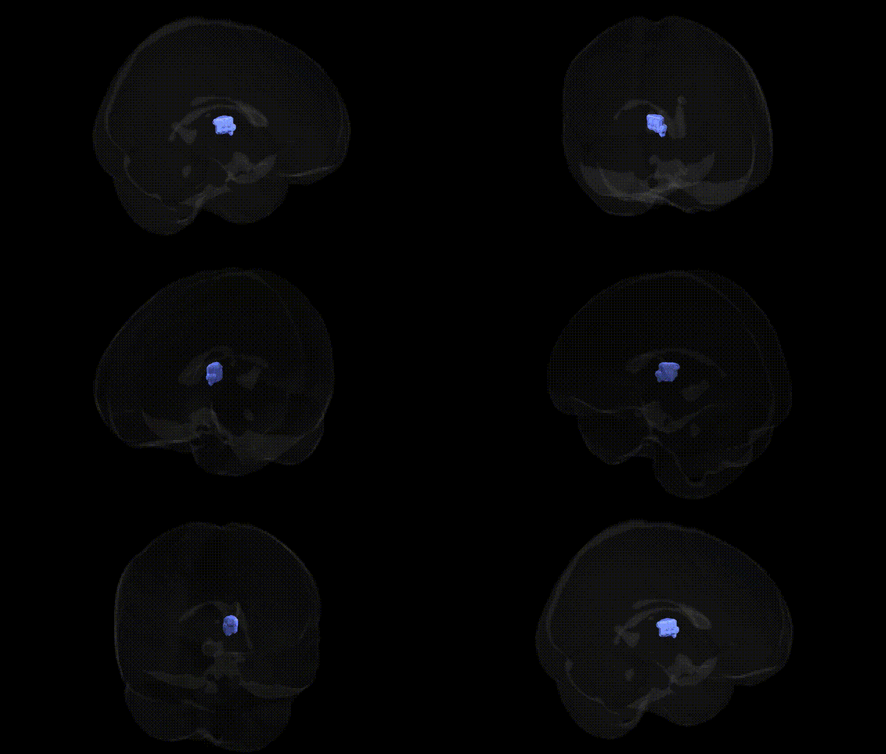
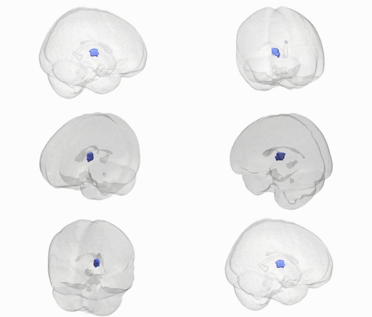
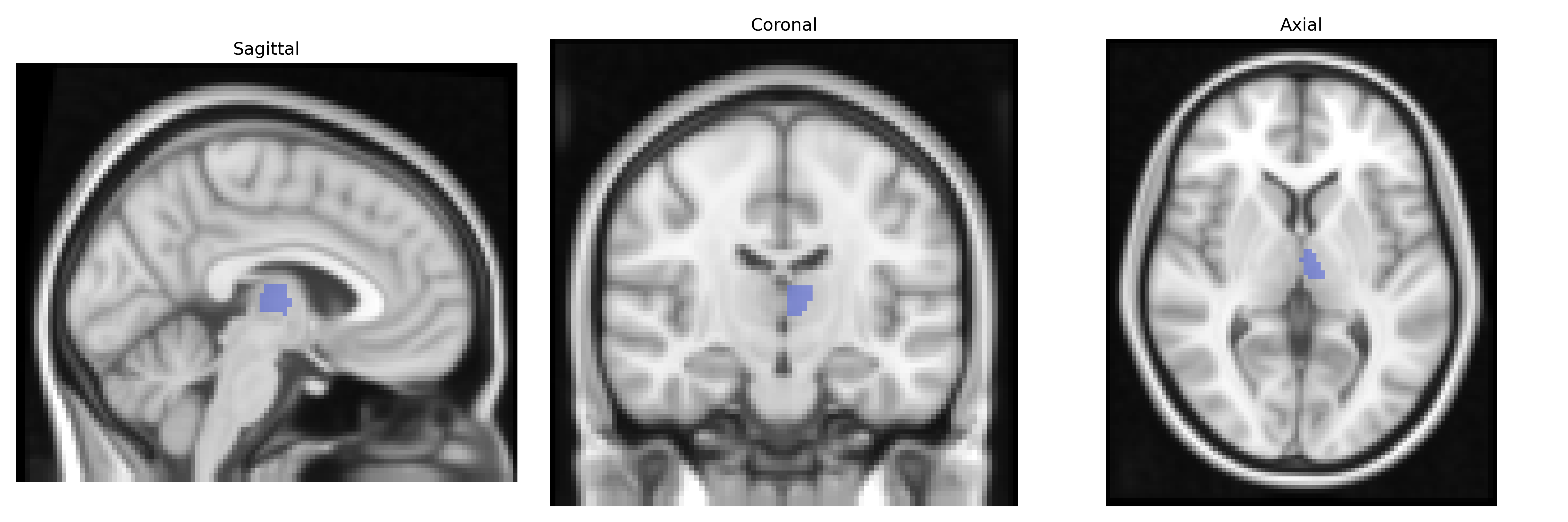
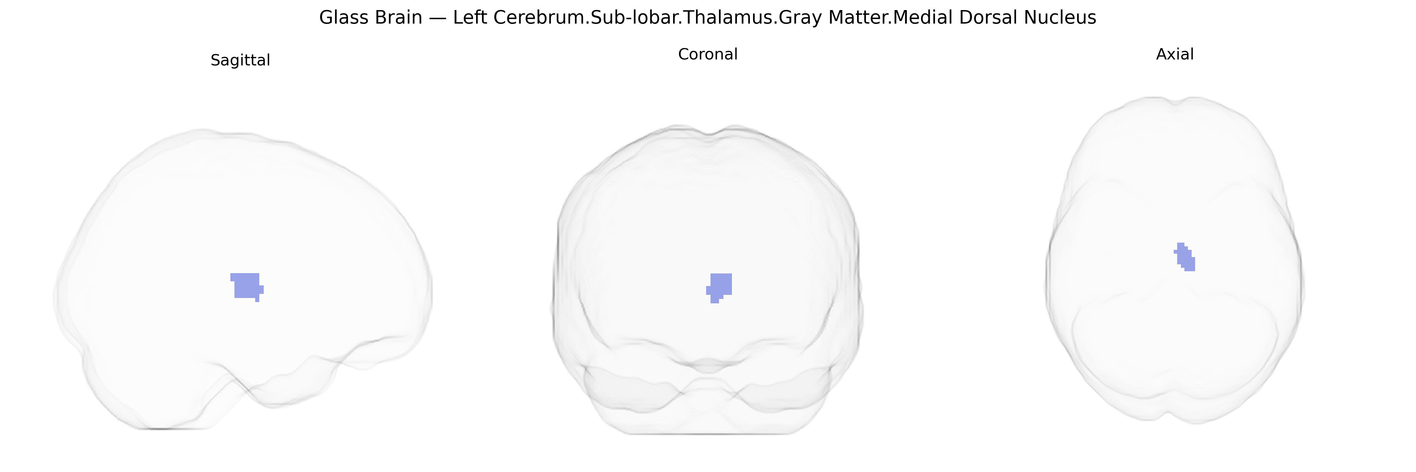
The bilateral Left Cerebrum.Sub-lobar.Thalamus.Gray Matter.Medial Dorsal Nucleus corresponds to the medial dorsal (mediodorsal) nucleus of the thalamus, a higher-order thalamic relay nucleus located within the subcortical gray matter of the diencephalon. This nucleus is heavily interconnected with the prefrontal cortex, limbic structures (including the amygdala), and basal ganglia, forming a critical hub for associative and limbic circuits. Functionally, it participates in executive processes, working memory, attention, emotional regulation, and aspects of decision-making, integrating multimodal information before projecting to widespread cortical territories. Pathophysiologically, the mediodorsal nucleus has been implicated in cognitive and affective disturbances, including those seen in schizophrenia and other neuropsychiatric disorders. Mediodorsal nucleus of thalamus
The medial dorsal (mediodorsal) nucleus of the thalamus, as defined in the Talairach 2 mm atlas, has been implicated in multiple genetic associations primarily through imaging genetics and neuropsychiatric GWAS. Variants in genes affecting glutamatergic and GABAergic signaling (such as GRM3, GRIN2A, GAD1) and synaptic plasticity (including CACNA1C, BDNF, and NRG1) have been linked to structural and functional alterations in this region, especially in schizophrenia, bipolar disorder, and major depressive disorder, where medial dorsal thalamic volume and connectivity with prefrontal cortex are often altered. Large-scale GWAS of subcortical brain volumes (e.g., ENIGMA consortium) have reported associations between common variants near genes involved in neurodevelopment and axon guidance (e.g., DCC, ROBO family, and cell-adhesion genes) and thalamic gray matter volume, which includes the medial dorsal nucleus as part of the thalamus-wide phenotype. ADHD and autism spectrum disorder GWAS and imaging-genetic studies have suggested that polygenic risk scores for these conditions correlate with thalamic volume or thalamocortical connectivity differences encompassing the medial dorsal nucleus, implicating genes involved in synaptic scaffolding (e.g., SHANK3, CNTNAP2) and dopamine signaling (e.g., DRD2, COMT). Moreover, GWAS of cognitive traits, such as general intelligence and working memory, have identified genetic variants associated with thalamic volume and thalamocortical connectivity, supporting a role for neurodevelopmental genes and neurotransmission-related loci in shaping the medial dorsal nucleus, which serves as a critical hub in fronto-thalamic circuits underlying executive function, emotional regulation, and susceptibility to psychiatric disorders.
Overview generated by GPT-4o (2026).
Region ID: 636
Hemisphere: bilateral
Atlas: Talairach labels 2mm
Full Quality Version: Download MP4
Full Quality Version: Download MP4
Wali Sidiqyar*, Gaurav Rudravaram*, Elyssa M. McMaster, Trent M. Schwartz, Adam M. Saunders, Kurt G. Schilling, Bennett A. Landman "Introducing SPINS: A Shared Public Visualization Library of Neuroanatomical Structures." Medical Imaging with Deep Learning- short paper
This resource is licensed under CC0 1.0 Universal (Public Domain).