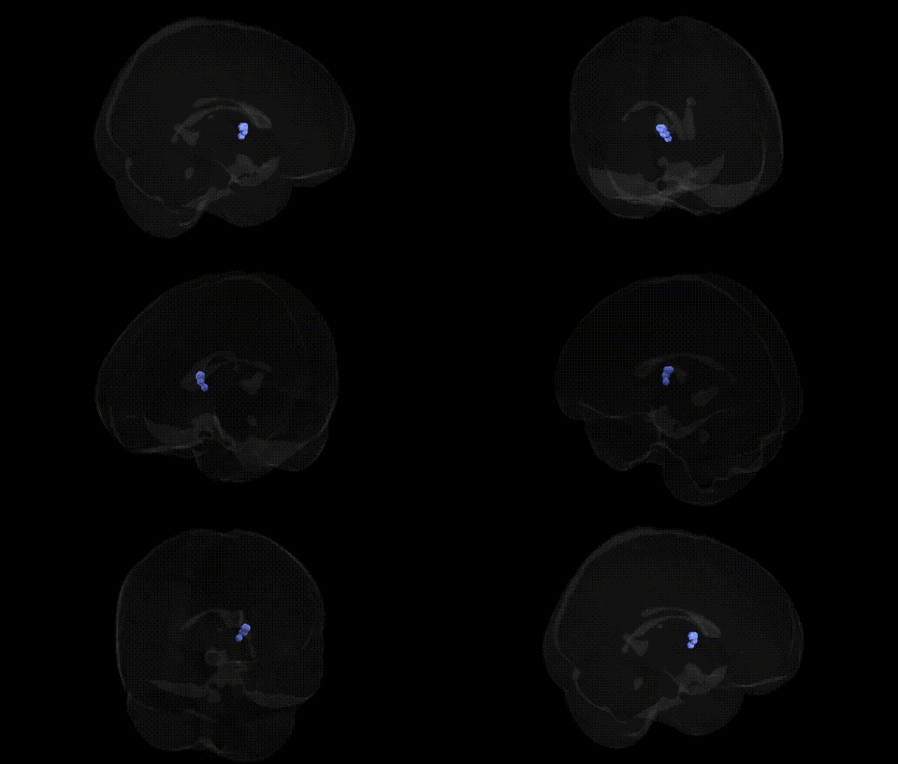
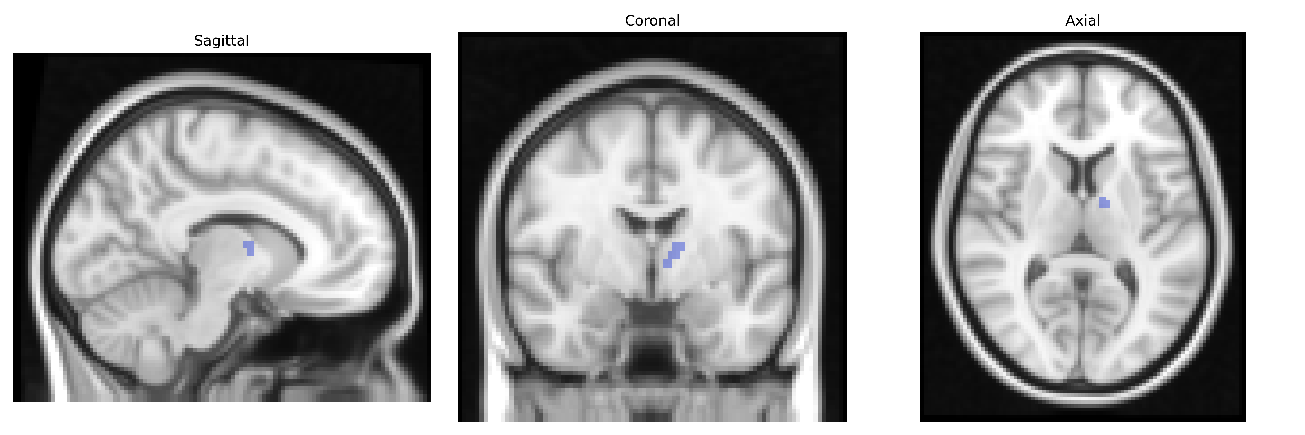
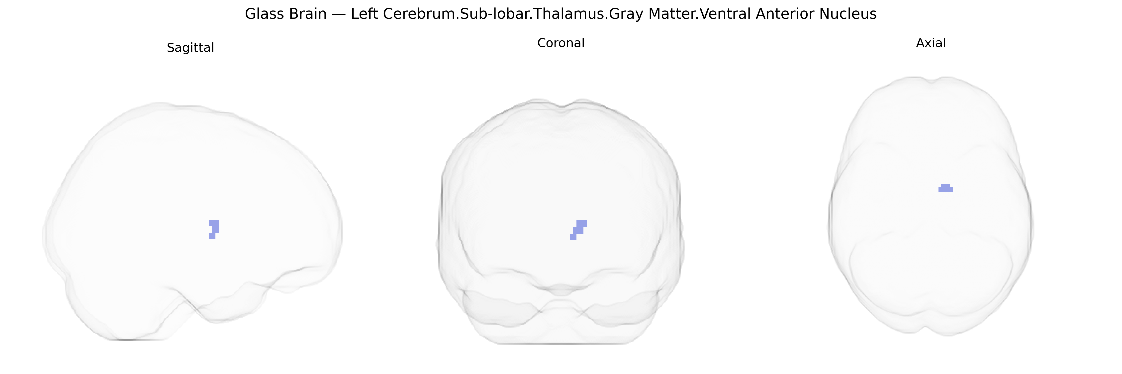
The bilateral Left Cerebrum.Sub-lobar.Thalamus.Gray Matter.Ventral Anterior Nucleus corresponds to the ventral anterior (VA) nucleus of the thalamus, a gray matter structure situated within the sub-lobar region of the dorsal thalamus. This nucleus is a key relay in cortico–basal ganglia–thalamocortical circuits, receiving major input from the globus pallidus and substantia nigra and projecting primarily to the premotor and supplementary motor cortices. Functionally, the VA nucleus is involved in the planning and initiation of voluntary movement, modulation of motor programs, and integration of basal ganglia output into cortical motor networks. It is also implicated in disorders of movement (such as Parkinsonian syndromes) and can contribute to neuropsychological and affective changes through its connections with frontal lobe regions. There is no direct link for the specific “ventral anterior nucleus” article; see the related structure: Thalamus.
The bilateral Left Cerebrum Sub-lobar thalamus gray matter ventral anterior (VA) nucleus, while not often isolated in genetic atlases, participates in cortico-striato-thalamo-cortical loops strongly implicated in neuropsychiatric and motor disorders with known genetic associations. GWAS and imaging-genetics studies of thalamic volume and microstructure consistently implicate variants near genes involved in neurodevelopment and synaptic function (e.g., BDNF, NRG1, GRM3, and broader polygenic architectures for schizophrenia, bipolar disorder, and major depression), with thalamic nuclei—including anterior and medial regions—showing structural and connectivity changes tied to polygenic risk scores for these conditions. The VA nucleus’ role in motor and premotor circuitry links it indirectly to genetic variants associated with movement disorders such as Parkinson’s disease and dystonia (e.g., loci near LRRK2, SNCA, and TOR1A), where thalamic relay dysfunction is a downstream consequence of genetically driven basal ganglia pathology. Thalamic relay regions related to the VA nucleus also show involvement in Tourette syndrome, obsessive–compulsive disorder, and attention-deficit/hyperactivity disorder, with GWAS highlighting genes affecting corticostriatal development and synaptic regulation (such as SLITRK, CNTNAP2, and dopaminergic pathway genes). In addition, large-scale GWAS of brain imaging phenotypes report significant heritability of thalamic volumes and shape metrics, with loci in neurodevelopmental and cell-adhesion pathways (e.g., MEF2C, DCC, and CELSR family genes) contributing to individual differences that are functionally linked to cognitive and motor traits engaging VA thalamic circuits, although explicit, nucleus-specific genetic associations for the VA region remain relatively sparse and are typically inferred from network-level rather than subnucleus-level analyses.
Overview generated by GPT-4o (2026).
Region ID: 646
Hemisphere: bilateral
Atlas: Talairach labels 2mm
Full Quality Version: Download MP4
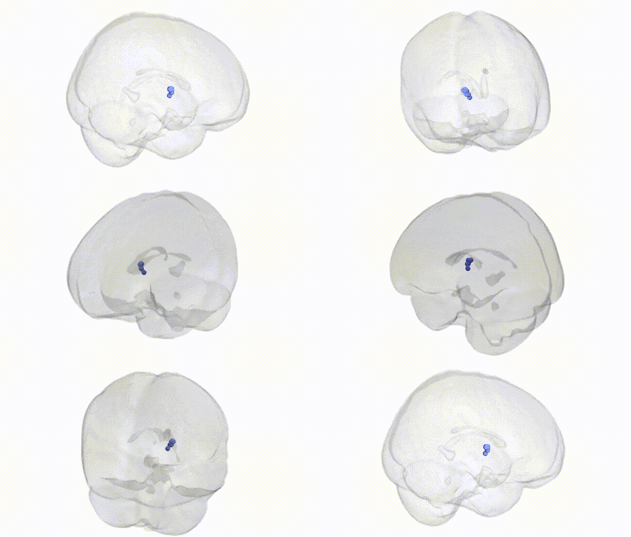
Full Quality Version: Download MP4
Wali Sidiqyar*, Gaurav Rudravaram*, Elyssa M. McMaster, Trent M. Schwartz, Adam M. Saunders, Kurt G. Schilling, Bennett A. Landman "Introducing SPINS: A Shared Public Visualization Library of Neuroanatomical Structures." Medical Imaging with Deep Learning- short paper
This resource is licensed under CC0 1.0 Universal (Public Domain).