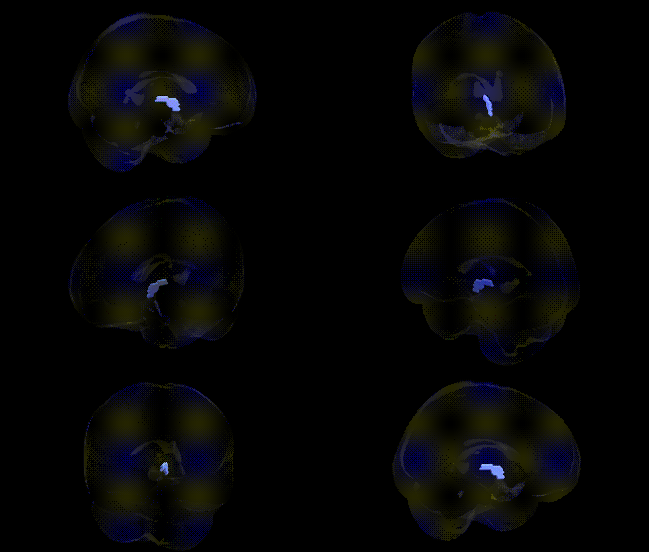
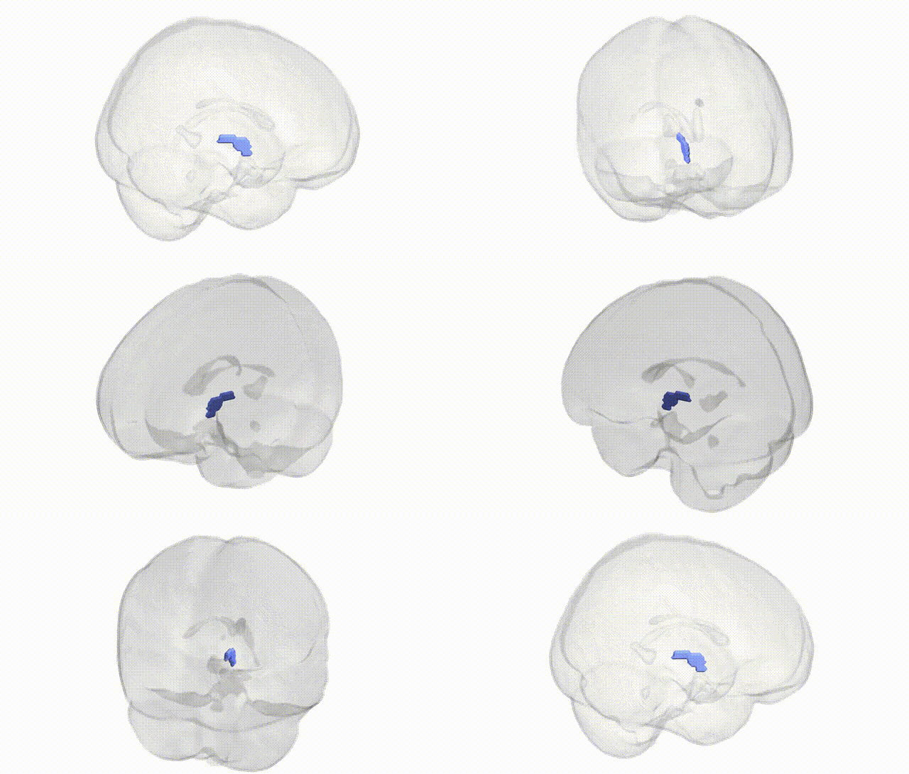
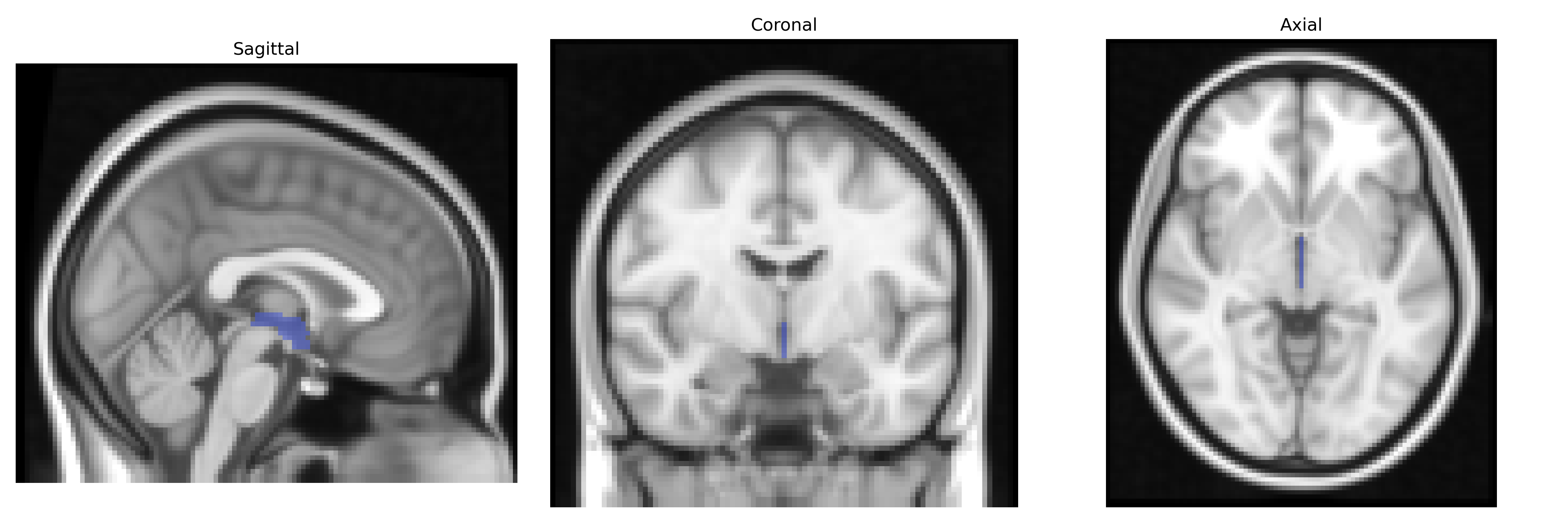
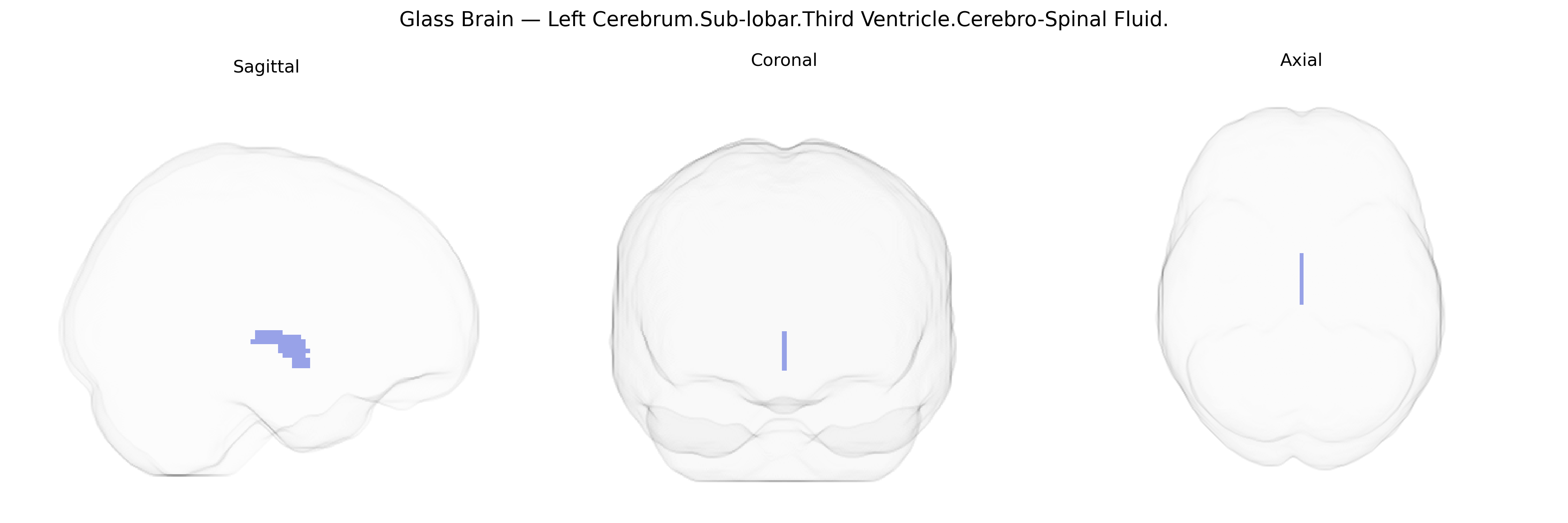
The bilateral Left Cerebrum.Sub-lobar.Third Ventricle.Cerebro-Spinal Fluid region corresponds to cerebrospinal fluid (CSF) contained within the third ventricle of the brain, a narrow midline cavity situated in the diencephalon between the two thalami. The third ventricle is part of the ventricular system that produces, circulates, and stores CSF, which cushions the brain, maintains intracranial pressure, and supports metabolic waste removal. Its walls are formed by key subcortical structures including the thalamus and hypothalamus, and it communicates with the lateral ventricles via the interventricular foramina (foramina of Monro) and with the fourth ventricle via the cerebral aqueduct. The CSF in this space reflects global ventricular morphology and can be altered in conditions such as hydrocephalus, atrophy, or mass lesions affecting the diencephalon. There is no direct link specifically for “Third Ventricle Cerebro-Spinal Fluid,” but the related ventricular cavity is described under Third Ventricle.
The bilateral Left Cerebrum.Sub-lobar.Third Ventricle.Cerebro-Spinal Fluid region in the Talairach 2 mm atlas corresponds primarily to cerebrospinal fluid space rather than neural parenchyma, so genetic associations relate indirectly to ventricular size and CSF volume rather than to the ventricle as a functional “tissue.” Large neuroimaging GWAS studies (e.g., ENIGMA, UK Biobank) have identified common variants influencing lateral and third-ventricle volume, with robust signals in and around genes involved in neurodevelopment, brain growth, and extracellular matrix (such as HMGA2, PTCH1, and loci near IGF1 and FOXO3), though specific third-ventricle–only associations are less frequently isolated from overall ventricular measures. Ventricular enlargement is a well-replicated imaging phenotype in schizophrenia, bipolar disorder, and major depressive disorder, and polygenic risk for these conditions correlates modestly with larger ventricular volumes, implicating shared genetic architecture affecting brain structure and neurodegeneration. In Alzheimer’s disease and other dementias, AD risk alleles (e.g., APOE ε4 and variants in CLU, PICALM, CR1) show associations with accelerated ventricular expansion, reflecting neurodegenerative atrophy rather than primary genetic effects on CSF space. Additional GWAS of intracranial and subcortical volumes have reported loci that influence global brain size and ventricular dimensions, but current evidence indicates that genetic effects relate to broader brain morphometry and pathology rather than to the third ventricle CSF compartment as a discrete, functionally specialized genetic target.
Overview generated by GPT-4o (2026).
Region ID: 362
Hemisphere: bilateral
Atlas: Talairach labels 2mm
Full Quality Version: Download MP4
Full Quality Version: Download MP4
Wali Sidiqyar*, Gaurav Rudravaram*, Elyssa M. McMaster, Trent M. Schwartz, Adam M. Saunders, Kurt G. Schilling, Bennett A. Landman "Introducing SPINS: A Shared Public Visualization Library of Neuroanatomical Structures." Medical Imaging with Deep Learning- short paper
This resource is licensed under CC0 1.0 Universal (Public Domain).