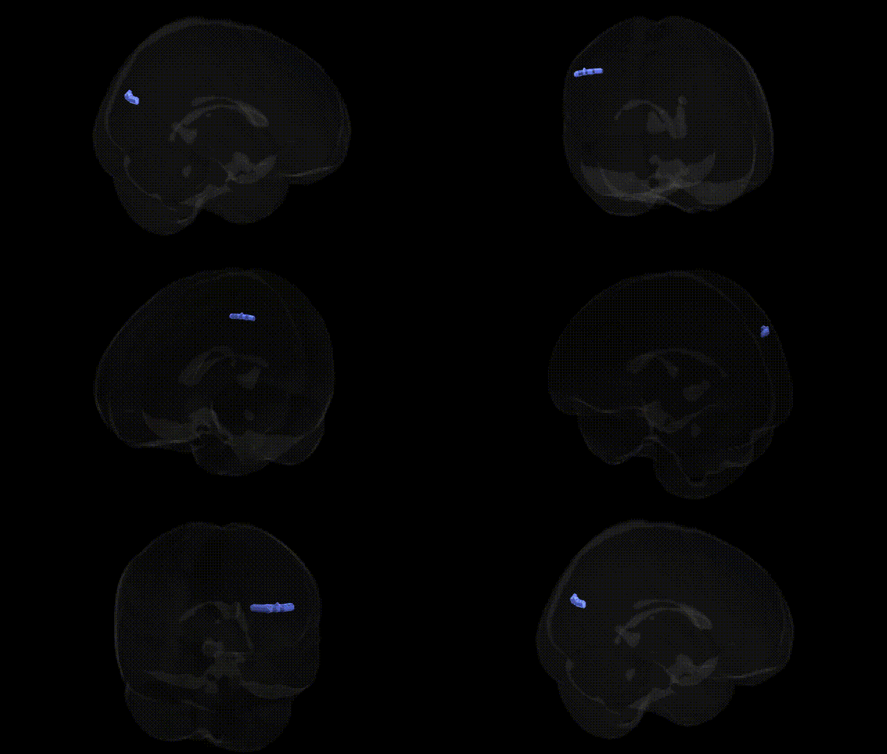
The bilateral Left Cerebrum.Temporal Lobe.Angular Gyrus.Gray Matter.Brodmann area 39 corresponds to cortical gray matter in the angular gyrus region, a multimodal association area located at the junction of the temporal, parietal, and occipital lobes. Brodmann area 39 is implicated in higher-order language processing (including reading, writing, and semantic integration), number processing, spatial cognition, and aspects of theory of mind and episodic memory retrieval. Functionally, this region serves as a critical hub for integrating visual, auditory, and somatosensory information, supporting complex symbolic manipulation and conceptual processing. Lesions in BA39 are classically associated with Gerstmann syndrome and various forms of aphasia, underscoring its importance in the neural substrates of language and cognition. Angular gyrus
The left angular gyrus (Brodmann area 39) of the temporal-parietal junction has been implicated in multiple genetic and GWAS-based neuroimaging and disorder studies, largely via measures such as cortical thickness, surface area, and gray matter volume. Imaging genetics work, including large consortia like ENIGMA and UK Biobank, has linked common variants in genes involved in neurodevelopment, synaptic function, and axonal guidance (e.g., BDNF, CNTNAP2, DCC, and loci near MAPT and KIAA0319) to structural variation in temporoparietal regions encompassing the angular gyrus, often in the context of language, reading, and higher cognitive function. GWAS of educational attainment, general cognitive ability, and reading-related traits (including dyslexia) report polygenic influences that map onto networks for semantic processing and number cognition, where left BA39 is a critical hub; several dyslexia-associated loci (such as those near DCDC2 and KIAA0319) have been linked to altered structure or activation in temporoparietal language areas that include the angular gyrus. Neuroimaging genetics in schizophrenia and bipolar disorder has associated risk variants (e.g., in CACNA1C, ZNF804A, and GRM3) with disrupted connectivity and microstructural changes in default-mode and language networks involving BA39. Additionally, GWAS of Alzheimer’s disease and mild cognitive impairment show that APOE and other AD risk loci are related to atrophy and hypometabolism in parietotemporal regions, including the angular gyrus, which is a key node of the default mode network and is vulnerable in early AD. Overall, genetic influences on this region appear highly polygenic and pleiotropic, contributing to variability in language, semantic processing, numerical cognition, default-mode network function, and vulnerability to neurodevelopmental and neurodegenerative disorders.
Overview generated by GPT-4o (2026).
Region ID: 958
Hemisphere: bilateral
Atlas: Talairach labels 2mm
Full Quality Version: Download MP4
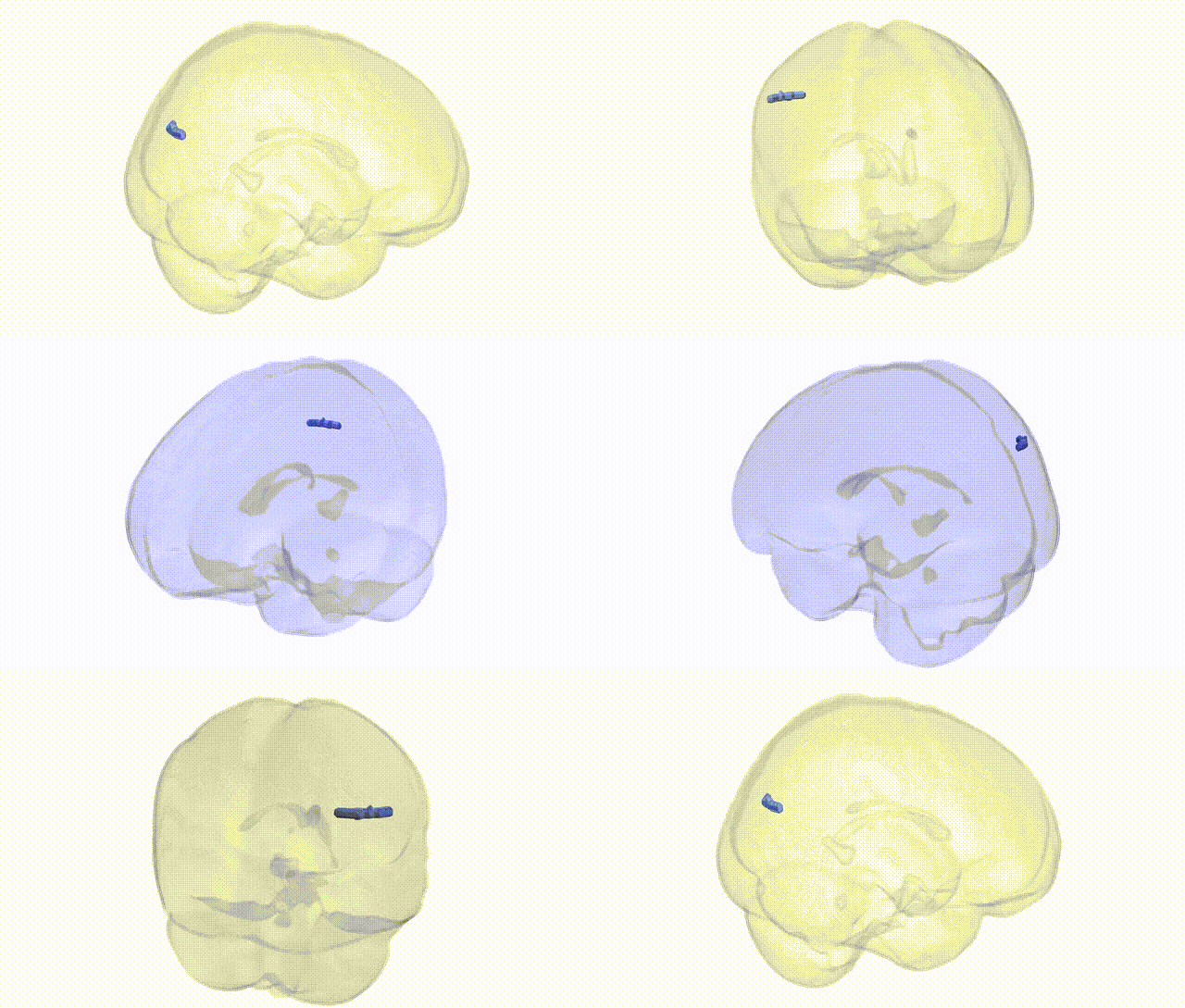
Full Quality Version: Download MP4
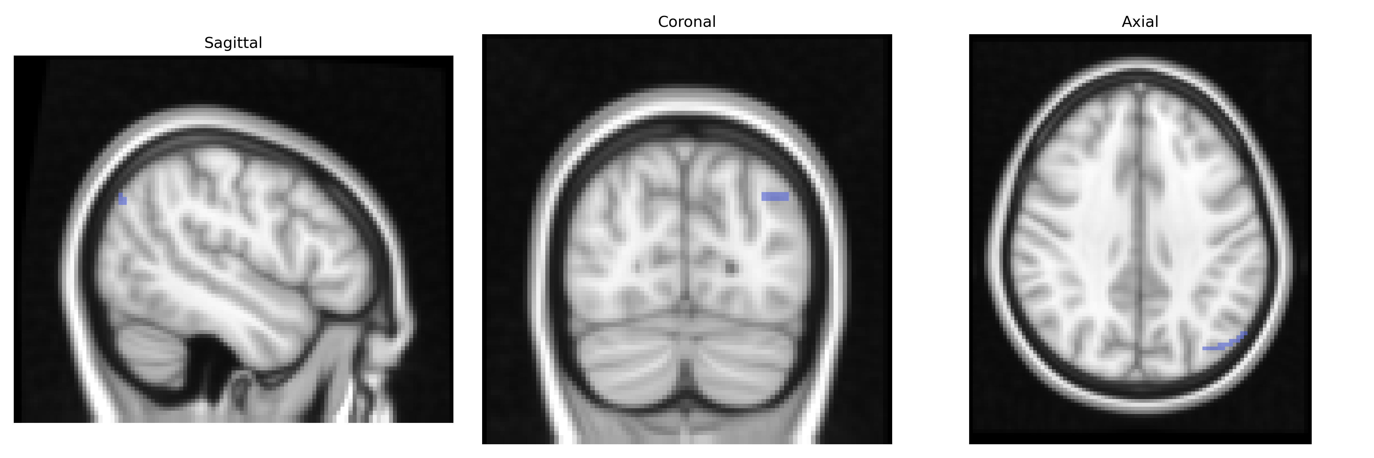
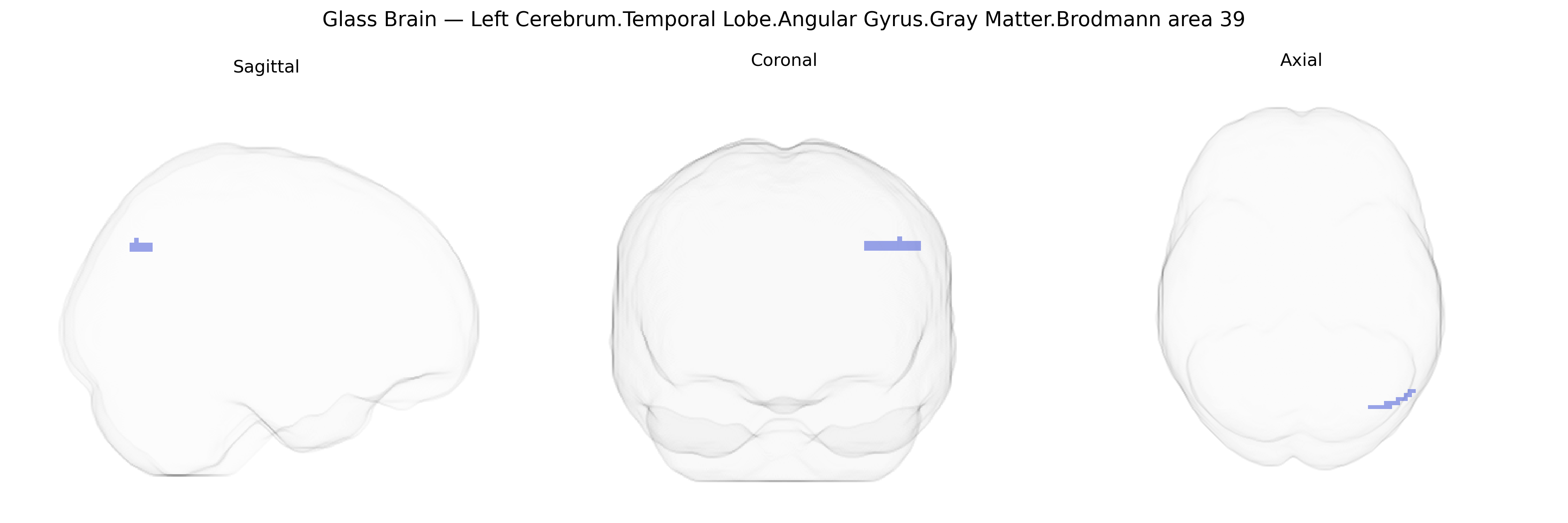
Wali Sidiqyar*, Gaurav Rudravaram*, Elyssa M. McMaster, Trent M. Schwartz, Adam M. Saunders, Kurt G. Schilling, Bennett A. Landman "Introducing SPINS: A Shared Public Visualization Library of Neuroanatomical Structures." Medical Imaging with Deep Learning- short paper
This resource is licensed under CC0 1.0 Universal (Public Domain).