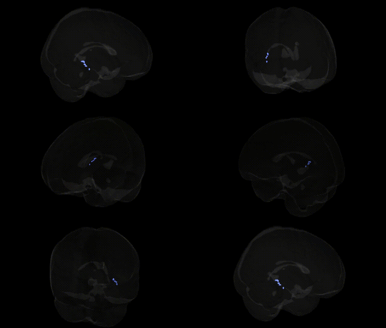
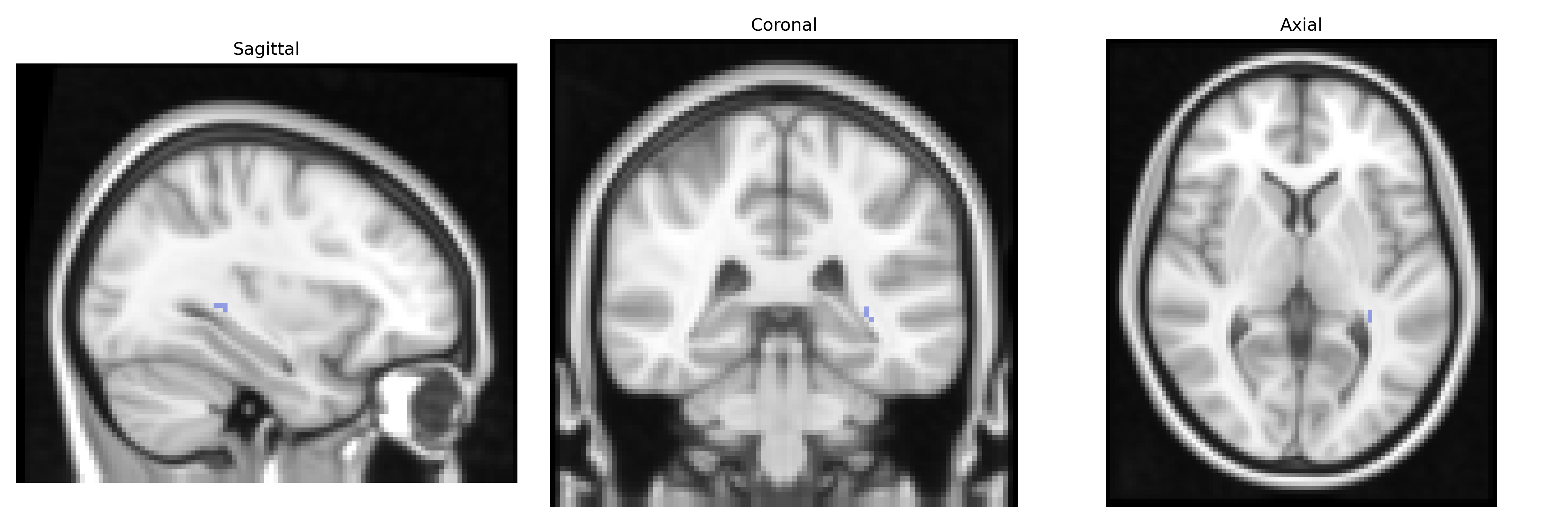
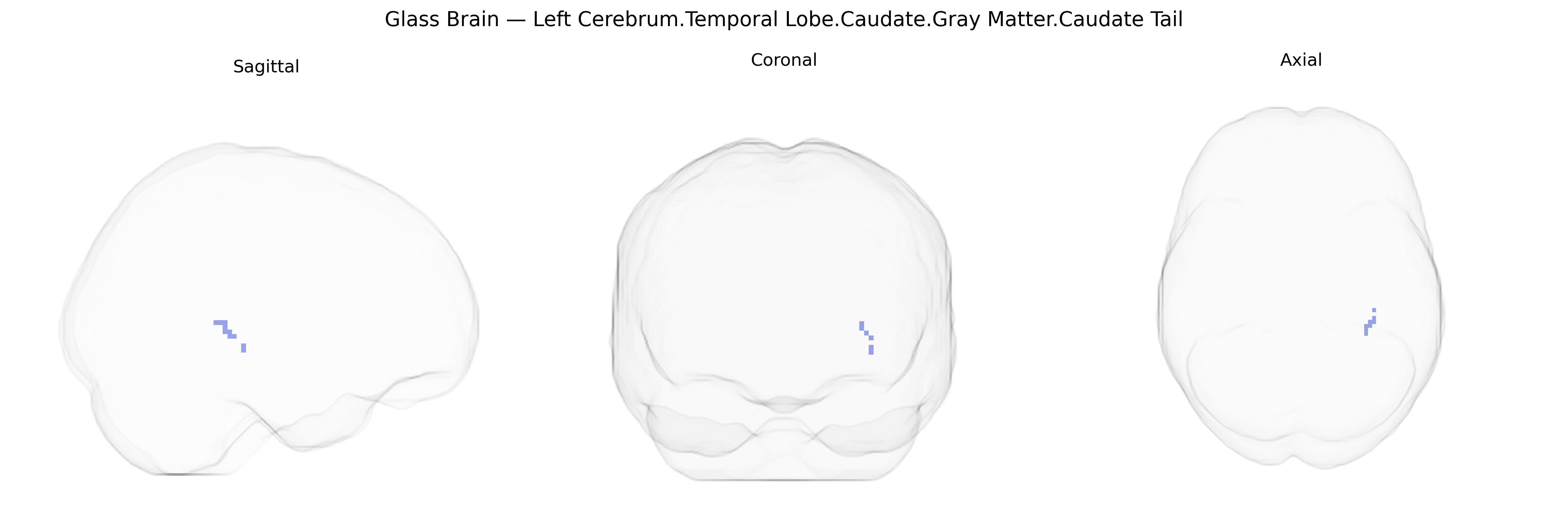
The bilateral Left Cerebrum.Temporal Lobe.Caudate.Gray Matter.Caudate Tail region corresponds to the posterior portion of the caudate nucleus, a C-shaped gray matter structure within the dorsal striatum of the basal ganglia, extending into the temporal lobe along the lateral ventricle. The caudate tail participates in cortico-striato-thalamo-cortical loops that integrate associative and limbic information, contributing to higher-order aspects of motor control, procedural learning, reward-based decision making, and certain components of executive and emotional processing. Although specifically focused on the tail segment, this region functions as part of the broader caudate circuitry involved in modulating behavior and cognition through dopaminergic and glutamatergic input. There is no direct link for the caudate tail; see the related structure Caudate nucleus.
The caudate tail within the temporal lobe—part of the striatal gray matter system—has been implicated indirectly in genetic studies that examine caudate volume and striatal circuitry more broadly, rather than this subregion alone. GWAS of caudate nucleus volume have identified common variants in and near genes such as HMGA2, DCC, and others affecting neurodevelopmental pathways, which influence striatal morphology including posterior (tail) portions. Striatal and temporal lobe networks incorporating the caudate tail are genetically linked to neuropsychiatric disorders such as schizophrenia, obsessive–compulsive disorder, Tourette syndrome, ADHD, and autism spectrum disorder, often via genes involved in synaptic signaling (e.g., DRD2, GRIN2A, CACNA1C), dopamine and glutamate transmission, and neuronal migration. Polygenic risk scores for schizophrenia, major depression, and bipolar disorder have been associated with altered caudate and temporal lobe structure and connectivity on MRI, suggesting that shared genetic architectures modulate this region’s morphology and function. In Parkinson’s disease and Huntington’s disease—both strongly genetic—striatal degeneration patterns encompassing caudate subregions, including the tail, have been linked to mutations in genes such as SNCA, LRRK2, and HTT, respectively. While no large GWAS explicitly targets “caudate tail” volume in the Talairach 2 mm atlas, convergent evidence from striatal imaging genetics, neurodevelopmental loci, and disorder-specific studies indicates that common and rare variants affecting corticostriatal circuitry and dopamine-related pathways are major genetic contributors to variability and vulnerability in this region.
Overview generated by GPT-4o (2026).
Region ID: 351
Hemisphere: bilateral
Atlas: Talairach labels 2mm
Full Quality Version: Download MP4
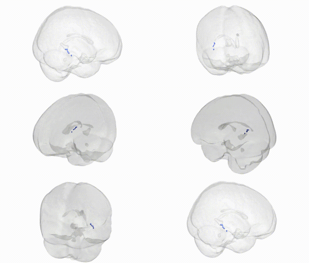
Full Quality Version: Download MP4
Wali Sidiqyar*, Gaurav Rudravaram*, Elyssa M. McMaster, Trent M. Schwartz, Adam M. Saunders, Kurt G. Schilling, Bennett A. Landman "Introducing SPINS: A Shared Public Visualization Library of Neuroanatomical Structures." Medical Imaging with Deep Learning- short paper
This resource is licensed under CC0 1.0 Universal (Public Domain).