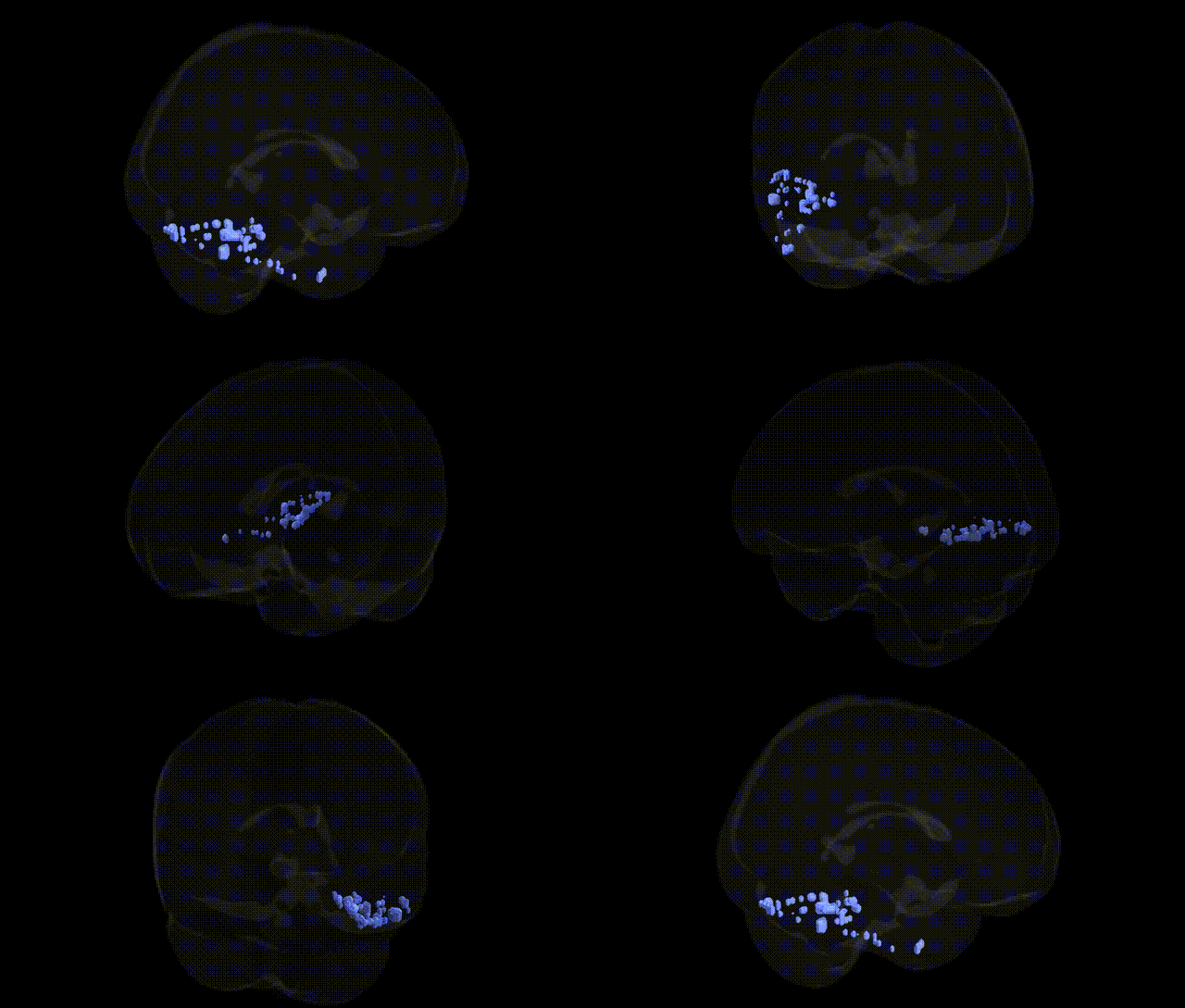
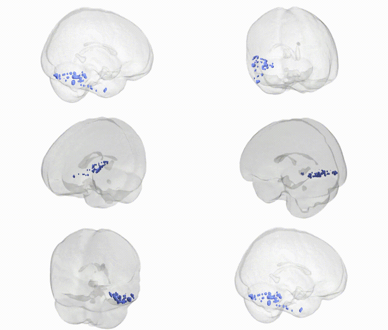
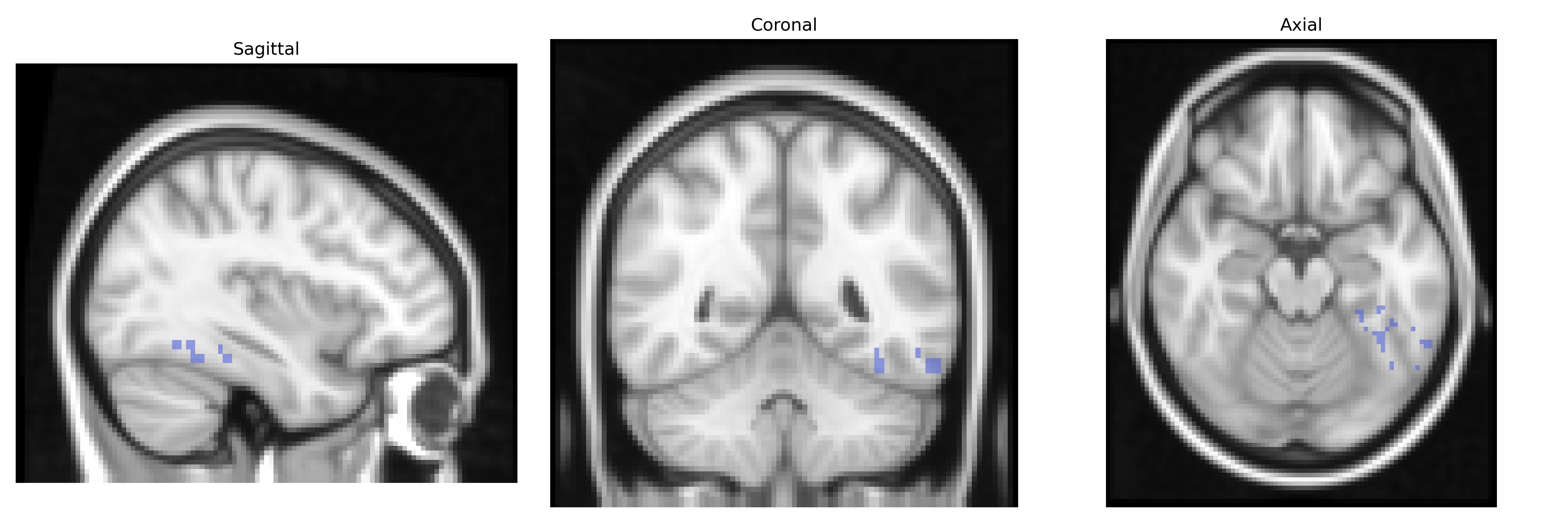
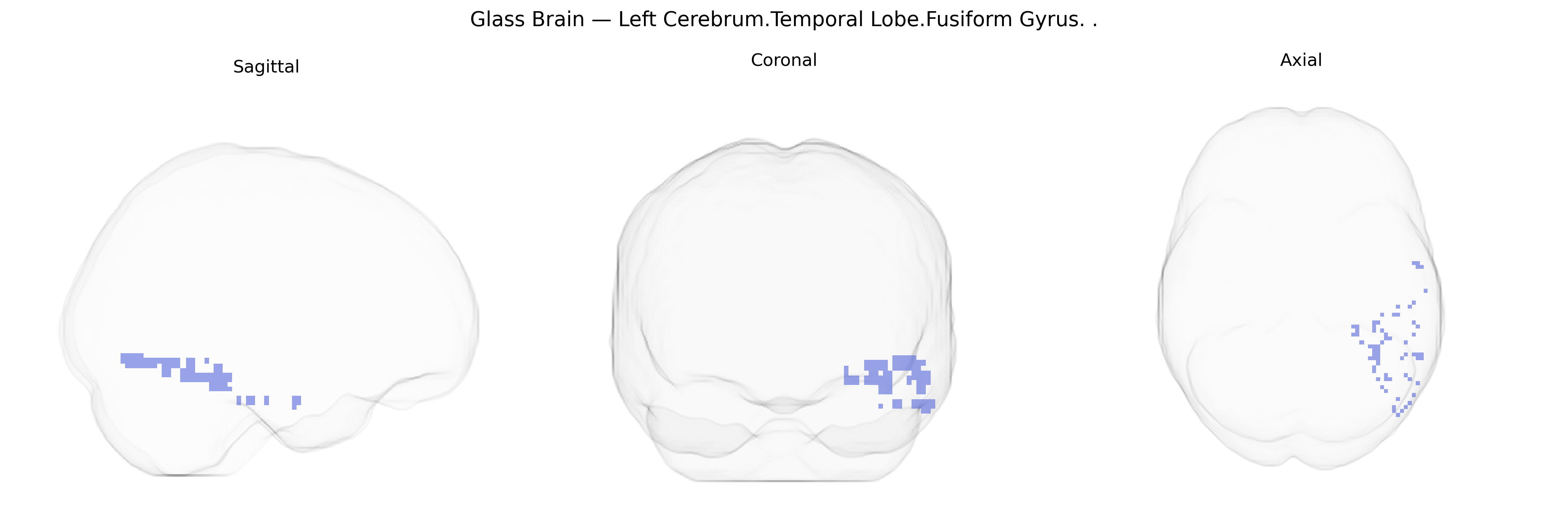
The bilateral Left Cerebrum.Temporal Lobe.Fusiform Gyrus, as defined in the Talairach 2 mm Atlas, refers to the fusiform gyrus located in the temporal lobe of the left cerebral hemisphere, with homologous territory in the right hemisphere. This elongated cortical region lies on the basal surface of the temporal and occipital lobes, bounded medially by the parahippocampal gyrus and laterally by the inferior temporal gyrus. It is a heteromodal association area critically involved in high-level visual processing, including object and face recognition, word and character form processing, and aspects of visual categorization. Functionally, it hosts specialized subregions such as the fusiform face area and visual word form area, and is richly interconnected with occipital visual cortices, temporal associative regions, and limbic structures, supporting integration of visual input with memory and semantic knowledge. Lesions to this region can result in deficits such as prosopagnosia and reading impairments, highlighting its role in complex perceptual and cognitive functions.
The bilateral left temporal fusiform gyrus, as defined in the Talairach 2 mm atlas, has been implicated in multiple genetically informed neuroimaging and GWAS studies, largely through its roles in visual word form processing, face and object recognition, and higher-order visual integration. Variants in KIAA0319, DCDC2, and other dyslexia-associated genes have been linked to structural and functional differences in left fusiform and adjacent occipitotemporal cortex, particularly in the “visual word form area,” while GWAS of cortical surface area and thickness (e.g., ENIGMA consortium) have identified polygenic influences across widespread loci (including regulatory variants near neurodevelopmental genes such as MEF2C and HMGA2) that contribute to interindividual variation in temporal fusiform morphology. In psychiatric genetics, large-scale imaging-genetics studies in schizophrenia and bipolar disorder have reported fusiform gray matter and activation differences associated with common risk alleles in CACNA1C, ZNF804A, and complement pathway genes (e.g., C4), though these effects are typically indirect and distributed across networks involved in social cognition and face processing. Autism spectrum disorder GWAS and rare variant studies implicate synaptic and chromatin-remodeling genes (e.g., NRXN1, SHANK3, CHD8), and imaging-genetic work has associated this polygenic risk with atypical fusiform response to faces and social stimuli, consistent with fusiform involvement in social perception. Additional links include reading and language-related traits, prosopagnosia, and broader cognitive measures for which polygenic scores correlate with fusiform structure or activation, underscoring that genetic influences on this region are highly polygenic, pleiotropic, and mediated through large-scale neurodevelopmental and cognitive networks rather than region-specific “fusiform genes.”
Overview generated by GPT-4o (2026).
Region ID: 121
Hemisphere: bilateral
Atlas: Talairach labels 2mm
Full Quality Version: Download MP4
Full Quality Version: Download MP4
Wali Sidiqyar*, Gaurav Rudravaram*, Elyssa M. McMaster, Trent M. Schwartz, Adam M. Saunders, Kurt G. Schilling, Bennett A. Landman "Introducing SPINS: A Shared Public Visualization Library of Neuroanatomical Structures." Medical Imaging with Deep Learning- short paper
This resource is licensed under CC0 1.0 Universal (Public Domain).